Perhaps the world would seem more attractive to us if we were able to see what remains hidden from us. Man is the most interesting and complex organism on the planet. It is capable of performing several functions simultaneously. Each organ within us has its own responsibilities and works harmoniously with each other. For example: the heart pumps blood , the brain develops a process that allows you to think. In order to understand our body well, we need to know what the location of the abdominal organs is.
…
What are internal organs
In order to imagine the structure of the human body, it is necessary to understand anatomy, the structure of tissues and cells, to know how the organs are located and what functions they perform.
Cellular and tissue structure are the structural units that make up more complex biological structures called organs.
Human organs have the following criteria:
- consist of various cells and tissues,
- are distinguished by their isolation,
- have a stable position within the body,
- develop during ontogenesis.
Organs grow along with the rest of the body tissues, but their size increases at different rates.
This is interesting! What does the structure and diagram of human skin look like?
The difference in growth rate is noticeable in the development of adolescents, when bone structures and body weight noticeably outpace the development of the heart and blood vessels, which is why young people and girls are often diagnosed with vegetative-vascular dystonia.
However, after some time, the speed of development levels out, and the child’s well-being returns to normal.
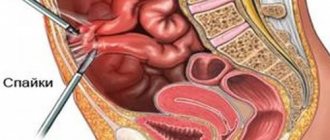
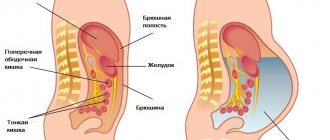
Description of the abdominal cavity
The human abdominal cavity is a container of organs and anatomical formations: stomach, gall bladder, spleen, intestines (jejunal, ileal, transverse colon, cecum and sigmoid), abdominal aorta. The location of these organs is intraperitoneal, that is, they are covered by the peritoneum, or more precisely, by its visceral layer, completely or partially.
Extraperitoneally (that is, in the retroperitoneal space) there are organs of the abdominal cavity: kidneys, adrenal glands, pancreas, ureters, the main part of the duodenum.
The partially visceral layer of the peritoneal lining flows around two spaces of the colon (ascending and descending), that is, these abdominal organs are located mesoperitoneally.
Among the organs that can be classified as intra- and mesoperitoneal, the liver can be distinguished. It is almost completely covered with a serous membrane.
Internal organ systems
The structural units of the human body are combined into the following systems:
- Digestive – provides mechanical and enzymatic processing of food, promotes the penetration of necessary substances into the blood through absorption and eliminates excess and undigested particles.
- Circulatory – responsible for transporting nutrients, removing toxins and exchanging gases.
- Respiratory – ensures the intake of oxygen into the body to obtain energy and the removal of respiratory metabolic products (carbon dioxide).
- Nervous – responsible for the regulation of most processes, sensitivity and motor activity.
- Sexual or reproductive – ensures the process of internal fertilization and gestation (in women).
- Excretory – ensures the removal of excess fluids, salts and metabolic products.
- Endocrine – provides hormonal regulation of vital processes.
Human anatomy is colorfully shown in specialized atlases in pictures. Anatomical atlases allow you to better examine and understand the human structure.
This is interesting! Basics of anatomy: human skeleton with the names of all bones
Digestive system
Digestion is an active process necessary to renew the necessary substances in the body and obtain energy. To maintain vitality and normal well-being, a person’s diet must include fats, proteins and carbohydrates.
Worth remembering! By refusing animal proteins, people limit the intake of essential amino acids, the synthesis of which does not occur in the human body and which are not supplied with plant proteins.
| Name | Location, department | Functional meaning |
| Teeth | Oral cavity | Intake of food, primary grinding and processing by the secretion of the salivary glands - saliva |
| Language | ||
| Salivary glands | ||
| Pharynx | Pharynx (throat area, fragment of breathing tube) | Preventing food from entering the respiratory tract by blocking the passage with the epiglottis, transporting food |
| Esophagus | Esophagus | Transporting a bolus of food into the stomach due to the mechanical work of the motor function |
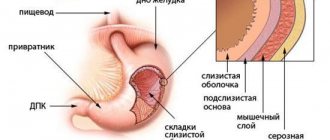
| Stomach | Stomach (under the ribs on the left side and under the xiphoid process) | Serves as a reservoir for food, performs chemical processing and suction functions |
| Small intestine | Small intestine (abdomen) | Enzymatic food processing |
| Colon | Large intestine (abdomen, pelvic cavity) | Absorption of fluid and formation of feces |
In addition to the digestive tract, auxiliary formations responsible for the secretion of enzymes are involved in the process of food processing:
- minor and major salivary glands (secretion of saliva),
- liver (bile),
- pancreas (digestive enzymes),
- gallbladder.
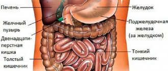
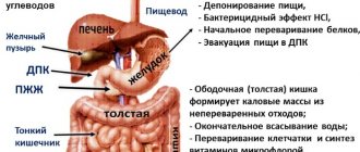
Layout of human internal organs (digestion):
The total length of the human digestive tract is within 10 m.
Anatomy of the cardiovascular system
The heart and blood vessels are responsible not only for blood circulation, but also for the supply of nutrients to all cells and tissues, the collection of waste products and the exchange of gases.
The main reason for the need for oxygen for human life is its ability to oxidize and release energy, which is why the supply of gases with the blood is so necessary for the functioning of the body’s biomechanism.
| Name | Where is | Functional meaning |
| Heart | Rib cage | Pumping blood |
| Systemic circulation | From the left ventricle of the heart to the right atrium | Nutrition of all cells and tissues |
| Pulmonary circulation | From the right ventricle to the left atrium (circulation of the lungs) | Gas exchange in lung tissues |
Systemic and pulmonary circulation:
The blood takes the products of cell metabolism and transports them to the structures of the kidneys and liver, where filtration and disinfection of toxins are carried out, as well as the return of purified blood to a new circulation.
This is interesting! Anatomy lessons: how many muscles are in the human body
Location of respiratory system structures
Respiration is a process necessary to oxidize nutrients in cells and produce energy. This fact explains the critical condition and imminent death of air-breathing living organisms when the supply of oxygen is stopped.
| Name | Location, department | Functional meaning |
| Nasal cavity | Upper respiratory tract | Warming air flows, preventing the penetration of large foreign particles and dust |
| Nasopharynx | Air transport | |
| Oropharynx | ||
| Larynx | Lower respiratory tract | |
| Trachea | Air transport, formation of mucous secretion | |
| Bronchi (bronchial tree) | Humidification and air transport | |
| Lungs | Gas exchange |
Location and internal structure of the human respiratory organs:
Suffocation that occurs due to compression of the upper airway or other obstruction of oxygen supply is called “asphyxia.” This is a dangerous condition for human life and health.
Important! In case of respiratory arrest, the victim must be provided with immediate pre-medical assistance, which consists of performing chest compressions and artificial respiration.
Location of the central nervous system and peripheral nerves
The nervous system is responsible for most of the regulatory processes of the body, ensures communication with the outside world by analyzing information received from receptors, movement and the ability to respond.
| Central nervous system (CNS) | Brain | Front | Finite | Olfactory, basal ganglia, cerebral cortex, lateral ventricles | |
| Intermediate | Epithalamus, thalamus, hypothalamus, third ventricle, metathalamus | ||||
| Brain stem | Average | Quadrigeminalis, cerebral peduncles, aqueduct of Sylvius | |||
| Diamond-shaped | Rear | pons, cerebellum | |||
| Oblong | |||||
| Spinal cord | |||||
| Peripheral nervous system | |||||
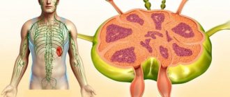
Layout of the human central nervous system and peripheral nerves:
The peripheral nervous system consists of nerves extending from the nerve trunk, distributed throughout the body to collect information and regulate processes.
Reproductive system
Unlike other functional structures, the reproductive system of men and women differs significantly.
Location of genital elements in men:
| Name | Where is | Functional meaning |
| Testicles | In the abdominal cavity, then descends into the scrotum | Sperm formation |
| Vas deferens | Abdominal cavity, urethra | Release of sperm during ejaculation |
The descent of the testicles is explained by the fact that the formation of sperm requires a temperature several degrees lower than the natural temperature inside the human body.
The location of the human genital organs in pictures (for men):
Also, the testicles and epididymis are responsible for the formation of the “male” sex hormone testosterone, which affects human behavior, growth and development.
Location of the internal organs of the reproductive system in women:
| Name | Where is | Functional meaning |
| Ovaries | Lower abdomen | Hormone synthesis, egg formation |
| Fallopian tubes | Transport of eggs | |
| Uterus | Abdomen behind the bladder | Gestation |
| Vagina | Small pelvis | Promotes the act of fertilization, participates in the process of excretion of fluids during menstruation |
Layout of the genital structures in women:
Urinary excretion in women, unlike representatives of the stronger sex, occurs through a separate opening of the urethra.
Location and structure of the urinary system
Urination is responsible for eliminating from the body excess fluids, salts and metabolic products that have been filtered in the kidneys.
| Name | Where is | Functional meaning |
| Kidneys | Retroperitoneal space near the spine | Filtration of blood, formation of urine |
| Bladder | Retroperitoneal in the pelvis | Urine accumulation (0.5–0.7 L) |
| Urethra | Urine excretion | |
Diagram of the human urinary organs in pictures:
On average, within 24 hours, up to 1500 ml of urine is formed in the human body, consisting of toxins, salts and excess fluid.
This is interesting! Biology lesson: how many pairs of chromosomes does a normal person have?
Organs of the endocrine system
Endocrine regulation occurs due to the production of a group of hormones by specialized structures.
The functions of hormonal regulation are:
- pancreas,
- adrenal cortex and medulla,
- pineal gland
- pituitary,
- epithelial body,
- thyroid,
- testicles,
- ovaries.
Diagram - photo with inscriptions:
Hormones are biologically active substances secreted by the body. The balance of hormones is involved in the functioning of all structures of the body, affecting growth, development, activity and other processes.
Differences in the structure of female and male bodies
The human structure, internal organs and systems in women and men differ according to several criteria. Even from kindergarten, children notice the difference in the structure of male and female bodies.
In addition to primary sexual characteristics, there are many other differences:
- The main difference between a woman and a man lies at the cellular level. Each cell has 2 copies of chromosomes. In men, the Y chromosome corresponds to the X chromosome, but in women there are no Y chromosomes; all their pairs consist of an X chromosome.
- The Y chromosome has about 25 genes, and the X chromosome has up to 1500. To equalize the level of genes in organisms, nature has designed it so that in each cell of a woman, one X chromosome is not active.
- Due to the unequal number of chromosomes, men are more likely to experience mental disorders and nervous breakdowns.
- According to statistics, the male body is 12% larger than the female (taller and wider). This is all individual and the data is taken as an arithmetic average.
- In childhood until puberty, girls grow faster only because they have more growth hormone, but its amount decreases on average by age 15.
- Thanks to female hormones, the body of the fair sex is protected from cardiovascular diseases and heart attacks. Therefore, heart attacks occur more often in men.
The human structure - the skeleton, internal organs and body parts cannot function alone. They all work smoothly, like clockwork, and depend directly on each other. Therefore, if your leg hurts, it can affect the functioning of your heart and vice versa. That is why doctors and pharmacists always warn about the dangers of self-medication, because you can treat the wrong organ that actually failed.
Article design: Mila Friedan
Normal anatomy
Let's start with the fact that there is quite a lot of material on the structure of the human body. As a result, some difficulties arose in the study of this science. That is why the human body was divided into parts, that is, systems.
It is the organ systems that are considered systematic (or normal) anatomy. The whole point is to divide complex parts into simpler ones. It is also important to note that this section of anatomy studies a person in a healthy state. This is the main difference between normal anatomy and pathological one.
Thoracic region
In this section you can see a photo of the location of the organs of the thoracic region. Let's look at the function of each of them:
- The heart is involved in pumping blood.
- The lungs saturate the blood with oxygen.
- The bronchi protect against foreign bodies and transmit oxygen to the alveoli of the lungs.
- The trachea moves oxygen to the bronchi and carbon dioxide in the opposite direction.
- The esophagus is necessary to deliver food to the stomach.
- The diaphragm plays an important role during breathing. Namely, control of pulmonary volume.
- The thymus produces white blood cells and performs a number of functions, including maintaining immunity and being responsible for growth and blood composition.
Lacrimal and muscular apparatus
Tears are a physiological fluid that is necessary to protect, nourish and maintain the optical functions of the external structures of the eyeball. The apparatus consists of the lacrimal gland, points, canaliculi, as well as the lacrimal sac and nasolacrimal duct. The gland is located in the upper part of the orbit. It is there that the synthesis of tears occurs, which then passes through the conducting channels to the surface of the eye. Inflammation of the lacrimal sac or tubules in ophthalmology is called dacryocystitis. It flows into the conjunctival fornix, after which it is transported through the lacrimal canaliculi to the nose. A healthy person secretes no more than 1 ml of this liquid per day.
The movement of the eye is provided by six extraocular muscles. Of these, 2 are oblique, and 4 are straight. In addition, the muscles that raise and lower the eyelid provide full work. All fibers are innervated by several optic nerves, which ensures fast and synchronous operation of the eyeball.
Hindbrain: pons and cerebellum
https://youtube.com/watch?v=zHEFeEQzT3k
The structure of the hindbrain includes the pons and the cerebellum. The function of the bridge is very similar to its name, since it consists mainly of nerve fibers. The cerebral pons is essentially a “highway” through which signals travel from the body to the brain and impulses travel from the nerve center to the body. Along the ascending pathways, the brain bridge passes into the midbrain.
The cerebellum has a much wider range of capabilities. The functions of the cerebellum are to coordinate body movements and maintain balance. Moreover, the cerebellum not only regulates complex movements, but also contributes to the adaptation of the motor system to various disorders.
For example, experiments using an invertoscope (special glasses that invert the image of the surrounding world) have shown that it is the functions of the cerebellum that are responsible for the fact that when wearing the device for a long time, a person not only begins to navigate in space, but also sees the world correctly.
Anatomically, the cerebellum follows the structure of the cerebral hemispheres. The outside is covered with a layer of gray matter, under which there is an accumulation of white matter.
Surgical anatomy
This type of such a vast science began its development only when the need for practical medicine arose. Who became the founder of surgical anatomy (it is also called topographical)? Quite a famous doctor Pirogov N.I.
This section studies the arrangement of organs and other elements in humans relative to each other. The following questions are also addressed here:
- structure in layers;
- lymph flow;
- blood supply (provided that the body is healthy).
It is important to note the fact that in all this, some factors are taken into account, namely:
- gender;
- age-related changes and so on.
Pathological anatomy
Just like physiology, pathological anatomy studies the changes that occur in the human body during a disease. Studies are carried out microscopically, which helps to identify pathological conditions:
- fabrics;
- organs.
It is definitely worth mentioning that the object of the study in this case is a person who died of an illness, that is, a corpse.
It is also important that all anatomical knowledge can be divided into two parts:
- Are common.
- Private.
The first group includes knowledge that reflects methods of research into the anatomy of pathological processes. The second includes the morphological manifestations of diseases (for example, tuberculosis, cirrhosis, rheumatism, and so on).