Ultrasound (ultrasound) is a safe and fairly accurate method for diagnosing pathologies of internal organs. This device consists of a screen on which images are displayed from a special sensor that emits and at the same time reflects echo waves. It is used in the diagnosis of disorders and damage to the uterus, liver, brain, i.e. soft tissues.
This research method has a number of significant advantages over other methods: it is safe, convenient, reliable, does not require long preparation, and is inexpensive.
Ultrasound examination of the digestive system
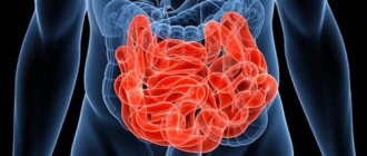
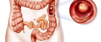
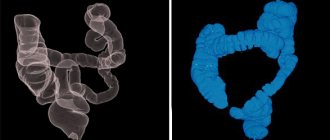
The small intestine is not convenient for echography. Due to the overlapping of its loops on each other, the image is distorted. However, it is quite possible to assess the functional status. Ultrasound of the gastrointestinal tract allows you to determine the condition:
- esophagus (cervical/cardiac);
- the entire stomach (including the wall structure);
- terminal zone of the small intestine;
- folds of the large intestine (the area from the cecum to the rectum).
Ultrasound diagnostics is not the main method for examining the gastrointestinal tract. The structure of the tract itself, as well as the peculiarities of its functioning, lead to low echogenicity of the structures. Basically, the functional state of those parts of the gastrointestinal tract where possible is assessed. As a rule, an ultrasound is performed:
- gatekeeper channel;
- gatekeeper's caves;
- lesser and greater curvature of the stomach;
- zones of connection with the duodenum.
Other areas of the gastrointestinal tract are poorly located. To study them, ultrasound remains an additional diagnostic method.
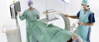
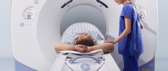
How the research is carried out
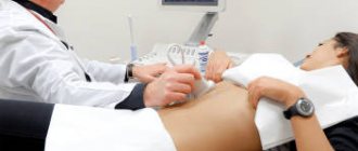
During the diagnosis, the patient lies on the couch. A special gel is applied to the area to be examined. This is necessary to displace air between the skin and the sensor, as well as to allow the device to slide easily. The specialist moves the sensor, examining the desired organ on the monitor. The patient is then asked to drink a small amount of water so that further examination will show internal changes.
When performing an abdominal ultrasound:
- the structure and size of organs and their location are assessed;
- the presence of free fluid is checked;
- the presence of neoplasms is excluded;
- Internal damage is examined.
In children, congenital organ pathologies are monitored using ultrasound.
With contrast
The use of contrast agents is common in radiology diagnostics. This method is also used in ultrasound studies, because allows for improved visualization of structures. Echo contrast compounds are a small amount of liquid in which gas bubbles are dissolved.
Ultrasound with contrast is performed to:
- Identify the difference between malignant and benign formations.
- Assess the blood supply to organs during the inflammatory process.
- Examine blood flow indicators in the vessels.
The substance is administered intravenously. After 10 or 15 minutes, it reaches the abdominal cavity, creating a contrasting area where the vessels pass. Bubbles in contrast at the border with blood enhance the reflection of ultrasonic waves. This makes the contents of the vessels visible on ultrasound.
The procedure is harmless to the patient, and the quality of tumor diagnosis is close to CT and MRI.
Advantages of ultrasound diagnostics
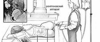
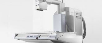
A comprehensive ultrasound of the abdominal cavity is safer than the x-ray method and is not limited to one projection. Unlike FGDS, there is no threat of infection of the organ being examined. In addition, unlike gastroscopy, ultrasound is able to recognize infiltrated oncological processes.
Ultrasound is highly informative in diagnosing esophageal reflux (the result is comparable to a contrast radiograph), inflammatory and erosive diseases of the gastric mucosa, studying its motor-evacuation function and oncological processes; in children, ultrasound often reveals thickening in the area of the pyloric muscle ring.
Diaphragmatic hernias and cysts are rarely diagnosed.
How is an abdominal ultrasound performed?
Ultrasound diagnostics allows you to visualize internal organs and tissues on the screen. To do this, the patient lies down on the couch. A hypoallergenic gel, safe for health, is applied to his stomach, which ensures a stable connection between the ultrasound sensor and the human body, prevents the penetration of air and allows the device to move unhindered over the skin. The doctor moves the sensor across the abdomen and records the data in an electronic protocol. The procedure takes from 20 minutes to an hour.
The main difficulty that specialists face when conducting ultrasound diagnostics of the abdominal cavity is gas bubbles that prevent the penetration of ultrasonic waves to the examined organ. Reducing gas formation allows you to obtain reliable data about the organs and tissues of the abdominal cavity.
Preparatory activities for diagnosis
It is imperative to prepare for the study in order to exclude gas formation and changes in the echo pattern of the study. Preparation includes:
- diet;
- abstinence from eating 8 hours before the procedure.
For children and patients with severe hunger pain, abstinence is limited to four hours. Preparation for the ultrasound diagnostic procedure begins two days before the examination.
Preparation for ultrasound involves eliminating foods that cause flatulence from the diet (baked goods, sweets, beans, peas, carbonated drinks, dairy products, etc.). Preparation also includes the above-mentioned regime of abstinence (refusal of water and food). The ultrasound procedure is always performed on an empty stomach.
Purgation
Often, during preparatory measures for an abdominal ultrasound examination, to ensure the greatest diagnostic accuracy, it is recommended to cleanse the intestines. If the ultrasound examination includes the retroperitoneal space, then this procedure will be mandatory. Cleansing should be done between 4 and 6 p.m. To do this, you need to use an Esmarch mug with pre-prepared raw cool water with a volume of about 1.5 liters. After the procedure, you must take sorbents.
Important! In the process of cleansing the intestines, it is prohibited to use drugs based on lactulose, for example, Duphalac, Prelaxan. Because they lead to unwanted bloating on ultrasound.
If for some reason the patient cannot cleanse the intestines, then he can take mild laxative medications. However, they can only be used if the patient is over 14 years old. You need to take laxatives in the evening before going to bed. You can also use microenemas to cleanse the intestines.
Contrast for ultrasound diagnostics of the gastrointestinal tract
Ultrasound examination is carried out in several stages. At the first stage, the gastrointestinal tract is studied using contrast, which is water. The patient should drink approximately 300-500 ml, slowly, in small portions, so as not to swallow air.
This stage allows you to evaluate:
- wall diameter;
- wall thickness (normally 2.5-5 mm);
- the amount of stomach contents (normally no more than 40 ml).
All deviations are attributed to SPO (hollow organ syndrome).
Deviations may indicate a hyperechoic structure of the contents, disruption of the layering of the organ walls, erosive pathologies, neoplasms (polyposis, malignant tumors), inflammatory diseases of the gastric mucosa.
At the second stage, the motor-evacuation functions of the stomach are assessed (also through contrast). With the development of a peptic ulcer or cancer process, food moves much more slowly. Reflux is diagnosed with a standard duplex scan.
What does an ultrasound scan of the gastrointestinal tract show?
Ultrasound diagnostics show the following:
- all sections of tissue of the esophagus, stomach, etc.;
- wall thickness, by which the presence of pathology can be determined;
- peristalsis of the large and small intestines;
- uniformity of organ walls;
- condition of lymph nodes, blood vessels. When examining the gastrointestinal tract, they also look at the condition of the liver, pancreas, gallbladder, etc.
Ultrasound is a fairly simple procedure and absolutely painless.
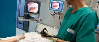
There is also an ultrasound scan in which a sensor is inserted into the patient’s oral cavity. This is an endoscopic method for examining the gastrointestinal tract. Do it on an empty stomach. The patient should be in a lying position on his left side. This type of study allows you to examine the walls of the esophagus and stomach in more detail and clearly.
It is important to know that an ultrasound should be performed not only if you have any problems or suspicion of a disease. Doctors recommend that even healthy people undergo ultrasound diagnostics 1-2 times a year for prevention. This measure will allow us to identify disorders in the early stages of occurrence and prevent further development of the disease.
Interpretation of the ultrasound picture
An ultrasound of the esophagus will reveal a hernia. It is recognized as a change in the direction of increasing the diameter of the organ from one and a half centimeters or more.
When registering the reverse flow of gastric contents after filling it with liquid up to half a liter, a diagnosis is made - exophagic reflux. With a cylindrical/cone-shaped expansion of the esophagus in the region of the heart, narrowing of its part leading to the stomach to three millimeters, the question of alakhasia arises. Esophageal cancer has characteristic signs: a violation of the layering of the walls, an increase in their thickness and the appearance of an uneven border.
Ultrasound allows you to diagnose impaired evacuation of food mass from the stomach, narrowing of the outlet section, as well as cancer of the organ. The leading, although nonspecific, echo trace of gastrointestinal lesions is SPPO (hollow organ syndrome, in particular thickening of its walls).
Loading…
Share with friends!
Possible results
The purpose of examining the abdominal organs with ultrasound is to determine their health status. The examination may show a normal result - if there are no abnormalities in the organs. But since an ultrasound of the abdominal organs is prescribed when a person has complaints, various changes are usually detected - signs of disease.
The doctor who conducts the examination gives only a description of what he saw. The final interpretation of the abdominal ultrasound results is made by the attending physician. He also makes a diagnosis, taking into account other studies.
Normal indicators
When describing the condition of the abdominal organs, the doctor follows a certain algorithm:
- size;
- outlines and contours;
- homogeneity of structure;
- fabric density;
- diameter of vessels and ducts.
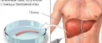
The sizes of the liver, gallbladder, and pancreas depend on the age of the subject, gender, height and body weight. Therefore, there is no strict number that the normal sizes must correspond to. Use an approximate range.
Table of normal sizes of abdominal organs:
| Organ | Dimensions |
| Liver | Length up to 18 cm, width up to 12 cm |
| Gallbladder | Length up to 10 cm, width up to 5 cm |
| Pancreas | Length up to 12 cm, width up to 6 cm |
| Spleen | Length up to 12 cm, width up to 8 cm |
Size indicators for men are 5-10 mm larger than for women - this is the norm. The contours should be clear and even. The normal structure of the tissue is homogeneous and fine-grained. Density is determined by the color of the organ in the image. The liver is taken as a reference point - it is gray in color. The pancreas and spleen should have the same color.
Defined diseases
Ultrasound reveals even minimal changes in the abdominal organs. This is especially important if you suspect cancer. Each disease has a specific set of ultrasound signs.
- Hepatitis is inflammation of the liver. The color becomes darker, the uniformity of the structure is preserved. The size increases, the contours are clear.
- Pancreatitis is inflammation of the pancreas. The color becomes darker, the uniformity of the structure is preserved. The size increases, the contours are clear.
- Cholecystitis is inflammation of the gallbladder. It increases in size and the wall thickens. With calculous cholecystitis, stones are visible in the cavity of the bladder - round white spots.
- Cirrhosis - normal liver tissue turns into scar tissue. In the picture the dimensions are reduced, the color changes to light gray or white. The structure is heterogeneous, lighter nodes are visible.
- Ascites on ultrasound is free fluid in the abdominal cavity. In the lower abdomen, dark areas are visible that change their position.
- With the help of ultrasound you can see cancer of any organs. The tumor has unclear contours; a white spot with uneven borders is visible in the middle of the gray tissue. Metastases also look the same - smaller, almost always round in shape.
- Cysts are cavities with air and fluid inside. The photo shows round dark spots with a white border.
- If an ultrasound shows enlarged lymph nodes of the abdominal cavity and retroperitoneal space, this is a sign of a cancerous tumor or tuberculosis of the spleen.
Since hollow organs, such as the intestines, are not accessible to ultrasound, not all diseases are detected. An abdominal ultrasound will not show appendicitis, colitis - inflammation of the intestines. Umbilical or inguinal hernias are not accessible to ultrasound waves.
Read more about pathologies of the abdominal organs in articles on the website dedicated to a specific organ.
A little anatomy
To understand where the retroperitoneum is located, you just need to know where the lumbar region of the back is located. Now we can accurately name the organs located in the retroperitoneal space:
- kidneys with ureters;
- adrenal glands;
- the aorta and inferior vena cava, which run along the spine.
There are organs that are partially covered by the peritoneum and are located in the abdominal cavity, and the other part is located retroperitoneally. Such bodies include:
- pancreas;
- duodenum;
- part of the large intestine: ascending and descending colon.
In addition to the organs, the retroperitoneal space is filled with fatty tissue that performs a supporting function.