X-ray of the esophagus and stomach is considered one of the simplest, inexpensive and effective measures for detecting functional disorders of the gastrointestinal tract (GIT). This study is also valuable for a number of diseases, allowing one to clearly visualize ulcers, neoplasms, and diverticula. The undoubted advantage of the method is its simplicity, the minimum number of contraindications and simple equipment that is available in any clinic. The study is carried out on an outpatient basis and does not require complex preparation or hospitalization. In some cases, X-ray examination of the stomach can be used - what is it and how safe is it? Contrast helps to more accurately determine some pathologies and contours of this hollow organ.
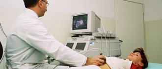
Combined system for radiography and fluoroscopy
X-ray of the stomach with barium
X-ray of the stomach with barium is a modern diagnostic method that makes it possible to identify pathological neoplasms in the gastrointestinal tract in the early stages.
What is a barium x-ray?
Indications for use
What does an x-ray show?
Is a stomach x-ray with barium harmful and how often can it be done?
Contraindications and restrictions
Preparing for an X-ray
How does the procedure work and how long does it take?
How is barium removed from the body?
X-ray examination of the hollow organs of the gastrointestinal tract (GIT) is performed with contrast. That is, before fluoroscopy of the stomach is performed, the patient takes an aqueous solution of barium sulfate orally. Barium does not transmit x-rays, allowing you to see and evaluate the condition of the walls of the gastrointestinal tract in detail.
X-ray of the stomach is one of the informative types of diagnostics that allows you to identify pathological changes. This alternative examination is carried out in cases where other diagnostic methods, such as ultrasound and FGS, are ineffective.
X-ray allows you to evaluate the structure of the mucous membranes, the location of organs, patency using a contrast agent, involves radiation, so it is important to know how to properly prepare for an X-ray of the stomach.
X-ray of the stomach is an instrumental method for examining the gastrointestinal tract, with the help of which informative information is obtained. During the procedure, the location of the gastrointestinal tract organs, sizes and changes in them are determined.
The stomach is a hollow organ, so to study it, radiography is performed with a contrast agent - a suspension of barium sulfate.
Indications
The need for a diagnostic study is determined after a preliminary examination of the patient and collection of anamnesis.
An X-ray of the stomach is prescribed in the following cases:
- patient complaints of pain in the epigastric region;
- disorders of the gastrointestinal tract (nausea, vomiting) not associated with poisoning;
- difficulty swallowing and passing food;
- belching, heartburn;
- a sharp decrease in body weight, blood in the stool;
- identification of compactions by palpation;
- suspicion of the presence of foreign bodies;
- condition after surgery on the gastrointestinal tract.
Radiography is a test in which a radiologist takes an image. Fluoroscopy allows you to monitor the gastrointestinal tract in dynamics and take pictures in different positions.
Both methods involve the patient receiving a dose of radiation, so it is important to perform the procedure without the need for a repeat study. The leading role here is given to proper preparation for x-rays.
If you conduct research right away, the results may be incorrect. Therefore, it is important to obtain information from your attending physician on how to properly prepare for the examination.
The radiation dose during one procedure is safe for humans.
Fluoroscopy of the stomach does not require special preparation. But to get the most truthful result, you should adhere to several rules:
- 2-3 days before the x-ray, you should not take medications other than Espumisan or activated charcoal. These products will help cleanse the gastrointestinal tract of mucus.
- On the day of the examination, smoking and chewing gum are prohibited.
- The subject needs to remove metal jewelry and dentures.
The patient is warned that the examination is carried out on an empty stomach. You should not eat food the morning before the diagnosis. You are allowed to drink some water.
Before radiography, the last meal is allowed no later than 7-8 hours before the examination. Before fluoroscopy with barium - at least 12 hours before the procedure.
Diet
A diet before undergoing an examination is prescribed to older people.
If the subject does not complain, this is not required. Doctors recommend eliminating heavy, difficult-to-digest foods from your diet.
3 days before the examination, you need to avoid foods that cause increased gas formation.
Nutrition rules
To ensure a clear result, 3 days before undergoing X-ray examinations of the gastrointestinal tract, you need to avoid foods that contribute to increased gas formation:
- all types of cabbage;
- Rye bread;
- legumes;
- potato;
- carbonated drinks;
- fatty, fried foods;
- dairy products;
- baking
It is recommended to eat porridges with water, steamed dishes made from lean meat or fish.
If a patient is hospitalized with a stomach disease, he is prescribed a special diet from the first days of his stay in the hospital. It is important to avoid taking dairy products before the examination.
When preparing a patient for a barium fluoroscopy of the stomach, the physician should identify the possibility of an allergic reaction. In case of allergies, another contrast agent containing iodine is selected.
A suspension of barium sulfate is not dangerous to health, except in rare cases of individual intolerance. This substance is capable of absorbing radiation without being absorbed into the gastrointestinal tract.
With the help of such a study, a more complete picture of the functioning of the system and clear pictures are obtained.
The patient should adhere to a diet for 3 days before the procedure.
During the examination, the patient is asked to drink two sips of the suspension, and during its passage the walls of the esophagus are examined. Then you need to drink the remaining barium (about a glass), and the radiologist examines the organs and takes pictures.
For patients who are prone to constipation, before undergoing the procedure, it is recommended to cleanse the intestines with an enema before bed and in the morning on the day of the x-ray.
The procedure algorithm is as follows:
- a radiologist examines the organs;
- the patient takes a barium suspension, and the specialist observes the filling of the stomach, the rate of filling and the structure of the mucosa.
The examination takes no more than 40 minutes.
About an hour after administration, barium enters the duodenum, from where it is completely excreted in a short time, and after 1-1.5 hours the stomach is freed from the contrast agent.
The radiologist takes pictures from different positions of the body: lying (on the side, back, stomach) or standing.
Using the Trendelenburg method, a picture of the stomach is taken in a position where the pelvis is elevated at an angle of 35-45 degrees. The doctor asks the patient to hold his breath for a while. This image allows you to determine the presence of a hiatal hernia.
During diagnosis, the following deviations can be identified:
- volume of the stomach (its contraction or increase);
- ulcerative lesions of the mucous membrane;
- polyps and neoplasms, predisposition to their development;
- inflammatory processes;
- violations of the lumen of organs;
- deformation of the walls or their displacement.
Contraindications
The X-ray procedure is not dangerous for the patient. In some cases, constipation and discoloration of stool may occur after taking barium. Normally, these phenomena disappear after 2 days. If it lasts longer, you should see a doctor.
There are contraindications for performing an X-ray of the stomach. These include:
- allergy to contrast agents;
- prolonged bleeding in the gastrointestinal tract;
- pneumothorax;
- serious condition of the patient;
- pathologies of the thyroid gland;
- pregnancy and breastfeeding.
According to indications, X-ray examinations are also performed during pregnancy, but the procedure is possible only in the 2nd and 3rd trimester.
Examination using a contrast agent is contraindicated in patients with diabetes, patients with severely impaired renal and liver function, and severe tuberculosis.
Conclusion
According to the requirements of the Radiation Safety Law, X-ray examinations are prescribed only after undergoing ultrasound, FGS and laboratory tests, i.e. non-X-ray methods.
If the examination is not very informative, an x-ray of the stomach is performed. A repeat procedure is permissible only after a year.
Main indications
As a rule, fluoroscopy of the stomach is prescribed for suspected diseases of the gastrointestinal tract. It can be:
- epigastric pain;
- dysphagia;
- periodic dyspeptic symptoms;
- regular bowel disorders;
- constant high acidity;
- blood in the stool, purulent or bloody discharge from the rectum;
- causeless and severe weight loss;
- anemia of unknown origin (origin);
- symptoms of obstruction of the stomach/esophagus.
X-ray of the stomach with barium can be used not only to diagnose pathologies, but also to assess the dynamics of the course of diseases, especially with peptic ulcers, pyloric stenosis, and tumor formations. Precancerous conditions (polyposis) or a tendency to tumors in some situations also become an indication for an R-scopy of the stomach and esophagus.
X-ray of the stomach has a minimal number of contraindications, which are relative. It is not performed on patients in shock, when it is impossible to stop bleeding from the stomach or esophagus, or in the first trimester of pregnancy.
X-ray of the stomach and gastroscopy are completely different research methods
X-ray of the esophagus and stomach is done only when it is impossible to prescribe fibrogastroduodenoscopy, or the data obtained from this study are insufficient for an accurate diagnosis. Such an examination is not used as a primary diagnostic measure.
What is a barium x-ray?
A barium X-ray is an examination that allows you to obtain not only a detailed image of the stomach, but also the entire gastrointestinal tract. The procedure is based on the introduction of barium sulfate. With the help of barium, you can carefully study the shape and relief of internal organs, as well as identify pathological changes.
The procedure occurs after the patient has taken the contrast agent orally. During the examination, the radiologist will examine the upper gastrointestinal tract and take a series of x-rays.
When is a stomach x-ray prescribed?
The procedure is prescribed after examination and medical history.
Indications and contraindications for radiology
Indications for fluoroscopy:
- signs of a stomach ulcer;
- dysphagia;
- control after gastric surgery;
- bowel dysfunction;
- blood in stool;
- profuse vomiting;
- sudden weight loss;
- pain symptoms after eating;
- malformations of the stomach;
- diverticula;
- pain in the gastrointestinal tract on an empty stomach.
Fluoroscopy should be performed if a blood test shows a sudden decrease in hemoglobin levels.
Diagnosis of gastric diseases using radiology (video author - Medical company ilaya).
Contraindications to the procedure are:
- pregnancy;
- continuous bleeding in the stomach and intestinal tract.
What does a barium x-ray show?
A standing barium x-ray of the stomach shows:
- size of the antrum of the stomach and esophagus;
- the condition of the folds and grooves of the upper part of the stomach;
- functioning of the gastrointestinal tract;
- structure and functioning of the duodenum.
Lateral position will show:
- pneumorelief of the antrum;
- wall displacement;
- gastric reflux.
In the Trendelenburg position, the patient's pelvis is elevated off the table by 35 degrees.
This situation shows:
- fiber condition;
- elasticity of walls;
- hiatal hernia.
X-ray examination reveals:
- polyps in the stomach;
- ulcerative and inflammatory processes;
- tumors;
- polyposis;
- deformation of the gastric walls (saccular protrusions, pathological narrowings);
- introduction of one section of the intestine into another;
- changes in motility in the gastrointestinal tract;
- the appearance of scars;
- pathologies of the digestive system;
- phlebeurysm;
- malabsorption syndrome;
- pathology of the duodenum;
- tissue lesions.
Today, x-rays of the stomach are precisely the diagnostic method that is considered the most accurate. Considering the fact that the stomach is a hollow organ, it is best to take an x-ray using a special contrast agent, most often barium sulfate. X-ray with barium makes it possible to timely detect serious diseases that can cost the patient his life.
This diagnostic method allows you to clearly see the location, shape and diameter of the lumen in the intestine, find out the degree of its elasticity and find out how much the tissue can stretch. The doctor evaluates the appearance of the organ and checks individual intestinal functions. X-ray with barium allows you to see in time the obstruction of the intestine, the anomalies that occur in it, for example, all this can be caused by polyps, diverticula and tumors.
Such a diagnosis will not cause any harm to health, since no mechanical impact is applied to the body and it will not be possible to injure the lining of the stomach or intestines. Barium sulfate is not absorbed into the blood and allows you to get clear photographs on the monitor, which are then examined by a specialist.
Using a contrast agent and a subsequent series of x-rays, it is possible to identify the following diseases of the digestive organ:
- gastritis and ulcers;
- benign and malignant neoplasms;
- inflammatory phenomena;
- decreased peristaltic function of the organ;
- diverticular phenomena;
- gastric obstruction;
- displacement and prolapse of the organ;
- other disorders and pathologies.
If you wish and have time, you can track the nature of the pathologies of the duodenum, since it is the next stage in the passage of the contrast agent through the digestive tract.
X-ray of the stomach refers to abdominal fluoroscopy, allows you to identify organ pathologies - peptic ulcers, polyps, inflammatory-dystrophic changes, signs of oncology. Also, during the diagnosis, the doctor has the opportunity to evaluate nearby organs - the duodenum and the esophagus. X-ray examination of the stomach is an affordable procedure that can be done at any diagnostic center.
Contrast radiography is the preferred method for examining the condition of the stomach. Without the use of barium, it will not be possible to study in detail the shape of the organ, reliefs and dystrophic changes.
X-ray with the introduction of barium sulfate diagnoses:
- folding of the membrane with gastritis or ulcers;
- neoplasms of various etiologies;
- cicatricial changes during pathologies or chemical burns;
- change in the lumen of the esophagus, narrowing or expansion;
- violation of the normal arrangement of organs.
Receipt of the results does not constitute the basis for making a final diagnosis. The patient will additionally need to undergo a series of studies, diagnostic and physical examination.
The examination shows the functioning of the ileocecal valve, which separates the layers of the intestine and is responsible for passing food between them.
Progress of the study
Most often, fluoroscopy of the stomach is performed with preliminary contrast. In this case, the patient’s preparation consists of taking 250 g of contrast - an aqueous suspension of barium sulfate is used, which the patient drinks immediately before all manipulations, in the presence of a doctor. This allows not only to more accurately determine all pathologies of the mucous membranes, but also the time elapsed until complete filling. After this, the radiologist can assess the size, shape and anatomical location of the stomach, its mobility, the degree of elasticity of the walls, and see contractions.
X-ray examination of the stomach and duodenum is performed in various positions of the patient - this allows for better visualization of organs and accurate diagnosis of all existing pathologies. Depending on the expected diagnosis, the doctor asks the patient to turn on the right or left side and take a certain position. Examination of the stomach in the Trendelenburg position is of great diagnostic importance - the patient lies on his back with the pelvis elevated by 45 degrees. This is the only way to accurately determine the presence of a hernia in the esophageal opening of the diaphragm.
A more detailed x-ray examination is performed using the double contrast method. Along with the barium solution, air is introduced into the human stomach, for which a gastric tube is used. This technique is often required in controversial situations, since the air straightens all the folds that abound in this organ. This allows you to visualize the mucous membrane in as much detail as possible in order to identify pathology.
Contraindications and restrictions
Reasons why fluoroscopy is contraindicated:
- exacerbation of chronic diseases;
- hemorrhagic manifestations of the gastrointestinal tract;
- stomach and intestinal bleeding;
- intolerance or allergy to barium;
- nonspecific ulcerative colitis;
- heart disease;
- pain syndrome under the xiphoid process;
- pregnancy;
- intestinal perforation;
- the patient is unconscious;
- complete disruption of the passage of contents through the intestines.
It is forbidden to take an x-ray after a recent intestinal biopsy, since taking a contrast agent can cause inflammation.
Evaluation of results
Everything that fluoroscopy shows is often assessed by more than one doctor. Often during the procedure, not only a radiologist is present, but also a gastroenterologist, who may be joined by a surgeon. Collegial interpretation of the results increases the accuracy of diagnosis, allowing you to see symptoms such as:
- changes in the position of the stomach and esophagus, prolapse and developmental anomalies, tumor formations, myocardial hypertrophy;
- changes in the lumen of the esophagus or the stomach itself caused by scars from ulcers, chemical burns, and also determine an increase in the tone of the stomach muscles;
- expansion of the esophagus and decreased tone of its walls;
- violation of the anatomical integrity of the walls of these organs;
- filling defects caused by polyps, foreign objects accidentally caught inside, papillomas, tumors.
X-ray image of the stomach
Regardless of how the study is carried out - with or without contrast - the diagnosis is made taking into account general clinical symptoms, laboratory results and other instrumental examination methods, if any have been carried out or planned.
How does the procedure work and how long does it take?
Features of the procedure for X-ray of the stomach with barium:
- On the day of the study, you should refrain from drinking and eating.
- Immediately before the x-ray, you need to get rid of various jewelry and removable dentures.
- Next, a survey X-ray of the chest and abdominal cavity is performed.
- The patient is given a contrast agent (barium sulfate). It has a rich chalk flavor and a dense consistency.
- The specialist takes the first x-ray, determining the relief of the walls of the esophagus.
- The patient takes various positions: lying on his back, on his side, and also using the Trendelenburg method.
On average, the procedure takes up to 40 minutes. If there is a need to take x-rays of the lower intestines, this may take up to a day.
Photo gallery
X-ray of the stomach with barium sulfate Violation of the lumen of the stomach Gastric ulcer on an x-ray
Photo gallery
Preparing for the study
No special or complex preparation is required for R-scopy of the gastrointestinal tract. In most cases, the study is scheduled for the morning. The last meal is allowed 10 hours before the procedure, and a diet is prescribed 3 days before the x-ray. It is advisable to completely remove from the diet foods that contribute to the formation of gases in the intestines:
- legumes;
- black bread;
- fatty foods;
- smoked meats;
- whole milk and cottage cheese;
- apples;
- cabbage;
- fruits.
Products that cause increased gas formation should be excluded
For three days, food should be easily digestible - low-fat steamed meats and fish, light soups (dairy-free), white (preferably stale) bread, natural yoghurts, eggs. If you are prone to constipation on the eve of R-scopy, a cleansing enema is necessary, and preparation for fluoroscopy of the stomach if a spasm of the pyloric region is detected requires probe rinsing immediately before the study. It is advisable to remove all jewelry in advance.
Along with the diet, you should stop taking medications or notify your doctor. It is advisable not to smoke 7-10 hours before the procedure, not to drink alcohol or carbonated drinks.
Complications of fluoroscopy
It should be noted that such a study practically does not cause complications or consequences. The X-ray itself does not have a negative effect on the body, since all manipulations associated with exposure to radiation are carefully recorded, and it is almost impossible to receive a dangerous dose of radiation. It is also important how fluoroscopy is done - the entire study takes minimal time, which rarely exceeds 10 minutes, and the power of the equipment is often adjusted individually.
The main consequences of fluoroscopy of the stomach and duodenum include digestive disorders: constipation, flatulence, bloating. Possible nausea and pain in the epigastrium associated with spasms of smooth muscles. All these consequences are caused by the contrast agent - barium. It also gives stool a gray tint with a whitish coating. In most patients, this goes away within 1-3 days and does not require medical intervention.
In the presence of severe pain, it may be necessary to prescribe antispasmodics. Other drugs are usually not used. To quickly restore normal digestion, you should drink at least 2 liters of pure still water per day. You can make compote from prunes - it will help prevent constipation. If there is severe bloating, you can take drugs such as Espumisan and activated carbon.
Activated carbon is an excellent adsorbent
Adequate physical activity after this study is important - it contributes to the rapid normalization of gastrointestinal motility and restoration of the normal digestive process. You should start your normal diet gradually. You should not eat heavy, fatty foods immediately after the procedure. It is better to prefer fermented milk products, fish, a sufficient amount of vegetables and herbs - all this will quickly eliminate the effects of barium intake.
Features of X-ray examination of the esophagus and stomach
This method of examining the gastrointestinal tract allows, using X-rays, to obtain a complete picture of the examined organs with all pathological changes in their shape, structure, and even evaluate them over time. But it is possible to take an x-ray of the stomach and other organs only under certain conditions that the patient will definitely need to fulfill in preparation for the study.
Due to the structural features of the digestive organs - the esophagus, stomach, small and large intestines, which have a hollow shape that does not block X-ray radiation, contrast is used to successfully carry out the procedure. As a contrast agent, a drug made from barium is used, diluted with water to the consistency of a kind of suspension.
In some cases, in order to straighten the folds of the mucous surface of the gastrointestinal tract, immediately before the start of the study, the patient is asked to drink a solution of crystalline soda, which increases visualization. Sometimes, for the same purposes, air is introduced under pressure or using a perforated tube, through which the patient simultaneously receives a barium suspension.