What diseases does MRI detect?
The MRI method is based on the effect of radio waves and a magnetic field on the body and the receipt of response signals, their subsequent conversion into three-dimensional graphic images.
Magnetic resonance imaging allows you to evaluate the structure of the organ and identify existing disorders.
Here's what the MRI shows:
- shape, size and location of the gallbladder, ducts;
- the internal structure of the organ (can be seen layer by layer in a section with a step of 0.5-1 cm);
- deformation, bends of the bile duct, narrowing of the bile ducts;
- foci of inflammation;
- contractility of the smooth muscles of the gallbladder and ducts;
- the presence of polyps, cysts;
- the presence and nature of the neoplasm (benign or malignant), metastases.
MRI is used when ultrasound and other instrumental diagnostic methods are not informative enough
Magnetic resonance imaging helps to identify:
- cholecystitis;
- dyskinesia (impaired contractility of the smooth muscles of the bladder and biliary tract);
- development of cholelithiasis (even small inclusions that are not detected on ultrasound are detected);
- polyps in the gallbladder;
- oncological damage to the organ, location and size of the tumor, the presence or absence of metastases.
Indications for the MRI procedure
Magnetic resonance imaging is used to identify possible pathologies, determine the location of organs in the abdominal cavity, the smallest changes in tissues, narrowing of the bile ducts and the presence of possible stones.
MRI is prescribed in the following cases:
- Suspicion of the presence of neoplasms. A study with a contrast agent allows you to diagnose the location of tumors, size and possible impact on neighboring organs.
- Suspicion of the presence of foreign bodies in the bile ducts (tiny stones), which cannot be diagnosed using other research methods.
- Presence of inflammation.
- Injuries to the abdominal organs.
- Jaundice of unknown origin.
- Suspicion of the presence of parasites in the abdominal cavity.
- Developmental pathology (normally, the gallbladder has the shape of a pear. For reasons beyond a person’s control, it can become deformed, curved, and even take on the shape of a boomerang).
- Liver damage (toxic damage, cirrhosis).
- Tracking the dynamics of existing treatment.
https://youtu.be/9CIzW4UGMfY
Indications
MRI is indicated to identify pathology or clarify the diagnosis in the following cases:
- for injuries and damage to the abdominal cavity;
- structural abnormalities of the gallbladder;
- clinical symptoms of cholecystitis (inflammation of the gallbladder) and cholangitis (inflammation of the ducts), cholelithiasis;
- suspected parasitic infection;
- jaundice of unknown origin;
- pain in the area of the right hypochondrium arising for unclear reasons;
- identifying abnormalities in the results of laboratory tests (increased activity of liver enzymes, high bilirubin content).
Also, MRI of the gallbladder allows you to evaluate how effective the treatment of a particular disease is, and to monitor the functional state of the biliary system after surgery to remove the organ.
Since the gallbladder and liver are structurally and functionally interconnected, simultaneous examination of both organs is often prescribed
What diseases and pathologies can the examination reveal?
Tomography of the gallbladder allows you to examine the full clinical picture and diagnose the following diseases:
- Gallstone disease is a pathology in which crystalline deposits form in the gallbladder and its ducts. In the early stages, it is difficult to determine the presence of stones: only when they begin to move through the ducts do they make themselves felt. A person experiences acute pain, especially when walking, as well as nausea, vomiting, colic, and a bitter taste in the mouth. People with large body weight are susceptible to the disease.
- Cholicystitis is characterized by an inflammatory process and occurs as a result of complications of gallstone disease. The image shows thickening of the walls of the bladder, which leads to disruption of the outflow of bile. Symptoms are: dull pain, vomiting, increased gas formation, loose stools. If treatment is not started in time, the disease will develop from acute to chronic.
- Polyps are round or oval growths on the mucous membrane of the bladder from the inside. They can be detected by doing an ultrasound, but it will not be possible to see the exact picture. MRI, computer diagnostics, endoscopic examination make it possible to determine the affected areas and the structure of tumors.
- Dyskinesia is a pathological change that leads to disruption of the contractile function of the gallbladder muscles. Despite the condition of the biliary tract, bile is stagnant. The causes of this phenomenon include disruptions in the gastrointestinal tract, hormonal disorders, endocrine pathologies, intestinal infections, hepatitis, and the activity of parasitic worms. Tomography is prescribed if other diagnostics have given controversial results.
- Biliary pancreatitis is an inflammatory disease of the pancreas. Occurs when there are abnormalities in the functioning of the liver and gallbladder and poor excretion of bile. In the acute stage, severe abdominal pain and indigestion and fever are observed. May cause the formation of pancreatitis.
Proper preparation and compliance with all standards and requirements during magnetic resonance diagnostics guarantee exceptional reliability of the results and the opportunity to obtain the most detailed information. Therefore, in some cases, MRI is the only possible method for making an accurate diagnosis of gallbladder pathologies.
Conducting research
To undergo an MRI of the gallbladder, a patient must have a referral from a doctor, a medical record, and the results of previous examinations (if any).
Preparation
Preparing for the procedure is quite simple. Necessary:
- a week before the examination, exclude fatty and fried foods from your diet;
- one day, stop consuming fermented milk products, carbonated drinks and high-fiber foods;
- to exclude flatulence, the day before the examination, take medications that reduce gas formation in the intestines;
- stop drinking alcohol and smoking on the eve of the examination;
- do not eat or drink later than 5 hours before the procedure (the study is carried out on an empty stomach).
If an MRI with contrast is planned, a contrast sensitivity test is performed immediately before the examination. To make sure there is no allergy, a small dose of contrast is injected subcutaneously and the reaction is observed. If allergic manifestations do not occur, examination is allowed.
During the tomography, the patient must lie still. Therefore, if a child or a person suffering from increased nervous excitability is being examined, sedatives must be taken the day before.
There are no age restrictions for magnetic resonance imaging. If necessary, the procedure is performed even on small children.
The examination of a very young child who will not be able to remain still is carried out using anesthesia.
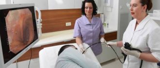
Procedure
The tomograph, which is a closed chamber, is equipped with a ventilation system, light, panic button and a two-way intercom for communication between the doctor and the patient.
Examination stages:
How to examine the gallbladder
- the patient lies down on the retractable tomograph couch;
- to ensure immobility, the arms and legs of the subject are fixed with special fastenings and belts;
- when performing MRI with contrast, the substance is administered intravenously using a syringe or catheter;
- in the presence of diseases of the cardiovascular and respiratory systems, sensors are installed that monitor the patient’s heart rate and respiratory activity during the examination;
- the retractable table is placed in the tomograph chamber, the examination begins, during which the doctor periodically gives the patient certain instructions (take a deep breath or exhale, hold your breath);
- the examination is completed, the table moves out of the tomograph chamber.
Phones, watches and other electronic devices, bank cards, metal objects (hairpins, jewelry, etc.) must be left outside the diagnostic room.
The procedure lasts about 30 minutes for a regular examination and about an hour for magnetic resonance imaging with contrast. In this case, the patient does not experience any discomfort or pain. The only complaint is that the tomograph makes quite a loud noise.
Features of patient preparation
Preparation for cholecystography begins 2-3 days in advance, when the patient needs to start following a slag-free diet, giving up flour, cabbage, and potatoes.
The general algorithm of actions for preparation for any method of drug administration includes:
- 14 hours before the procedure - avoid fatty foods and smoking;
- after a light dinner - refusal to eat until cholecystography is performed, you are only allowed to drink water;
- It is recommended to perform an enema the day before the procedure.
When administering contrast orally, 12 hours before cholecystography, the patient takes Bilitrast in the dose prescribed by the doctor.
When preparing for intravenous cholecystography or cholangiography, the medicine is administered in the treatment room.
The final stage of preparing the patient for the procedure: come on an empty stomach to the X-ray room at the appointed time.
Preparing children for the procedure is carried out in the same way as preparing adults.
results
The tomograph registers the received signals and transmits them to a computer, where they are converted using a special program into a three-dimensional image of the organ being examined. At the end of the procedure, the specialist deciphers the received images and prepares a conclusion. The patient can pick up the issued examination results within an hour after the end of the examination. Then the results must be provided to the attending physician - he will make a final diagnosis and prescribe appropriate treatment.
To make an accurate diagnosis, a specialist must decipher the received images.
Scintigraphy of the gallbladder
An examination that studies the anatomy and physiology of the biliary tract, motility of the gallbladder, and the degree of patency of the bile ducts is called scintography. According to the technique, a radioactive isotope is injected intravenously into the patient’s body. The drug is metabolized by liver cells and released into the biliary system. Scanning is performed at intervals of 10–15 minutes for 1–2 hours.
Using dynamic scintography, the movement of bile from the gallbladder is monitored. Unlike ultrasound, scintography does not detect stones in the biliary tract. And in patients who took alcoholic drinks before the study, a false-positive result may appear.
As a rule, during scintography, the condition of the bile and liver is assessed.
Features of MRI with contrast
In some cases, for more accurate visualization of pathological processes, it is advisable to perform cholangiography (MRI with contrast). The contrast agent is administered intravenously using a syringe or through a catheter. Moving along the bloodstream, the compound penetrates the organ under study and accumulates in pathological foci, including neoplasms.
Previously, products containing iodine were used as a contrast. However, recently contrast diagnostics have been carried out with gadolinium-based drugs (Primovist, Magnevist, Omniscan). This component extremely rarely causes allergic reactions, does not accumulate in the body and does not have side effects on internal organs.
MRI with contrast allows you to get a clearer picture of pathological changes
Dyskinesia
This disease is associated with poor flow of bile from the bladder. This pathology occurs due to insufficient muscle contraction and there are no spasms. Diagnosis of the disease is difficult. It includes several research methods. A computer study is also prescribed to clarify data on the condition of the gallbladder.
When dyskinesia occurs in a person, the following signs may be present in the body:
- Pathological processes of the genital organs.
- Menopause period.
- Disruption of the endocrine system. Disorder of the internal secretion of the body.
Contraindications
Contraindications to MRI of the gallbladder and ducts are:
- the presence of metal devices and implants in the patient’s body (artificial joint, pacemaker, fixed hearing aid, clips on blood vessels, insulin pump, metal dentures, ammunition fragments);
- body tattoo made using metallic dyes;
- obesity of 2 and 3 degrees (standard closed tomographs are designed for patients weighing up to 120 kg, and open devices that allow obese people to be examined are not available in every clinic);
- fear of closed spaces - claustrophobia (examination can be carried out, but only with the use of anesthesia or in an open type apparatus);
- mental disorders, epilepsy;
- alcohol or drug intoxication;
- severe and extremely serious condition of the patient, acute cardiac and respiratory failure;
- severe liver disease (for MRI with contrast, since the contrast agent can provoke an exacerbation of the pathology, up to the development of acute liver failure);
- kidney disease (only for contrast diagnostics);
- pregnancy (routine examination can be carried out in the second trimester if there are serious indications; MRI with contrast is prohibited at all stages of pregnancy and during breastfeeding).
After removal of the gallbladder, tomography can be performed because the clips installed during the operation are made of a titanium alloy inert to magnetic waves.
Before conducting the examination, the doctor must make sure that the patient has no contraindications
Gallstone disease
This disease is associated with the formation of stones in the gallbladder or its ducts. They are crystallized clusters. Typically, this disease occurs and develops in those people who are overweight. MRI of the gallbladder allows you to accurately diagnose this disease. The disease occurs in several stages, namely, the active development of stones and their growth. There are also periods of calm.
When the stones are in the bladder, they do not cause any concern to the person. Therefore, patients often have no idea that they have them in their body. Acute pain occurs when stones move into the ducts. A person feels pain in the upper abdomen and right hypochondrium. The patient's condition worsens, nausea and vomiting appear, and a taste of bile appears in the mouth.
Advantages of the method
Examination of the organs of the biliary system using MRI has a number of advantages, including:
- high accuracy and information content;
- safety;
- painlessness;
- no radiation exposure (unlike radiography and computed tomography);
- minimal time investment (the procedure itself lasts on average half an hour, and after another 60 minutes you can get results);
- the possibility of conducting repeated examinations (for example, to monitor the effectiveness of treatment);
- no age restrictions.
Often patients try to figure out which method is more informative - MRI or CT. The main difference between the two methods is the type of radiation used. MRI is based on the effects of a magnetic field and radio waves. They are absolutely safe for the body, so if necessary, the procedure can be repeated after a short period of time.
Computed tomography is based on exposure to x-rays. Due to harmful radiation exposure, CT scanning is not allowed to be performed again shortly after the first procedure. In addition, it is extremely harmful to be exposed to radiation in the presence of a malignant tumor. This can accelerate and aggravate pathological processes. Nevertheless, for the diagnosis of some pathologies, it is advisable to perform a CT scan. The optimal diagnostic method in each case should be selected by the doctor.
Magnetic resonance imaging makes it possible to identify structural and functional disorders at the earliest stages of the development of pathological processes, when changes cannot be detected by other instrumental methods. Early and accurate diagnosis ensures timely initiation of treatment and helps prevent the worsening of the disease and the development of complications.
The structure of the bile ducts
The biliary duct system is a branched network of tubular formations. The initial small ducts originate inside the liver and form branches similar to the crown of a tree. The intrahepatic tracts from the right and left lobes of the gallbladder unite into 2 large “vessels”, and at the exit from the liver they form the common bile duct.
The mouth of the extrahepatic bile ducts is located at the junction of the lobar ducts. The united hepatic tract merges with the cystic tract, through which bile flows from the gallbladder. Together they are united into the common bile duct. This is the bile duct that leads to the duodenum.
At the confluence of the biliary system and the initial part of the intestine there is the sphincter of Oddi, a muscular valve that regulates the passage of bile. In front of it, the bile duct communicates with the pancreas through a separate duct, forming the ampulla of Vater. Before “entering” the intestine, bile and pancreatic juice mix to digest food.
Throughout the biliary system, the ducts have different diameters. Their walls consist of 4 layers:
- internal lined with epithelium;
- muscle consists of individual strips of muscle tissue arranged in a spiral;
- on top of the smooth muscles there is a connective tissue membrane that covers the muscles and fills the space between them;
- the outer layer is formed by loose connective tissue, saturated with blood vessels.
Inside the walls there are glands in which mucus is synthesized, and the cavities of the bile ducts contain beneficial microflora that perform a bactericidal and protective function.
https://youtu.be/Oz2kj0MB68U
What is MRI?
MRI stands for magnetic resonance therapy. This study allows specialists to clearly see the insides of the human body, which is very important for complex diagnostics.
The MRI procedure is based on a constant magnetic field and radiofrequency energy. During the study, special equipment is used, including a powerful computer, which allows you to create high-quality graphic images of internal organs.
During the procedure, the patient is in a special scanner. The magnetic field, in turn, aligns protons in the tissues of the human body. Radiofrequency energy also affects these protons, causing them to create signals that the sensor in the scanner analyzes. Graphic images are formed during computer processing of the data and signals that arrive at the scanner receiver.
Difference between MRI and computed tomography (CT)
Magnetic resonance therapy receives data from special sensors of the device, and then models it into a three-dimensional image. A CT scan also produces a three-dimensional image that is generated by a computer. But what is their difference?
There are two points that distinguish these two diagnostic methods. The first is the nature of the beams used in the study. While CT uses X-rays, magnetic resonance therapy uses electromagnetic beams. The second important point is the fact that this or that method is suitable for diagnosing a certain type of pathology.
Thus, computed tomography is most often prescribed in the following cases:
- To study the condition of joints, teeth and individual bones.
- Computed tomography allows you to see recent bleeding, so it is prescribed for previous injuries.
- For diagnosing diseases of the spine - scoliosis, hernias, osteochondrosis, etc.
- To detect tuberculosis, pneumonia and other diseases of the chest area.
- Research of the thyroid gland.
- For examination of the stomach and intestines.
- To identify diseases in the genitourinary system.
- Study of blood vessels, including detection of atherosclerosis, various types of aneurysms, etc.
MRI is prescribed in the following cases:
- To detect tumor formations in soft tissues.
- To study the condition of the brain and spinal cord.
- As a diagnosis of the pelvic organs.
- For studies of patients who have suffered a stroke, as well as those suffering from multiple sclerosis.
- To assess the condition of muscles and ligaments.
In this case, it is wrong to say that any of the above diagnostic methods is more effective and accurate. They just have slightly different areas of application, so the attending physician decides whether to use CT or MRI based on the problem area of the body.