X-ray of the abdominal cavity (survey)
Survey radiography of the abdominal cavity is a method of non-contrast X-ray diagnostics of the condition of the abdominal cavity.
For the abdominal organs, this method is not very informative, since the soft tissues of the body are mostly X-ray negative, i.e. with such a study, the radiologist will not see images of the abdominal organs. Based on survey X-ray data, the doctor can only judge the presence and distribution of gas, liquid, as well as stones or foreign bodies in the abdominal cavity and digestive tract. The method is effective in diagnosing ruptures and perforations of the genital organs of the abdominal cavity. The images will show free gas in the abdominal cavity, which normally should not be there. Plain radiography is the main method for diagnosing intestinal obstruction (the images will show shadows of distended intestinal loops with levels of liquid and gas, the presence and location of which can be used to judge the presence and location of obstruction). Survey images can also show kidney and gallstones, the presence of blood and ascitic fluid in the abdominal cavity.
For what diseases is an abdominal x-ray performed?
- cholecystitis;
- nephrolithiasis, urolithiasis;
- pancreatitis;
- blockage of the intestinal lumen by a tumor;
- intussusception;
- abscess;
- diverticulitis;
- appendicitis;
- injuries;
- inflammatory bowel diseases;
- intestinal ischemia;
- small bowel obstruction
Most often, the examination is performed without special preparation (for emergency indications); a cleansing enema (1.5 liters) is routinely administered the night before and on the day of the examination 2 hours before the scheduled time. You can eat.
X-ray of barium passage through the small intestine
Diagnosis of diseases of the small intestine remains a serious problem to this day. Diagnostic difficulties arise due to the large extent of the small intestine, the presence of numerous bends in it and the difficult accessibility of this part of the digestive tract for instrumental diagnostic methods, as well as the low information content of the diagnostic methods used.
X-ray of barium passage through the small intestine - X-ray visualization of the progress of contrast through the small intestine. Radiography reveals diverticula, strictures, obstructions, tumors, enteritis, ulcerations, malabsorption and motility of the small intestine. X-ray contrast examination of the small intestine is carried out after ingestion of a barium suspension solution (barium sulfate). As the contrast moves through the small intestine, targeted radiographs are taken at intervals of 30-60 minutes. X-ray is completed after all its parts have been contrasted and barium has entered the cecum.
Radiography of the passage of barium through the small intestine requires preparation of the intestine for the study: the use of a slag-free diet and laxatives in order to accelerate the passage of barium during the study.
The drug "Fortrans". The package of the drug is dissolved in 1 liter of boiled water or mineral water without gas. Preparation includes taking 3-4 sachets (dissolved in 3-4 liters of water) depending on weight. Take once in the evening, on the eve of the test, starting at 18:00, 1 liter of the drug with an interval of 1 hour. Take 2 liters of solution the evening before the examination and 2 liters on the day of the examination 3-4 hours before the procedure.
Recommended diet during the day:
Recommended: white bread, boiled meat, fish, butter, cheese, pasta.
Not recommended: dairy products, vegetables, apples, brown bread, spicy seasonings, coffee, chocolate, alcohol.
To achieve the best image quality, your stomach should be empty. On the day of the examination, it is advisable not to eat or drink anything (including any medications, especially antacids), and refrain from chewing gum and smoking.
Return to RADIOGRAPHY section
Study of the passage of barium through the small intestine
a) Purpose
. Examination of the small intestine with the best contrast (planned).
b) Equipment and methodology
. Overview photo. Administration of one glass of liquid barium orally => a series of photographs at appropriate time intervals. If contrast has not reached the colon within 4 hours => late images (eg after 24 hours).
c) Risk of Schwartz test
(passage of barium through the small intestine): • Radiation exposure: 300 mrem (3 mSv). • Barium peritonitis: examination is contraindicated if perforation/failure is likely. • Deposition of barium precipitates during stasis can lead to functional obstruction.
d) Interpretation
: • Crohn's disease: assessment of changes in the mucosa and the number of strictures, the presence/nature of fistulas. • External intestinal fistula: characteristics of the intestinal segment bearing the fistula, identification of distal obstruction. • Partial intestinal obstruction: signs and nature of the transition point, additional areas of obstruction. • Bleeding from the gastrointestinal tract with an unidentified source: identification of an altered segment of the intestine, tumor.
e) Difficulties
: • Overlay of images of contrast-filled colon segments => difficulty recognizing pathological changes. • False positive results (air bubbles, intestinal contents).
Abdominal x-ray
X-ray of the abdominal organs - radiography - is a traditional diagnostic method of clinical medicine, based on localized irradiation with a minimal dose of X-rays, which results in projection images of the internal structures of the body.
The most common types of x-rays include plain abdominal x-rays and contrast x-rays.
It should be noted that X-rays cannot be reflected by the parenchyma of internal organs and soft tissues, so there is no complete “picture” of many organs in the images. However, a plain abdominal x-ray may show some organ damage; the presence of foreign objects, pathological formations (tumors, cysts, hematomas) and calculi (stones) in the kidneys, gall and bladder; the presence of gases and liquids (including blood) in the intestines and abdominal cavity and their location, as well as the pathological accumulation of metabolic waste in the large intestine.
A survey X-ray of the abdominal cavity has long proven its diagnostic effectiveness and is mandatory in cases of suspected intestinal obstruction, violations of the integrity (perforation) of its walls or perforation of other abdominal organs.
Contrast radiography is performed using a contrast agent, which fills cavities that are not visible with a regular plain x-ray.
Barium sulfate (barium sulfate in powder form) used as an X-ray contrast agent is practically insoluble in water, alkalis and basic acids, including hydrochloric acid, but it absorbs X-rays. Before contrast radiography, a barium suspension is taken orally (or administered through a probe): 80 g of powder per 100 ml of water. The drug is not absorbed from the gastrointestinal tract and does not enter the blood (if there is no violation of the integrity of the intestinal walls); is evacuated from the body with feces. If an abdominal x-ray with barium is performed to examine the colon, a suspension (per liter of 0.5% aqueous solution of tannin 750 g of barium sulfate powder) is introduced into the rectum using an enema.
An abdominal X-ray with barium provides a detailed image of pathologies of the gastrointestinal mucosa (ulcerations), perforations of hollow organs, narrowing of the lumens in the intestines and tumors.
Instead of barium sulfate, air or nitrous oxide can be used in contrast radiography, and when examining the bladder, drugs with sodium amidotrizoate (Triombrast, Verografin, Visotrast) can be used.
In addition to classical radiography, fluoroscopy is performed if necessary. This is when, with the help of additional special equipment, you can not only capture a static image of internal organs on film, but also observe them in a dynamic state at a given moment in time. Such an x-ray of the abdominal organs gives the doctor the opportunity to visualize the processes occurring in the organs (contractions, stretching, displacement, etc.).
Home X-ray Abdominal and pelvic organs X-ray of the abdominal organs: how and when it is done, what it shows
X-ray picture of the intestine is normal
A person feels healthy when all his organs work harmoniously. There are no useless organs or systems in the body. Wrong mode
, uncontrolled use of medications, sedentary lifestyle and frequent
adversely affect the condition of the human body. Sometimes the target of the disease is
Diseases of the digestive system are very unpleasant. Every day a person needs to receive nutrients from food. In many cases, the process of eating food gives people great pleasure.
If a malfunction occurs in the digestive system, a person
, weakens, loses weight, he has
, appears
. If ailments of this kind persist for a long time, you should consult a doctor.
After analyzing the complaints and examining the patient, the doctor may prescribe an X-ray of the intestine. The method will allow you to detect the cause of the ailment and make an accurate diagnosis. The localization of the pathological process and the need for medical procedures or even surgical intervention will also be determined.
It is very important to promptly identify intestinal pathology, since colon cancer is currently in third place among other types of cancer. If in previous years it affected people in the middle and older age groups, now it is increasingly occurring among young people. Early diagnosis of cancer helps to completely cure this deadly disease.
X-rays were discovered in 1895 by Wilhelm Conrad Roentgen.
X-ray radiation is a type of invisible electromagnetic radiation, the source of which is the cathode-anode ray (
) a tube. X-rays are penetrating, and different substances absorb them differently. When X-rays pass through the human body and are recorded on sensitive material (
) you can get an image of the internal organs. The photo comes out in black and white. The color intensity of the organ in the image will depend on its density. Yes, on
the bones appear light and the lungs appear dark. Later it turned out that X-ray irradiation in large doses is unsafe and can cause
, genetic mutations and
Despite the possible adverse effects on the human body, X-ray radiation is successfully used in medicine. The benefits of its use significantly outweigh the harm. Currently, diagnosis of most diseases is unthinkable without X-ray examinations.
Currently, x-rays are the main method for diagnosing intestinal diseases. During its implementation, modern technologies and equipment are used, which can significantly reduce the time of the study, discomfort and radiation exposure from the use of this procedure.
There is a different method for x-ray examination of each part of the intestine. When choosing it, the patient’s complaints and condition, medical history, and the results of other types of examination are taken into account. X-ray diagnostics can be carried out without the introduction of contrast agents or with their use.
The main types of X-ray examination of the intestines are:
- Radiography. An X-ray examination method with minimal radiation exposure. Allows you to obtain a linear image of an organ or area of interest on film or digital media. It is the first and most accessible method, quite accurate and informative. The procedure does not take much time.
- X-ray. A type of x-ray examination that allows you to observe the functioning of an organ. The method is lengthy, with a high total radiation exposure.
- Computed tomography (CT) method. A type of x-ray examination with a high radiation dose. Uses special X-ray equipment and software that allows you to create images in multiple projections and sections. Computed tomography also reproduces a three-dimensional image of an organ or area covered by a pathological process. The method is very accurate, but expensive.
X-ray diagnostics of the intestine is most often carried out using contrast agents due to the anatomical features of this organ. Being hollow, it is poorly visible on x-rays. The intestine is the longest and most mobile organ. Its length is on average six meters. It starts after
and ends with the anus. Anatomically, it consists of two sections - thin and thick. For effective X-ray examination of each of them, there is an optimal technique. Increasingly, double contrast is used to increase the information content of radiographs.
DETAILS: Proper preparation of a child for an abdominal ultrasound
The main methods of X-ray examination of intestinal sections with the introduction of contrast are:
- duodenography (x-ray method for examining the duodenum);
- radiocontrast enteroclyster (x-ray method for examining the small intestine);
- radiography of the passage of barium sulfate through the small intestine;
- radiography of the passage of barium through the large intestine;
- irrigoscopy (x-ray method for examining the large intestine).
The use of contrast agents not only makes the x-ray image clearer, but also reveals features that are not visible on a regular x-ray. The intestine is a hollow organ and needs additional “illumination”.
The use of contrast agents in X-ray diagnostics of the intestine makes it possible to study the relief of its mucous membrane, determine the shape and contours of its sections, and evaluate the tone, elasticity, and peristalsis of its walls.
Barium sulfate is the main contrast agent used in the X-ray diagnosis of intestinal pathologies. In addition to this, air is often used to increase the information content of X-ray studies. This technique is called “double contrast”.
Contrast radiography is a group of X-ray examination methods using contrast agents. These techniques significantly increase the information content of X-ray images. They are widely used in x-ray diagnostics of diseases of the gastrointestinal tract, since the abdominal organs are difficult to read on conventional x-rays.
Based on their ability to absorb X-rays, contrast agents are divided into the following groups:
- high-contrast substances (these include substances that have the property of absorbing x-ray radiation to a greater extent than biological tissues);
- low-contrast substances (these are substances that absorb x-rays to a lesser extent than biological tissues).
- insoluble substances (barium sulfate, sulfobar);
- water-soluble iodine-containing substances (ionogenic - triombrast, urografin; non-ionic - omnipaque, iopromide);
- fat-soluble iodine-containing substances (iodolipol, duroliopaque);
- alcohol-soluble iodine-containing substances (etiotrast, iopanoic acid).
- nitrous oxide;
- carbon dioxide;
- oxygen;
- room air.
In X-ray diagnostics of intestinal diseases, barium sulfate and air are most often used as contrast agents. Barium sulfate is non-toxic, is not absorbed into the blood and does not cause
. Less commonly, when performing X-ray examinations of the intestines, iodine-containing substances are used as contrast. They are used if there are contraindications to the use of barium sulfate preparations. Iodine-containing substances are toxic and can cause allergic reactions.
- a type of instrumental examination of the condition of the large intestine. This procedure is performed using a colonoscope. A colonoscope is a special probe with a video camera that transmits an image to a monitor. During the examination, air is pumped into the large intestine.
The advantages of the colonoscopy method are:
- the method does not carry radiation exposure;
- the method is very accurate and informative;
- Duration of the procedure is 15 - 30 minutes;
- allows you to examine the condition of the mucous membrane, motility, diagnose the slightest changes and formations in the large intestine along the entire length (up to 2 meters);
- it is possible to collect biological material for research;
- allows you to remove foreign bodies, small polyps and tumors during the procedure;
- coagulates minor bleeding;
- allows you to take pictures;
- is an excellent preventative method for colon cancer.
- preliminary bowel preparation is required for 2 - 3 days;
- soreness;
- in some cases anesthesia is necessary;
- discomfort after the procedure;
- complications are possible (in 1% of patients - perforation of the intestinal walls, bleeding, abdominal pain and a feeling of fullness).
Colonoscopy is a good alternative to x-ray examination of the large intestine (
). Colonoscopy is superior to irrigoscopy in its ability to diagnose the smallest tumors. For preventive purposes, it is recommended to carry it out once every five years for all people over 40 years of age.
Method
creates a three-dimensional image of the organ under study in multiple planes and projections. The principle of image acquisition is computer processing of data obtained from the interaction of the influence of a strong magnetic field on the cells of the body and sound waves of different frequencies.
MRI of the intestine is called virtual colonoscopy because of its high information content. The method is high-tech, precise and expensive.
The advantages of the MRI method are:
- no radiation exposure;
- the procedure is painless;
- the method is very accurate;
- re-use possible;
- can be carried out after surgery.
- impossibility of use in patients with metal implants;
- restrictions on use in pregnant women;
- younger children;
- intolerance to contrast agents or contraindications to their use.
X-ray of the abdominal organs: how and when it is done, what it shows
Abdominal X-rays are classified into plain and contrast. X-rays of internal organs using X-rays are not highly informative. Parenchymal and air tissues do not reflect radiation, therefore a clear picture is not visualized on radiographs. To detect the pathology of hollow structures and low-density formations, they need to be completely contrasted (injected with a contrast agent).
Purpose and characteristics of the procedure
It should be noted that this procedure does not apply to preventive examination methods. It is used only if there are suspicions of serious illness. In addition, this research method is indicated when a person is unable to undergo other alternative diagnostic methods due to various pathologies.
The small intestine is an important organ in which many useful elements that are vital for humans are absorbed. Any deviations from its operation are a reason to suspect an inflammatory process or other pathology. The barium component, contrary to popular belief, does not harm the body and is excreted unchanged.
The suspension is a white thick liquid that has a chalky taste. The passage of barium through the small intestine lasts almost 24 hours. Moreover, this method allows you to examine not only the small and large intestines, but also the stomach. Most often, complex diagnostics are performed.
- diarrhea that does not go away for a long time;
- the appearance of blood in the stool, which may indicate the presence of a tumor;
- a sharp decrease in body weight for unknown reasons;
- abdominal pain of unknown etiology.
The procedure requires careful preparation. If the examination of the large intestine occurs by introducing a contrast agent into the anus, then for the small intestine an oral method is provided.
Indications
An X-ray of the stomach with barium is done in the following cases:
- suspicion of an ulcerative process;
- detection of a neoplasm;
- protrusions or other deformations of the gastric walls;
- inflammatory processes in the stomach;
- state of dysphagia (functional impairment of swallowing);
- abdominal pain;
- severe constant heartburn;
- sudden involuntary release of gas into the oral cavity from the stomach or esophagus with a sour odor;
- the appearance of scarlet blood in the stool;
- decrease in red blood cells;
- sudden weight loss without objective reasons.
An X-ray of the intestines with barium is prescribed if the following symptoms are present:
- pain in the epigastric region;
- detection of mucous and purulent discharge, as well as blood impurities, in feces;
- chronic increase in intervals between acts of defecation, hardening of stools, feeling of incomplete bowel movement;
- frequent diarrhea with changes in stool color (black, tar-like);
- rapid weight loss not due to dieting.
Features of the technique
Barium passage is not carried out for preventive purposes. Diagnostics is used only for obvious disorders of the intestines, and if it is not possible to examine the organ by other methods. Barium passage is a highly informative technique, without which it is sometimes difficult to make a diagnosis.
The advantages of the study include less pain compared to colonoscopy (and this diagnosis applies only to the large intestine). With the help of a barium passage, the entire gastrointestinal tract, including the small intestine, is assessed. The method is inexpensive, but takes a long time.
Contraindications
X-ray of the stomach with barium is contraindicated in the following cases: disturbances in the functioning of the hematopoietic system, a pathological condition associated with clouding of the lens of the eye, affecting visual acuity, oncopathology of the bronchopulmonary system, carrying a child at any stage, endocrine pathologies of the thyroid gland.
There are the following contraindications for performing an X-ray examination of the intestine:
- intestinal perforation;
- unconsciousness of the patient;
- general serious condition of the patient;
- nonspecific ulcerative colitis;
- a combination of segmental or total dilatation of the colon against the background of signs of systemic toxicity;
- severe diseases of the cardiovascular system;
- complete disruption of the passage of contents through the intestines;
- internal bleeding;
- severe pain in the epigastric region;
- pregnancy.
Important!
The reliability of the results depends on the correct preparation for the procedure. To ensure a comprehensive examination, the digestive tract is cleared of large volumes of feces, strictly following the prescribed diet.
You should follow a slag-free diet for two days before the test. Fatty and fried foods, coffee, fresh vegetables and fruits, as well as foods that promote gas formation (rye bread, legumes, baked goods, pearl barley and oatmeal porridge, milk, etc.) are excluded from the diet. Instead of fresh bread, you can eat Lenten cookies and crackers made from wheat buns.
It is recommended to drink plenty of fluids (up to 2 liters per day, in the absence of contraindications). Drinks such as fermented milk drinks, dried fruit compote, and weak green or herbal tea are allowed.
The day before the test, breakfast and lunch should be light. The last meal is the day before at 19:00, after which you are allowed to drink weak tea and water.
Radiography of the barium passage must be carried out! on an empty stomach, in the morning before the procedure it is forbidden to have breakfast.
To conduct a study, the doctor must first study existing information about your disease.
We ask you to bring your medical history or outpatient card with you to the study.
If there is no data on the course of the disease in the medical history or outpatient card, this information can be provided in any form - in extracts, research forms, copies of medical documents. The more the doctor knows about the patient's situation, the more effective the diagnosis will be.
You must also bring with you dry and wet wipes (for hygiene after bowel movements) and a clean sheet. You can ask for a sheet in advance from the nurses in the department where you are being treated.
Preparation
In order for the procedure to be successful and give reliable results, it is necessary to properly prepare for an X-ray of the stomach with barium. This is a mandatory condition, if not met, the radiologist may refuse to perform the diagnosis. Preparation for an x-ray of the stomach should begin 3 days before the scheduled examination and include the following.
It is necessary to adhere to a certain diet, which involves excluding from the diet foods that cause increased gas formation (legumes, sauerkraut, black bread, fresh fruits and vegetables, whole milk). On these days you need to eat low-fat foods, steamed or boiled/baked.
12 hours before the scheduled x-ray examination, you must completely stop eating. The procedure is performed exclusively on an empty stomach. Some patients are advised to cleanse the intestines with an enema and gastric lavage.
Drinking alcohol and smoking before the examination is strictly prohibited. Immediately before the procedure in the X-ray room, the patient takes off clothes with metal fittings, jewelry, removable dentures, etc., which can negatively affect the quality of the images.
An X-ray of the small intestine will also be most effective if the patient prepares for it correctly. The subject must adhere to a strict diet three days before the procedure, which will prevent flatulence and fermentation in the intestines. The doctor who sends the patient for such an examination, as a rule, explains what needs to be excluded from the diet and gives the patient a reminder.
If the patient suffers from chronic constipation, then he will need to use laxatives and try to drink more clean water without gas. Colon cleansing must be done in the evening the day before the scheduled x-ray. This is done in the classical way using a series of enemas, which are also repeated on the day of the examination, or using special pharmaceutical preparations - microenemas.
The day before the test, you should completely give up your usual food. Allowed to drink are broths, herbal teas, clear fruit juices. Before the procedure, it is recommended to refrain from smoking and alcohol for at least 7 days.
What happens after the diagnostic process
The main complication that occurs after an X-ray with barium is digestive disorders and problems with stool. As a rule, the patient's condition stabilizes within 2–3 days. But if the consequences of an X-ray of the stomach with barium have become unpredictable, then the patient should urgently seek medical help.
After the procedure, the patient may experience minor complications:
- mild nausea, which will go away on its own after some time;
- constipation caused by the astringent properties of barium sulfate solution;
- unusual white stool color that is caused by the color of the barium mixture.
Usually, no complications or problems appear after an X-ray of the small intestine with contrast, so the doctor allows the patient to return to a normal lifestyle and diet. In some cases, for example, if perforation of the intestinal wall occurs, the patient may be hospitalized in a hospital.
After analyzing the image on the monitor and the picture recorded in the photographs, the results are interpreted by a radiologist. Most often, it will take him no more than an hour to draw up a conclusion, but in difficult cases, the doctor will decipher the results for a longer time, and may even involve related specialists.
After this, the images, along with the diagnostician’s conclusion, are given to the patient. They must be transferred to the attending physician who referred the patient for examination - then he will rely on these data, establishing the final diagnosis, developing treatment tactics, and deciding on the need for surgical intervention in the small intestine.
The small intestine is the part of the digestive tract that bears the main burden of processing food and sucking nutrients from it: fats, proteins, carbohydrates. It is in the small intestine that these substances break down into fatty acids, amino acids and monosaccharides, after which they enter the vascular system, and then into all tissues and organs of the human body.
In order to assess the condition of the small intestine, its motility, the health of its walls, the presence of narrowings or dilations, pathologies, tumors, inflammatory processes and various diseases, the attending physician prescribes the patient special diagnostic measures, one of which is an x-ray of the small intestine.
The procedure is carried out using a contrast agent containing barium, or filling the intestine with gas - only in this way is it possible to most accurately visualize a hollow organ belonging to the digestive tract on an x-ray.
More fresh and relevant information about health on our Telegram channel. Subscribe: https://t.me/foodandhealthru
Carrying out
An X-ray of the esophagus and stomach begins with a general image of the abdominal cavity. After this, the patient is asked to drink prepared barium sulfate. The initial targeted photograph is taken after two sips of the drug. At this moment, the relief of the walls of the esophagus with barium is determined. Then the patient is allowed to finish the rest of the drug. During the examination, the doctor may press on the patient's abdomen to promote better distribution of the contrast.
During the process, various pictures are taken in different positions - lying on your back as standard or with the pelvis raised at an angle of 45°, lying on your side, standing. At the same time, at the command of the radiologist, the patient must hold his breath. Modern X-ray rooms are equipped with a special table that rotates while taking pictures.
As a rule, to examine the upper part of the gastrointestinal tract, it is enough for an adult to take 250-300 ml of dissolved barium sulfate (in some cases the dose may be increased). If radiography is prescribed for a child, then the required amount of suspension is calculated based on the age category. For children, as a rule, 100 ml of barium paste is sufficient.
Bowel examination
X-ray of the small intestine with barium is performed in stages. The procedure begins with the patient drinking 0.5 liters of barium suspension. If the study is carried out with double contrast, the drug enters the body through a special tube that is inserted into the patient’s mouth. Along with the contrast, air or inert gas is supplied.
After this, wait at least 2 hours - during this time the barium has time to reach the small intestine. As contrast fills the small intestine, the radiologist takes a series of pictures, asking the patient to assume different body positions. And after relieving themselves, they take the last control shot. After filling them with contrast, the diagnostician examines the various segments of the small intestine in detail on the monitor for half an hour.
The contrast procedure allows you to evaluate intestinal motility and its mucous membrane. While the contrast agent is still present in small quantities, the relief of the inner wall of the intestines is examined, and when there is a lot of contrast, the shape, size, contours and functionality of the intestines are assessed. With maximum barium filling, it is possible to identify inflamed segments, ulcerative processes and identify neoplasms.
If the passage of barium is disrupted, the radiologist gently presses on the anterior wall of the peritoneum to distribute it evenly. During a procedure with contrast, as the substance is distributed in the intestinal lumen, an experienced radiologist can draw conclusions about the presence of pathological processes. If the barium suspension is distributed in the form of flakes, then this is a clear sign of impaired absorption. And if the contrast fills the lumen unevenly, this may indicate oncopathology.
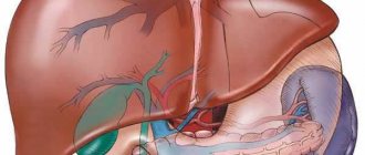
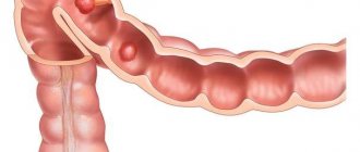
Small intestine: structure and functions
Anatomically, this section of the intestine is located between the stomach and the large intestine, and together with the large intestine makes up the entire intestine. The name “small intestine” is due to the peculiarities of its structure - it has thinner walls and a narrower lumen than the large intestine.
The small intestine, in the totality of all its sections, is the longest tubular organ of the digestive tract - in the body of a living person its length is 4-5 meters; after death, the walls, muscles and ligaments relax, and the total length of the small intestine can reach 7 meters.
The beginning of the small intestine is the pylorus of the stomach, followed by the duodenum, about 25-30 centimeters long, followed by the jejunum and ileum, which make up 2/5 and 3/5 of the total length of this section of the intestine, respectively. The small intestine ends at the ileocecal opening - at this point it passes into the large intestine.
Along the entire length of the small intestine, its lumen tends to narrow from 4-6 to 2.5 centimeters.
The main function of the organ is to convert beneficial substances contained in ingested food into soluble compounds so that they can subsequently be absorbed by the intestinal walls. Processing occurs under the influence of pancreatic enzymes and intestinal juice, as well as bile.
As a result, proteins, fats and carbohydrates are broken down into amino acids, fatty acids and monosaccharides, respectively.
Next, these substances, along with water and salts, are absorbed into the blood and lymphatic vessels, through which they are carried to all tissues and organs in the human body. In addition, the small intestine also takes part in the hormonal regulation of the body.
Its specific cells - neuroendocrine - produce various hormones, including: serotonin, gastrin, histamine, secretin, cholecystokinin.
Along the entire length, the walls of the small intestine have the same structure. They are formed by four layers:
- mucous inner lining;
- submucosa;
- muscle layer;
- outer serous membrane.
results
What does a stomach x-ray show:
- abnormalities in the structure of the digestive tract;
- acute expansion or narrowing of the lumens of the stomach/esophagus;
- malformations of certain organs of the gastrointestinal tract;
- hypertonicity/hypotonicity of the muscular wall of the stomach;
- tumors, papillomas, foreign bodies;
- reduction or radial arrangement of shell folding;
- cicatricial changes at the site of tumors, ulcers, chemical burns.
What does an intestinal x-ray show:
- pathological narrowing;
- saccular protrusions and elasticity of the walls;
- the introduction of one section of the intestine into another with the possible development of gastrointestinal obstruction;
- intestinal motor function;
- the presence of ulcerative and inflammatory processes;
- tumors, polyposis.
An examination can show how the bauhinium valve functions. This is the structure that separates the small and large intestines and is responsible for passing food between them. If it has pathological changes, then the food gets back access, and this poses a danger to the patient’s life.
Interpretation of the results of an X-ray examination with barium
https://youtu.be/ukgHLpQZiWs
When interpreting the results, the radiologist can identify the following diseases and pathologies:
- Foreign objects in the gastrointestinal tract. The presence or penetration of sharp objects into the walls of organs is clearly visible in the photographs.
- Intestinal obstruction is diagnosed when barium spreads in steps or jerks.
- Diverticulum. Stretching and enlargement of the walls of the organ with subsequent rupture appears on the image as a protrusion or unevenness.
- Esophageal dyskinesia (impaired peristaltic function of the esophagus). Characterized by darkening.
- Hernia in the esophageal part of the diaphragm. Possible lowering of the lower part of the esophagus into the abdominal cavity due to loss of rigidity by the diaphragm. It looks like part of the stomach is lifted through the diaphragm and the esophagus is displaced.
- Esophagitis. Swelling of the mucous membrane of the esophagus, decreased tone and impaired peristal function. Characteristic of internal burns. It appears as a ragged, uneven area.
- Esophageal ulcers. Characteristic points may be visible on the lower edges of the esophagus. They can often be accompanied by the presence of the same ulcers on the walls of the stomach.
- Formations inside the esophagus with benign signs. They have a fuzzy shape and a homogeneous structure.
- Fibroids and types of tumors that develop on the inner wall of the esophagus have unclear shapes and ragged edges.
Reviews
Reviews from patients generally agree that this procedure is not the most unpleasant, but its main disadvantage is the radiation exposure.
X-ray of the stomach, esophagus and small intestine with barium is an important and informative examination method. For it to be beneficial, you need to properly prepare for it, and during the implementation process you need to clearly cooperate with the radiologist.
An intestinal X-ray is a diagnostic procedure that allows you to identify the pathology of this organ by irradiating it with X-rays. An X-ray image can be displayed either on a screen, which allows you to monitor the procedure immediately at the time of its implementation, or printed on a special film and then studied by a radiologist.
What does the passage affect?
First of all, a passage is a means of evaluating a text by a search robot. If you want your page to appear in the search results for some query, when writing text, take into account the principles of division into passages:
- Do not enter words from the key phrase too far apart from each other.
- Do not separate words with tags, periods, question marks, or exclamation marks.
Passages influence the formation of the snippet. A snippet is a description of a page that is displayed in the search results list under the link to the site.
When forming it, the search engine takes information from the page that seems most relevant to the query, and where the keys are most densely located. Proper placement of words in a passage increases the chance that Yandex or Google will take this particular part of the text for a snippet. And a good snippet increases click-through rates. We wrote a separate article about how a snippet affects a site’s search results, read it.
Do not enter a bunch of identical keywords in one sentence, this can lead to pessimization of the page.
The inclusion of key phrases is like coal dust in the website promotion engine. Read about other fuels and the engine itself in our article about optimization of articles.
Why is radiographic examination performed?
X-rays may be taken with or without contrast. Barium is used as a contrast agent for X-rays. It is not harmful to the body, is not absorbed in the intestines and is easy to remove from the body. An X-ray of the large intestine with a contrast agent is called irrigoscopy.
An X-ray can only be prescribed by a doctor, most often a highly specialized specialist - a gastroenterologist, oncologist.
X-ray diagnostics are performed for:
- frequent constipation or diarrhea of unknown etiology;
- suspected Crohn's disease, diverticulitis, peptic ulcer;
- suspected adhesions, formation of fistulas, polyps;
- chronic inflammatory process in the intestines;
- ingestion of solid non-edible objects into the gastrointestinal tract (stomach, and then intestines) - a frequent indication for x-rays for children;
- suspected oncological tumors;
- patient complaints of abdominal pain, bloating, indigestion, changes in the consistency and color of stool;
- examination of a patient with congenital intestinal anomalies.
Pros and cons of x-rays
X-ray of the intestine with or without a contrast agent has a number of advantages: safety (radiation doses are minimal, the substance does not enter the blood), efficiency (detects even small changes in the structure of organs in 75% of cases, shows neighboring tissues).
Modern drugs have many regimens. You can adjust the beam delivery (dose and area). X-rays of the intestines with contrast (barium) are allowed even for children.
The disadvantages of the procedure are minor. After a barium X-ray of the stomach, diarrhea may occur. Sometimes there is intolerance to the drug. After administration of the substance, other diagnostic studies cannot be carried out, since the picture may be distorted (the contrast is displayed after a couple of days).
The barium diagnostic method can be used to examine children
Sometimes x-rays of the colon are done with double contrast, which involves introducing a small amount of air into the rectum to straighten it and create the most accurate picture possible. This may cause some discomfort (a feeling of fullness).
Preparation for the procedure
X-ray of the intestines requires careful preparation for the examination, and if everyone is accustomed to the fact that an X-ray of an arm or leg can be done at any time, then with the organs of the gastrointestinal tract things are more complicated. The intestine is a hollow organ; in order to examine it, it is necessary that there is nothing in the small and large intestines, not even food. Preparation for x-rays begins two to three days before the expected examination.
Basically, you need to give up fresh vegetables (cabbage, legumes), carbonated and alcoholic drinks, fatty foods and heavy dishes (fried meat), as well as flour. You need to carefully follow the drinking regime and drink at least two liters of water a day.
You should also start taking laxatives as prescribed by your doctor. They will help the intestines prepare for x-rays and will have time to clear it of stagnant feces. In the evening, on the eve of the x-ray, you need to do two cleansing enemas. A repeat enema is done in the morning on the day of the x-ray. People who smoke will have to give up this habit three hours before the procedure. Before going for an examination, you need to empty your bladder. On the morning of the x-ray, you can only drink water and not eat.
Video
X-ray of the intestine: the essence of the examination
The essence of the examination boils down to the following features:
- The procedure will be carried out using an X-ray machine. She will scan the necessary areas and areas of the gastrointestinal tract and identify all areas susceptible to a crisis.
- The passage of barium through the small and large intestines makes the picture of the study more informative and complete. The hollow intestine, when scanned without a barium mixture, appears unclear.
- X-rays show only dense areas of tissue. Barium for intestinal X-rays is illuminated in white, which makes it possible to highlight the relief of the entire intestine.
A video from the Virus-1 channel will help you understand and study in more detail the entire process of examining the gastrointestinal tract with contrast.
When preparing for a fluoroscopy procedure, the patient must do the following:
- A few days before the procedure, you must begin to follow a special diet that excludes foods containing flour, milk, baked goods and sweets. Sweet and carbonated water should also be excluded.
- Do not eat fruits, vegetables, beans, legumes, nuts. These products provoke gas formation and complicate the fluoroscopy process and negatively affect the identification of the disease and the passage of barium through the intestines.
- The day before the fluoroscopy, the patient should begin taking medications that empty the intestines. One of these drugs is Fortrans. This is one of the few drugs that allows you to comprehensively cleanse the small and large intestines. It is taken orally, after dissolving the packet with the powder mixture in a small amount of water. The course of admission consists of 4 packages. They must be taken within one hour. Then the maximum effect of the drug is achieved.
- On the day of the procedure, the patient should not be allowed to eat. Fluoroscopy is prescribed, in most cases, in the early morning.
Performing fluoroscopy
X-ray of the small intestine suggests the following procedure:
- Arriving at the hospital, the patient must remove all metal objects and preferably wear comfortable clothing.
- The patient takes a place on a special X-ray table, the limbs and torso are fixed.
- The first photo is taken. Next, he is asked to drink barium, or it is supplied to the body through a tube with a special supply of contrast along with air.
- The radiologist takes pictures every 45 minutes for five hours while the barium begins to spread through the small intestine.
- If the passage of barium through the intestines is impaired, then the radiologist can press on the patient’s abdomen to restore the passage and distribute the contrast evenly throughout the small intestine.
- After receiving eight images from different points, the X-ray review stops. The last picture is taken after the patient has emptied his body.
The patient is warned that barium will leave the body naturally within a few days, and therefore the stool will be white.
X-ray examination of the colon is performed slightly differently:
- The patient takes a position lying on his side, the lower part of the body is without clothes, the upper leg is pulled up to the stomach, the lower one is simply bent at the knee.
- The rectum is examined with a sigmoidoscope.
- Using the Bobrov apparatus, a barium mixture is slowly fed into the large intestine, which must first be diluted. You need to dilute with warm water 35 degrees.
- While the fluid enters the body and spreads through the large intestine, the patient is asked to turn from side to side, or this is done using a special table that rotates the patient relative to the x-ray.
- The radiologist makes sure that barium does not leak out of the anus, otherwise the procedure will be pointless.
- When the barium reaches the small intestine, the procedure is stopped. Throughout the procedure, a picture is displayed on the screen or pictures are taken.
Features of diagnostics with barium sulfate
Features:
- If the procedure involves examining only the large intestine, then barium will be injected directly into the organ being examined.
- If a comprehensive examination of the large and small intestines is carried out, then the barium sulfate solution will need to be drunk immediately before the procedure.
- A barium suspension solution is prepared immediately before the procedure. The mixture consists of the components of barium sulfate, aluminum gel and air; in addition, the solution may contain sodium citrate, sorbitol, and an antispasmodic. The dosage ranges from 400 to 650 grams.
Before starting to take the mixture, the doctor must make sure that the patient has completed all the necessary preparations:
- removed all metal jewelry, hairpins, pins and other objects that could interfere with the operation of the X-ray machine;
- took off all his outer clothing and underwear and put on a robe;
- took the position indicated by the doctor.
Depending on the nature of the study, the patient may:
- Vertical position. This speeds up the process of passage of the barium solution through the gastrointestinal tract.
- Horizontal position. It will allow you to examine the stomach as accurately and efficiently as possible.
Sometimes a Trendelenburg position may be required, in which:
- the patient’s pelvis is higher than the torso and elevated by 45 degrees;
- the diaphragm, stomach and intestines are examined as accurately as possible;
- The barium solution travels slowly and fills all parts of the intestine;
- mechanical indirect massage of the anterior abdominal wall evenly distributes barium sulfate throughout the esophagus and stomach.
Then the composition is taken in sips, the order of which is:
- A small initial sip. Allows you to check the quality of the solution and its speed of movement through the esophagus.
- The rest of the dose. After this, the barium solution completely fills the entire gastrointestinal tract.
The x-ray is taken within 40 minutes, while the doctor records from 6 to 10 images. The last picture will be taken after the barium solution has been evacuated from the body.
After the procedure, the patient may experience minor complications:
- mild nausea, which will go away on its own after some time;
- constipation caused by the astringent properties of barium sulfate solution;
- unusual white stool color that is caused by the color of the barium mixture.
What does an intestinal x-ray show?
X-ray with or without contrast can show different parameters; in the first case there will be more of them, so X-ray with barium enema is a more informative research method.
X-ray will help establish the following indicators:
| X-ray without contrast | X-ray with contrast |
| Position of the intestines relative to other organs | Appearance of the intestinal walls |
| Is the intestine enlarged, is there inflammation? | Presence of wounds, ulcers, diverticula or polyps |
| Diameter of the intestinal lumen | Presence of a tumor, its location |
| Elasticity of the intestinal walls | Elasticity of the intestinal walls |
| Functional ability of the mucosa | Intussusception |
| Intestinal obstruction, the location where the obstruction is located | |
| Performance of the Bauhinium valve |
additional information
The barium passage involves refusing to eat on the day of the study. In addition, you need to prepare carefully using the following simple rules:
- 3-4 days before the test, do not consume foods that can increase gas formation in the intestines, namely legumes, cabbage, and fruits.
- Stick to a slag-free diet. Avoid soda, chips and any fast food.
- The day before the passage, take a special drug to cleanse the intestines. Most often this is Fortrans.
After the patient drinks about 500 ml of barium, he must wait until the suspension reaches the small intestine. It may take up to 5 hours. When the goal is achieved, the doctor begins to take pictures using special X-ray equipment.
If barium in the small intestine changes its structure and becomes like small particles, this indicates a malfunction of the organ, or rather poor absorption. Dark areas in the image indicate the presence of tumors and other similar changes in the mucous membrane.
This procedure is not suitable for everyone. It is not performed during pregnancy or severe stomach pain. You should tell your doctor about the presence of all accompanying symptoms.
Price
X-rays of the intestinal tract will vary in cost depending on which part of the intestinal tract is examined and whether a contrast agent is used or not. You can find out how much this procedure costs for different cities in the table.
| City | Price for x-ray without contrast | Price per x-ray with contrast |
| Kyiv | 120-895 UAH | 350-1055 UAH |
| Moscow | 2600-7800 rub. | 5400-10100 rub |
| Saint Petersburg | 2500-4500 rub. | 3500-5700 rub. |
| Nizhny Novgorod | 513-1700 rub. | 1200-2500 rub. |
Video