Blood flukes, which have many types, are the causative agents of schistosomiasis, a group of diseases that rank second after malaria in the number of deaths. Suffice it to say that in Africa, up to 200 thousand people die every year from schistosomiasis.
To date, the characteristic features of blood flukes, their morphophysiology, as well as the routes of infection have been well studied, however, only treatment at a mild or moderate stage of the disease can provide a favorable outcome.
What to do in such a situation? To get started, we recommend reading this article. This article describes in detail methods of controlling parasites. We also recommend that you consult a specialist. Read the article >>>
In severe cases of schistosomiasis, surgical intervention is required.
A vaccine against these parasites has not yet been invented, so preventive measures play a huge role.
What is a liver fluke?
The liver fluke is a flatworm that belongs to the class of digenetic flukes. It parasitizes the bile ducts, as well as the liver of humans and other warm-blooded animals.
Liver flukes are various types of worms, including:
Giant liver fluke.
Cat fluke, etc.
The giant fluke and hepatic fasciola are dangerous to humans. These two types of flukes cause a disease called fascioliasis.
The giant fluke has quite impressive dimensions; its body can reach 76 mm in length and 12 mm in width.
The liver fluke has a flat, leaf-shaped body. There are two suckers on the head of the parasite. The liver fluke can reach 20-30 mm in length and 12 mm in width. Immediately after the parasite's short esophagus there are two loops of intestine that end blindly. Worms are hermaphrodites; inside their body there are unpaired ovaries, a small uterus and two branched testes. The parasites release large oval eggs covered with a yellow shell into the external environment.
Outbreaks of parasitic infestations can be both widespread and sporadic. The statistical coverage of the population affected by fascioliasis ranges from 2.5 to 17 million people. The main symptoms of human infection with the liver fluke are: fever, urticaria, nausea, pain in the right hypochondrium.
The permanent host of the liver fluke is most often domestic animals (cattle, small cattle, horses, rabbits, donkeys and sheep); humans are less often infected. The liver fluke can parasitize the body of wild animals, including kangaroos, beavers, deer, squirrels, etc.
The next liver fluke that parasitizes the human body is a trematode of the genus opisthorchis, which provokes a disease called opisthorchiasis.
An adult is a flat parasite whose body can reach 18 mm in length and 2 mm in width. The anterior end of the worm's body is pointed, the suckers are located on the peritoneum and on the upper part of the body (oral sucker). The digestive system of the parasite is not closed; there is an excretory canal at the rear end of the body.
Trematodes have both male and female reproductive organs. For reproduction, one individual parasitizing the body is enough.
This parasitic invasion is widespread throughout the world, but the palm belongs to Russia. In some regions, the number of infected people reaches 75%, which is explained by food traditions (consumption of dried, lightly salted or frozen fish). Cases of opisthorchiasis are registered in Belarus, Kazakhstan, Ukraine, Western European countries, etc.
Another worm is the liver fluke, which can parasitize the human body - the lanceolate fluke. This fluke causes a disease called dicroceliosis.
The parasite reaches 15 mm in length and 5 mm in width. Just like other liver flukes, lanceolate flukes are hermaphrodites. They lay eggs that have already developed miracidium. The larva hatches after it enters the body of the first intermediate host.
It should be noted that dicroceliosis is a rare disease among humans. Medicine knows only isolated cases of parasitic infestation by this liver fluke. The final host of the lanceolate fluke is most often small and large cattle.
Let's remember biology
The parasites belong to a type of flatworm, a class of digenetic flukes.
Classification
The genus name Fasciola comes from the Latin word fascia, meaning ribbon. The body of the helminth really resembles a flat braid.
This group includes the following types:
- eastern (Chinese) fluke;
- sheep liver fluke;
- giant fluke;
- cat fluke;
- hepatic fasciola.
Flatworms of the family Fasciolidae (liver fluke and giant fluke) are the cause of the disease fascioliasis, which occurs with inflammation of the gallbladder and hepatic colic.
The giant fluke (Fasciola gigantica) is a large worm. Its length is 33-76 mm, width – up to 12 mm.
Big sizes
Fasciola gepatica has a leaf-shaped body, its length is 20-30 mm, width - 12 mm. There are two suckers on the worm's head. The worm's pharynx passes into the esophagus, from which two branching intestinal loops extend.
Anatomy
Fasciolas are hermaphrodites, each individual has an ovary, a small uterus and testes. Worms release oval-shaped eggs covered with a yellow shell into the external environment. There is a larva inside. There is a cap on one of the poles of the egg.
The final hosts of the fluke are numerous herbivores (odd-toed ungulates, small and cattle) and humans.
Life forms
Intermediate hosts are different species of mollusks that live in fresh water bodies (small pond snail).
The main source of infection is herbivores, which release parasite eggs into the external environment along with feces. When eggs fall into the water, helminth larvae (miracidia) hatch after 1-1.5 months, which penetrate the body of freshwater mollusks.
Small pond snail
In the body of intermediate hosts, asexual reproduction of the larvae occurs and the next stage of development, cercariae, is formed.
Wet pasture
When the water temperature exceeds 9 degrees, they come out and attach themselves to underwater rocks and algae, where they transform into adolescaria. This life form can remain viable for a long time (up to 5 months) and can also withstand prolonged drying. Adolescaria remain alive in dry hay for several months.
Animals often ingest helminth larvae when eating grass or drinking water from ponds with standing water.
She has no choice
It is dangerous for humans to drink unboiled water and eat watercress, which grows in natural bodies of water.
Care must be taken when preparing
Fascioliasis is common in Africa, South America and Asia. Outbreaks of the disease are common in Portugal and France.
The first signs and symptoms of liver fluke
The first signs of a liver fluke are associated with the incubation period of the development of the parasite.
There are three stages of parasitic infestation:
Symptoms of the liver fluke during the incubation period
The incubation period can last from 7 days to 2 months. It depends on how many parasites have entered the body. During this time, the infected person does not experience any signs of illness.
Symptoms of liver fluke in the acute stage
This stage is also called the migration stage.
It is characterized by general toxic and allergic symptoms, including:
- Increased body temperature. The mark on the thermometer may remain at the level of low-grade fever, or may rise to 39-40 °C. The temperature can rise in waves, or it can be remitting, when daily fluctuations exceed 1-2 degrees.
The patient experiences weakness, headaches, and general malaise.
The most common allergic reaction is urticaria, which is accompanied by severe itching. The development of Quincke's edema is possible.
Dyspeptic disorders are characteristic, including: pain in the right hypochondrium, nausea and vomiting.
The liver increases in size and jaundice develops. When palpated, the organ responds with pain.
Chest pain is a sign of developing myocarditis of an allergic nature. Possible increase in blood pressure and increased heart rate.
The number of eosinophils and leukocytes in the blood increases.
The acute phase can last several weeks; upon completion, the symptoms of the disease disappear.
Symptoms of liver fluke in the chronic stage
Chronitization of the parasitic disease occurs 3-6 months after the invasion.
In this case, symptoms indicating damage to the liver and biliary tract come to the fore:
The patient experiences frequent paroxysmal pain in the right side.
During an exacerbation of the disease, jaundice develops.
The liver remains constantly enlarged in size.
The longer the untreated infestation persists, the worse the prognosis. The formation of liver cirrhosis, hepatitis, and anemia is possible.
Secondary infection is dangerous, which can cause liver abscess, purulent cholangitis and cholecystitis.
Medicine knows of cases where liver flukes developed in the mammary glands, in the brain, in lung tissue, in the larynx and under the skin. Prolonged stress of the immune system makes a person susceptible to diseases of various etiologies.
Symptoms of caused diseases
The presence of a helminth in the body is manifested by signs of intoxication:
- fever;
- nausea with vomiting;
- frequent diarrhea;
- soreness in muscles and joints;
- loss of appetite;
- weakness;
- headache.
Opisthorchiasis and other pathologies caused by parasites can also be accompanied by symptoms in the form of sore throat, cough, runny nose, and swelling of the larynx.
The nature of the symptoms is influenced by the location of the fluke in a person. When it enters the liver, the patient experiences:
- skin rashes;
- yellowing of its integument;
- itching;
- soreness under the ribs on the right side.
Routes of infection with liver fluke
Infection with liver fluke and giant fluke occurs when eating plants (garden and wild) that were watered with water from fresh water bodies, provided that there were infective worm larvae in it. Infection is possible by drinking unboiled water or by swallowing water while bathing.
Infection with the liver fluke, which causes opisthorchiasis, occurs when a person eats infected fish that has not undergone high-quality heat treatment.
Infection with the liver fluke of the genus Fluke lanceolata occurs through accidental ingestion of infested ants, for example, during the consumption of berries, meadow grasses, vegetables from the garden, etc.
Thus, the route of infection with the liver fluke is food. That is, in order for any liver fluke worm to begin parasitizing in the human body, it will need to get into the gastrointestinal tract.
Methods for diagnosing pathology
The problem is that diagnosing diseases caused by the liver fluke is difficult. Worm eggs are found in human feces only 3-4 months after infection. Due to difficulties in making a diagnosis, treatment tactics may be chosen incorrectly.
To begin with, the doctor examines the medical history, conducts a visual examination, prescribes a series of immunological tests, blood tests, ultrasound of the abdominal organs, and radiography. Additionally, examination of the pancreatic ducts, bile ducts, magnetic resonance imaging, and computed tomography is indicated.
The final diagnosis is made after detection of parasite eggs in biological masses:
- contents of the duodenum;
- feces.
However, it must be remembered that so-called transit eggs can also enter the human body. At first it is not possible to determine them, so to obtain reliable data it is necessary to conduct a repeated stool examination.
The doctor needs to obtain information about the gastronomic preferences of the sick person, his place of residence, his occupation and visits to exotic countries.
Treatment of liver fluke
Treatment of liver fluke consists of two stages. The first stage is preparatory, and the second is aimed at directly eliminating the parasite from the human body.
The first stage of treatment for liver fluke
Treatment of this parasitic infestation is carried out in a hospital setting, although outpatient management of the patient is possible if the patient is in satisfactory health. If the disease is diagnosed in the acute phase, the patient is transferred to a gentle diet and prescribed desensitizing drugs. To eliminate allergic manifestations, medications such as Suprastin, Tavegil, Zyrtec, Zodak, etc. can be prescribed.
As for diet, when getting rid of liver fluke, table number five according to Pevzner is considered optimal. Spicy, smoked and fried dishes are being removed from the menu. It involves steaming, boiling and baking foods. After completing the second stage of treatment, the patient is recommended to consume more foods rich in fiber, which will enhance intestinal motility and improve bile discharge.
To remove toxins from the body, the patient is prescribed Smecta, Polyphepan and other sorbents. To reduce body temperature, you can take Ibuprofen or Butadione. In addition, these drugs act as anti-inflammatory drugs. Glucocorticosteroids are prescribed against the background of myocarditis.
The second stage - antiparasitic treatment of liver fluke
Antiparasitic therapy begins after the symptoms of the acute phase of the disease subside.
Drugs prescribed for the treatment of liver fluke:
Chloxyl (taken for 3-5 days).
Praziquantel (Biltricide). The drug is taken once.
Triclabendazole. Depending on the severity of the infection, a single or double dose of the drug is possible.
Choleretic drugs are prescribed to quickly remove dead parasites from the human body, in addition, they prevent stagnation of bile. These may be drugs such as Holiver and Holosas.
Control diagnostics with stool analysis and duodenal intubation are carried out 3 months later and another six months after treatment.
Taking antibiotics is necessary in case of purulent complications. Surgical intervention with drainage of the liver, bile ducts, etc. is not excluded.
The third stage is restoration of the body after treatment of liver fluke
Since the liver fluke disrupts the functioning of the digestive organs and the functioning of the body as a whole, patients will need to undergo a rehabilitation period.
It may include the following activities:
Normalization of the functioning of the gallbladder and liver with the help of a course of choleretic drugs (Holiver, Holosas, Allohol, Holagol, etc.).
Improving liver function, protecting and restoring it with the help of hepatoprotector drugs. These can be products such as Ursosan, Galstena, Silymarin, etc.
Normalization of digestive processes with the help of enzyme preparations (Pancreatin, Creon, Panzinorm, etc.).
Improving metabolism with multivitamins.
The earlier the disease is detected, the faster and easier it is to achieve a full recovery. If the parasite infestation is highly intense, or if a secondary bacterial infection is added, then the prognosis worsens significantly. Even the death of the patient is not excluded.
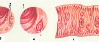
Structure and habitat
The photo clearly shows how the female is located in the male’s body.
Depending on the species, the size of the parasites may vary, but all blood flukes are characterized by a developed abdominal sucker and another sucker, the oral one, through which they literally suck blood and with it nutrients from the human body.
Blood flukes have a special structure, in addition to the gynecophoric canal: their digestive system consists of the anterior section (mouth) and the esophagus with two branches passing into the middle section.
In this case, the anus is absent, that is, the digestive system ends blindly, and waste products are excreted through the bladder and excretory opening.
Blood flukes have well-developed muscles, but rather weak sensory organs, although the larvae distinguish light and strive for it, which is what one of the methods for diagnosing schistosomiasis is based on. Their type of metabolism can be either oxygen or oxygen-free.
As already mentioned, blood flukes are widespread in regions with tropical and subtropical climates: Africa, Latin America and the Caribbean, the Middle East, China, and Southeast Asia.
Statistics show that the blood fluke, the causative agent of schistosomiasis, is found in more than 200 million people in countries where the risk of epidemics is high, and approximately 700 million more people may be infected.
In Europe and North America, schistosomiasis is diagnosed mainly in people who have visited these regions.
Prevention of liver fluke
Prevention of liver fluke is a set of measures that will prevent the development of the disease:
Under no circumstances should you drink raw water from reservoirs.
Vegetables, fruits, garden herbs, and berries must be thoroughly washed before eating.
You should avoid eating raw fish.
An important role in the prevention of liver fluke is assigned to public services. They are obliged to prevent pollution of water bodies from the ingress of fecal matter into them, and to combat the proliferation of mollusks. No less important are preventive measures for deworming livestock, as well as conducting sanitary and educational work among the population.
Doctors have the task of timely identifying patients who are carriers of liver fluke and providing them with quality treatment. If you suspect a parasitic infestation, you should contact an infectious disease specialist.
Education: in 2008, received a diploma in the specialty “General Medicine (Therapeutic and Preventive Care)” at the Russian Research Medical University named after N. I. Pirogov. I immediately completed an internship and received a diploma as a therapist.
13 effective spices for weight loss
5 best products for male power!
It is well known that parasites in the human body strive to exist as inconspicuously as possible. However, our body is very difficult to deceive, and it gives signals to a person that an infection has occurred. The main thing is to be able to recognize these signals and interpret them correctly.
Ascariasis is a type of helminthiasis caused by roundworms belonging to the class of nematodes, family Ascarididae (roundworms). They parasitize the intestines of humans and animals. There are several types of these parasites that affect various representatives of vertebrates: cats, dogs, birds, pigs.
The symptoms of toxocariasis are so diverse that doctors practicing in various branches of medicine - neurologists, ophthalmologists, hematologists, gastroenterologists, and therapists - can encounter its manifestations. Although researchers called humans an “ecological dead end” of toxocariasis, because it is unusual.
Anthelmintic drugs are used to treat various types of helminthiasis - damage to the body by parasites. These agents can be either universal (broad-spectrum) or selectively acting on any type of helminth. Until recently, the pharmaceutical industry could not offer parasite expulsion to patients.
In case of pinworm infection, all close relatives of the infected person are subject to preventive treatment. Testing for the presence of pinworms often gives a false negative result. The ease of infection with enterobiasis through household items and shared bedding, contact with animals and handshakes, as well.
LIVER FLUKE, flatworm of the class of flukes. It parasitizes the internal organs of large and small ruminants and is also found in humans. The leaf-shaped body of the worm reaches 30 mm in length and 13 mm in width. In the mature stage, the fluke lives in the bile ducts of the liver and can lead to their blockage. Flukes are held in the walls of the ducts by two suckers - oral and abdominal. From the effects of the host animal's bile, the body of the liver fluke is protected by a multilayered dense shell - the cuticle. Like most flatworms, liver flukes are hermaphrodites. An adult, while in the body of a host animal, produces a large number of eggs daily, which enter the host’s intestines along with bile and are then released into the external environment. Further development of the eggs occurs in water, where cilia-covered larvae emerge from them. In a short time, the larva must find an intermediate host - a small pond snail. A change of two generations of the parasite occurs in its body, as a result of which tailed larvae are formed, which again go out into the water. They actively swim, and then settle on the coastal vegetation of the reservoir and become covered with a dense shell (turn into a cyst), maintaining viability in this form for a long time. Domestic animals, eating grass, swallow the larvae along with it. In the intestines of these animals, the membranes dissolve, the larvae enter the bloodstream, are carried through the bloodstream into the vessels of the liver and penetrate the bile ducts, gradually turning into an adult parasite.
A person can become infected with liver fluke by drinking water containing its larvae.
Published by: Parazitolog in Helminths February 21, 2019
The liver fluke is one of the most famous parasites. Another name for this parasite is cat fluke. The liver fluke has a complex life cycle and is characterized by frequent changes of hosts. The fluke can parasitize not only human organs, but also fish and herbivores, which act as a secondary host.
Carrier of the parasite
The development cycle of the liver fluke includes the following stages:
- An egg released with the feces of the previous owner into the aquatic environment.
- A larva wandering in search of mollusks living in the water.
- Transformation into a sac with embryos (sporocyst) inside the mollusks.
- A larva with a tail (cercaria) that has left the mollusk and is freely floating in the vastness of the reservoir.
- Adolescaria is a larva that has emerged onto land and is covered with a protective shell. She waits for her main owner - a sheep, pig, horse, cow, etc. Often such an owner turns out to be a person.
- The adult is the liver fluke itself, developing inside the liver of the new host and laying eggs.
All intermediate stages are harmless with respect to human infection. Only adolescaria can be the beginning of the development of the parasite in the liver.
A mature individual grows up to 3 mm in length, and it is this that causes devastating consequences for human internal organs. The development cycle of the liver fluke in the animal body is no different from the maturation of individuals inside the liver in humans. There is only one difference: in humans this process is more unnatural.
Diseases of the internal organs occur from mechanical damage to the walls of the liver. And since the development cycle of the liver fluke includes the throwing of eggs, this also increases the risk of acquiring pathologies of internal tissues. The eggs of the parasite are so small that they are able to penetrate the blood and wander for a long time with its current. At high concentrations, blockage of small vessels can occur.
The development cycle of the liver fluke and the bovine tapeworm is the same in terms of the possibility of infection. The cysts of both parasites live on plants near water bodies or drift freely in the water. Where livestock lives and goes to water, parasite eggs are sure to exist. This means that it is much more likely to become infected while swimming in such a pond or relaxing on its shore.
If we consider the development cycle of the liver fluke separately, its sequence can be briefly described as follows:
- domestic animal (not necessarily just cattle);
- water;
- shellfish;
- cyst;
- mammal.
That is, if one of the conditions is removed from the chain, the parasite dies. If a mollusk called a pond snail lives in a pond, you can confidently judge that the plants in the area are infected.
The specialized literature provides many variations describing the development cycle of the liver fluke. The pictures depicting people affected by the parasite are terrifying. The effects of the tapeworm are destructive to the liver. If the case is advanced, the organ must be replaced, and not every person can afford this expensive procedure.
If the parasite is detected in time by certain symptoms, disability can be prevented. The treatment ends positively and is carried out in a hospital. Pills alone cannot remove the parasite from the body. However, often, if negative health factors are excluded, the parasite can die under the influence of immunity.
The huge parasite, the bovine tapeworm, is known for its nasty suckers and hooks with which it clings to the walls of the human intestine. The body of the parasite can reach 3 m. When removing it, they try to pull out the head, since if it remains inside, the worm will continue to grow again.
You can become infected with bovine tapeworm from livestock while caring for it. But meat is also dangerous to health, since parasite cysts live in muscle fibers. They get here when cows eat contaminated grass. The eggs of the parasite open, and the larvae penetrate the muscles of the animal through the blood ducts.
The next stage of helminth development is a condition called finna. Finna is a small, pea-sized vesicle with a small hole in which undeveloped suckers and hooks are located. This is the head of the future tapeworm. With it, the parasite attaches itself to the intestinal wall, after which it begins to build up its body.
Human and animal feces are carriers of tapeworm or fluke eggs. Therefore, isolation of latrines and their subsequent treatment can stop the spread of infection. Periodic cleaning of the bottom of reservoirs from silt and shellfish is also used. Livestock should be taken for grazing only in uninhabited areas.
Meat must undergo long-term temperature treatment. And the safest cooking method is boiling. Livestock should be constantly checked for parasites and treated. At the same time, it is necessary to observe the rules of personal hygiene.
Fluke class
The class of flukes includes about 4,000 species that parasitize the internal organs of humans and various animals.
A representative of the class is the liver fluke, which lives in the liver of cattle. It has oral and ventral suckers. With their help, the worm is kept inside the host's liver. The fluke feeds on blood, sucking it through the oral sucker.
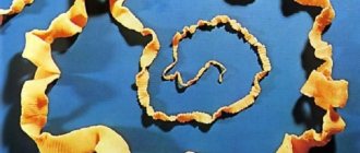
Once in the water, microscopic larvae equipped with cilia develop from the eggs. They penetrate the body of the small pond snail mollusk, in which they grow, multiply, and tail larvae appear.
These larvae leave the mollusk, actively swim in the water, then attach to plants, cast off their tail, and become covered with a thick shell—a cyst is formed. With grass or water, the cyst enters the cow's intestines, where it develops into an adult worm. A person can become infected with liver fluke if he drinks water from a dirty pond.
Another parasite that can infect humans and mammals (dogs, cats, wolves, etc.) is the cat fluke.
Unlike the liver fluke, its life cycle occurs with the participation of two intermediate hosts: a freshwater gastropod - bithinia - and freshwater cyprinid fish. A person or animal becomes infected with cat fluke when they eat raw, insufficiently salted or cooked (fried) fish.
How do you get infected with tapeworm?
If we understand the entire developmental cycle of the liver fluke, the routes of human infection will be clearer and they can be excluded. So, consuming raw water, especially from a reservoir, is dangerous. It is not recommended to have a picnic on the shore, in a place where pets can be walked. Unwashed hands after swimming in a pond can cause cysts to enter the body.
You can also get infected from pets. Cysts can cling to the fur of cattle, birds, and dogs, which they wait on plants near a pond. A person can also be a carrier, but he is not dangerous to others.
Similar to the previous parasite, bovine tapeworm can be infected through food. However, the Finn is much larger and cannot be swallowed so easily. Therefore, cases of infection are quite rare and occur with absolute carelessness of a person. Raw meat is the main cause of infection.
We invite you to familiarize yourself with Fleas in cats - causes, symptoms and treatment
The parasite can only penetrate unnoticed with food. Those who love rare meat run the risk of acquiring an unpleasant parasite for many years. The parasite pulls out all the beneficial substances from food, preventing the body from getting what it needs. And when the tapeworm reaches enormous sizes, shock may occur and the intestines may become blocked.
What is a liver fluke
A dangerous parasite that infects the liver and causes fascioliasis is the liver fluke. Let's look at its life cycle, routes of infection and methods of destruction.
Helminth refers to a parasitic form of flukes that live in the organs of vertebrates (animals, humans) and invertebrates. Another name for the parasite is cat fluke, since a cat is the most suitable host for the worm.
The adult infects humans and cattle.
The fluke has a leaf-shaped body and reaches a size of 3-5 cm. It can get into the organs of herbivores and fish, which act as a secondary host.
Main characteristics of the parasite:
- there is a special cover on the body that protects it from digestion by the host’s juices;
- many fastening devices: hooks, suction cups, etc.;
- simple structure of the digestive system;
- high fertility and asexual reproduction;
- regressive development of sensory organs and nervous system.
The parasite is characterized by a complex life cycle with transformations and frequent changes of hosts. This leads to its dispersal and protects the main carrier from excessive overpopulation and death. Most often, human infection occurs when drinking unboiled or untreated water.
Main types
Three types of liver fluke can parasitize the human body:
- Fasciola vulgaris. It has a leaf-shaped body up to 2–3 cm in length. Causes a serious helminthic infection - fascioliasis, which is characterized by cholecystitis and hepatic colic. Infection can occur by drinking dirty water, fruits and vegetables.
- Asciola gigantea. The body of the individual has a tongue-like shape up to 7 cm and two suction apparatuses: oral and abdominal. In some parasites, a third sucker is also observed, located at the posterior end of the body.
- Chinese liver fluke. It is the causative agent of helminthiasis called clonorchiasis. It received its name because it is most active in eastern countries such as China, Korea and Japan. The length of an adult worm can range from 10 to 25 cm. The parasite needs to visit two intermediate hosts. The first is a freshwater snail, the second is a freshwater fish. Ultimately, when a person or animal eats unfried or undercooked fish, fluke larvae penetrate the consumer’s body.
Developmental cycle of the liver fluke
During their existence, these helminths go through several stages.
The stages of development of the liver fluke have a certain sequence:
- the first larval stage is the miracidium, which emerges from the egg and floats in the water until it penetrates the body of the mollusk;
- after this implementation, a sporocyst is formed, which begins to reproduce by parthenogenesis;
- The further stage of the life cycle of the larvae of these helminths is redia, the structure of which already contains the rudiments of organ systems. Also in their body there are germ cells, from which a new larval form - cercariae - is parthenogenetically hatched;
- A special feature of the structure of the cercaria is the presence of a brain, an eye and a powerful tail, which gives it freedom of movement. At this stage, the larva emerges from the mollusk, trying to quickly get out of the water;
- The cercaria on land transforms into a cyst, losing its tail, and attaches to coastal plants. This stage is called adolescaria. So the fluke can stay for a very long time until it enters the body of a person or animal that feeds on grass;
- After successful introduction of the larva into the host's body, it is transformed into a sexually mature individual - marita. At this stage, the trematode lays eggs (thousands of eggs per day!), then they are released along with feces into the external environment. If they get into the water, the helminth development cycle repeats.
Frequently asked questions to the doctor
Is a patient with fascioliasis contagious?
Hello! My husband recently became ill and was told that hepatic fluke eggs were found in his stool. I think I picked up this nasty thing when I was on a hike: I said that I drank raw water from the lake. Tell me, could I get infected from him? I’m very worried, I wash my hands twenty times a day, I see these eggs everywhere.
Good afternoon No, it is impossible to become infected from a sick person. Even if it excretes helminth eggs with feces, the larvae that appear at this stage of development survive only in the body of the intermediate host (mollusk).
Life cycle of the liver fluke
Fasciola is characterized by frequent transformations and changes of hosts. The life cycle of the liver fluke is represented by the following chain: final host; egg; miracidium; intermediate host (pond snail); sporocyst; maternal redia; daughter redia (cysts); cercariae; adolescary; adolescaria in the external environment.
The liver fluke begins to develop from an egg from which a miracidium emerges. The larva has a nerve ganglion, excretory organs and a light-sensitive eye. The germ cells are located in the back. The front part of the body has a gland that produces an enzyme that dissolves living tissue and penetrates the intermediate host.
At this stage, reproduction occurs without fertilization using germ cells. The sporocyst bursts and redia emerge, which parasitize the same host. Rediae have a number of formed organs: mouth, digestive tube and pharynx, opening for the release of eggs.
Each cyst contains germ cells from which the next larval generation, cercariae, is formed. The cercarium has suckers, a gut, an excretory system, and a nervous system. The larva has a long muscular tail. The cercarium emerges from the mollusk and moves in the water.
Free-swimming cercariae attach to plant stems and objects in water, becoming covered with a membrane. This stage is called adolescaria. The future fluke has a spherical shape.
If the larva is swallowed by an animal from among the definitive hosts, then the fasciola shell dissolves in the intestines of the host and the helminth enters the liver, where it develops to a sexually mature state. Infestation of animals occurs when they eat grass in water meadows and when drinking water from contaminated reservoirs. Humans are infected through contaminated vegetables.
Internal structure of the liver fluke
In humans, the causative agent of fasciola can be either Fasciola vulgaris or giant fasciola. Both have a specific and almost identical structure and functioning, which is due to their parasitic lifestyle. Let's consider the internal structure of the liver fluke.
The parasite has a leaf-shaped body, 2-7 cm in size and grayish-yellow color. Lives in the bile ducts, liver and pancreas of vertebrates. With the help of oral and abdominal suckers, it is attached and held on the walls of the ducts.
The fluke is characterized by simplification and specialization in the structure of some organs. This is due to her parasitic lifestyle.
The specialization includes suckers, spines and other formations on the body of the worm, powerfully developed genital organs and several complex life cycles. Morphological simplification is expressed by the absence of sensory organs in sexually mature individuals, which act as endoparasites
The main vital systems of a worm:
- digestive - the mouth opening is connected to the muscular pharynx (sucking apparatus). Behind the pharynx there is a branched esophagus and blindly ending intestines;
- nervous - is a peripharyngeal nerve ring, from which three pairs of nerve trunks arise (the lateral ones are the most developed). The nerve trunks are connected by bridges, which makes them look like a lattice;
- excretory – developed protonephridia deep in the parenchyma. Thermal cells have channels with cilia, which select tissue fluid and dissimilation products from the parenchyma. Cilia move fluid through the channels and excretory pores, removing it from the body;
- sexual – the helminth is a hermaphrodite. The male reproductive system is represented by a pair of testes, two vas deferens, which merge into the ejaculatory duct and the cirrus. The female reproductive system is more complex: ovary, vitelline, spermatheca, ootype (fertilization and formation of eggs occurs in it), uterus and genital opening. In some species, fertilization occurs in the spermatheca. In most cases, insemination is cross-insemination, but there are also cases of self-insemination.
Stages of disease development
The faces of the liver fluke, which enter the human stomach with food and water, penetrate the liver.
Liver damage
Migration routes:
- with blood flow, through the portal vein;
- penetration into the liver through Glisson's capsule.
The larvae enter the bile ducts. The main organ of helminth fixation is the abdominal sucker, which ensures a stable position of the parasite in the liver. The worm also has semi-suction cups of the reproductive and excretory systems.
On the ventral and lateral surfaces of the body there are small spines and single suckers, which are also intended for attachment to the liver. After four months, the formation of the reproductive system ends.
Helminth eggs
Pathological effects of fasciolae on the body:
- mechanical damage to liver tissue;
- accumulating in the bile ducts, disrupting the circulation of bile;
- helminth waste products have a toxic effect on the body and often lead to the development of allergic reactions (especially in the early period of the disease);
- constant irritation of the nerve endings of the bile ducts leads to the formation of visceral reflexes, which affect the functioning of the digestive system and pancreas;
- impaired bile circulation is a favorable environment for infection (microabscesses often form in the liver tissue).
Blockage of the common bile duct
In the late stage of the disease, the lumen of the common bile duct expands, and the chronic inflammatory process leads to thickening of the walls. In some cases, purulent cholangitis develops.
Intermediate host of the liver fluke
As soon as the larvae enter the body of the final host, and this can be an animal or a person, they continue their development to sexually mature individuals. Acceptable environments for the survival of the parasite's offspring are animal and human excrement. With them, the worm eggs end up in water bodies, repeating their life cycle. This stage is called aledoscaria.
The worm can exist in an aquatic or humid environment for a long period of time, maintaining its viability.
The pond snail is an intermediate host of the liver fluke. The parasite larva invades the snail's body, where it lives and develops at its expense. An already grown individual leaves its carrier and attaches itself to the stems of aquatic and coastal plants using suction cups and spines on its body. At this stage, the helminth is covered with a protective shell - a shell.
Miracidium of the liver fluke
The fasciola egg begins to develop only when it gets into the water, where, under favorable conditions, after 25-30 days, a larva emerges - the miracidium. Miracidium has a nerve ganglion, a light-sensitive organ “eye”, and excretory organs. In the back part there are germ cells.
The anterior end of the body is equipped with a gland that produces an enzyme capable of dissolving living tissue upon penetration into an intermediate host. The miracidium is covered with cilia, thanks to which it actively floats in water. It does not feed, but exists due to the nutrients accumulated in the egg.
The parasite then develops into the next larval stage, the sporocyst. Sporocysts resemble a shapeless sac, devoid of any organs, including the nervous and excretory systems. This larval stage is capable of reproduction.
In the sporocyst, a new larval generation, the redia, develops parthenogenetically (i.e., without fertilization) from the germ cells. The sporocyst bursts, and the redia come out of it, but continue to parasitize the same host.
A number of organs are already being formed in redia: the mouth, pharynx, digestive tube and the opening for the exit of individuals of the new generation. Inside the redia, the next larval generation, the cercariae, is also parthenogenetically formed from the germ cells.
The body of the cercarium contains many organs characteristic of marita. It has suckers, intestines, nervous and excretory systems, but unlike the adult form, it is equipped with a long muscular tail, which provides forward movement. The cercarium leaves the mollusk and actively moves in the water.
Next, free-swimming cercariae attach to some object, such as plant stems, and become covered with a membrane. In this larval stage, called adolescaria, the fasciola is spherical in shape. If adolexaria is swallowed by an animal from among those that are the final hosts of fasciola, then the shell dissolves in the intestine of the host and the parasite penetrates the liver, where it reaches a sexually mature state.
Livestock most often become infested by eating grass in flooded meadows and by drinking water from reservoirs where Adolescaria may be present. People usually become infected through vegetables (most often lettuce) that are watered with water from reservoirs containing adolescaria.
It is interesting to note that in the life cycle of Fasciola there is a free-living stage - the miracidium, which is morphologically close to ciliated worms, which serves as one of the proofs of the origin of flukes from ciliated worms.
Diagnosis of liver fluke
Based on the presence of fluke eggs in feces, liver fluke is diagnosed. The parasite can be detected in healthy people after drinking contaminated water or food. Eggs begin to be excreted in feces 3-4 months after infection.
In the acute stage of fascioliasis, the diagnosis is made on the basis of painful symptoms.
The following methods are used in the diagnostic process:
- collecting anamnesis, that is, epidemiological data: swimming or drinking water from stagnant bodies of water, eating unwashed vegetables, fruits, as well as fish, meat or animal liver;
- clinical signs of pathology: early symptoms and signs of chronic fascioliasis;
- laboratory tests depend on the stage of the disease. In the early phase, serological methods are used, that is, studying the blood for antibodies - ELISA, RNGA reactions. In advanced stages, copro-ovoscopy or duodeno-ovoscopy is performed.
Based on the results of the diagnostic procedures performed, the doctor makes a final diagnosis and prescribes a treatment regimen for the helminth.
How to get rid of fascioliasis?
Treatment is carried out in a hospital.
Modern drug
Therapy includes:
- bed rest;
- diet (limitation of fats and coarse fiber);
- anthelmintic drugs: praziquantel, chloxyl, bithionol.
Parasites don't stand a chance!
In the acute period of the disease, specific treatment is prescribed after the elimination of allergic manifestations and normalization of body temperature. Three months after the course of treatment, duodenal contents and feces are examined three times (with an interval of seven days) for liver fluke eggs.
Routes of infection with liver fluke
The causative agent of fascioliasis is indiscriminate in its choice of hosts: it can develop in both animals and humans. The routes of infection by the liver fluke are related to its life cycle.
The parasite is a hermaphrodite, that is, at any stage of development, the larva can produce its own kind in large quantities. The helminth develops in the external environment, as its larvae enter there after birth. As a rule, these are bodies of water or wet areas. Worms attach themselves to plants, entering the body of a new victim.
There are risk groups that have an increased chance of contracting fascioliasis:
- nationalities whose traditional cuisine includes dishes made from raw meat and fish;
- hunters, fishermen and people working with the land or in nature;
- children playing with earth or sand, relaxing in camps located in nature;
- sellers in meat and fish stores.
The liver fluke enters the animal body with contaminated grass or water. A person becomes infected in the same way by eating dirty vegetables, fruits, and herbs. Another source of infection is water with worm larvae. There are also cases where fasciola has been introduced into the human liver when eating undercooked fish.
The routes of infection for humans and animals are similar.
Fluke eggs are not dangerous to humans. They can enter the body with water or food, but their further development in the human intestine is impossible. The larvae are excreted from the body in excrement. But their life cycle does not end there.
The future helminth ends up in sewer water and develops to the next stage, ending up in water bodies where they are eaten by animals. Therefore, it is very important to drink only purified water, thoroughly wash food before consumption and heat treat it.
Treatment of trematodes with folk remedies
Traditional medicine will help speed up the elimination of parasitic individuals from the body, but only as an additional treatment to drug therapy. Various decoctions and infusions of medicinal herbs will help to cope with helminthic infection.
Recipe No. 1. A tablespoon of dry tansy inflorescence is poured with a glass of boiling water and infused for 2-2.5 hours. Antiparasitic medicine is taken 1 tbsp. l. 3 times a day 15 minutes before meals. The course of treatment is 7-10 days.
Recipe No. 2. Grind a teaspoon of dry cloves into powder. The medicinal mass is poured into a glass of boiled water and infused for 10-12 hours. Take a tablespoon 2 times a day - morning and evening.
Recipe No. 3. Brew a tablespoon of dry wormwood in 200 ml of water. The healing agent is infused for 3-4 hours, after which 1 tbsp is taken. l. 3 times a day. To improve the taste, you can add a little sugar or natural honey to the medicine. The course of treatment is determined by the consulting parasitologist.
Recipe No. 4. A herbal collection of medicinal plants will help cope with the flat parasite. The healing composition of the antiparasitic treatment includes: pumpkin seeds, tansy, buckthorn bark. All ingredients are taken in equal proportions (1 tbsp) and 250 ml of water is poured. The healing mass is brought to a boil over low heat. The medicinal composition is boiled for 3-4 minutes, after which it is removed from the heat and infused for 2-3 hours. It is necessary to take the folk remedy half an hour before meals, 2 tbsp. l. 3 times a day. The course of treatment is 5-7 days.
Recipe No. 5. Garlic with milk. To prepare the medicine, you need to chop 5-6 cloves of garlic and boil the entire healing mass in 250 ml of milk. The medicine is taken in two doses - morning and evening. The course of treatment is 2-3 days.
Liver fluke - affected human organs
Entering the human digestive tract with food, helminths penetrate the abdominal cavity, and then into the liver and bile ducts, where they attach to tissues. If you notice signs of a liver fluke, you should not put off visiting a doctor! You need to know that these worms begin to suck out nutrients and actively parasitize the body, causing severe harm to various internal organs.
Human organs further affected by the liver fluke (through the bloodstream) include the lungs, skin and even mammary glands. With the long-term presence of the parasite in the body, severe diseases of the gastrointestinal tract, blood vessels, heart, and nervous system develop, and an oncological process may begin.
The first signs of a liver fluke are associated with the incubation period of the development of the parasite.
Drug therapy for invasion
The basis of drug antiparasitic therapy is a course of drugs to combat helminths.
To expel the liver fluke from the body, the drug Praziquantel, Biltricid, Triclabendazole is prescribed.
Along with them, it is necessary to take medications to stimulate the normal functioning of the gallbladder and pancreas.
Triclabendazole
Triclabendazole is a broad-spectrum anthelmintic; the main active ingredient kills parasites at any stage of their development. The dosage regimen is determined by body weight; 10 ml of the drug is prescribed for each kilogram of weight. Against the liver fluke, tablets are taken twice a day with a mandatory interval of 12 hours.
Praziquantel
Praziquantel is effective against tape parasites, tablets are taken three times a day. The dose is determined as follows: for each kilogram of body weight, take 0.25 mg of the active substance. Sometimes, to eliminate helminthic infestation, it is enough to take the drug for 2 days.
Biltricide
Biltricide is also made based on the active ingredient praziquantel. Antihelminthic therapy lasts from 1 to 3 days, 25 mg of medication is prescribed per kilogram of weight. Treatment is taken three times a day.
The course of therapy includes:
- drugs against allergic reactions;
- choleretic tablets;
- enzymatic agents.
If symptoms of hepatitis or toxic myocarditis occur, the patient is given steroid hormonal drugs.
Before starting drug treatment, the doctor will prescribe the sick person to follow a diet for several days; diet table No. 5 according to Pevzner is well suited. Six months after completing the course of therapy, the patient must again submit a sample of bile and stool for testing. If helminths are detected, a repeat course is prescribed.
Prevention of liver fluke
To minimize the risk of infection with liver fluke, it is necessary to adhere to preventive recommendations.
Prevention of fascioliasis consists of the following rules:
- maintaining cleanliness in everything. Wash your hands after using the toilet and before every meal. It is necessary to thoroughly wash salad herbs, vegetables and fruits, if possible, pour boiling water over them or blanch them before use;
- Particular attention should be paid to the heat treatment of products. Boil or stew fish caught from a pond, even if it is intended for pets (cats are carriers of fascioliasis). Do not eat raw meat or liver;
- Avoid drinking unboiled or untreated water from stagnant bodies of water. It is not recommended to swim in stagnant water;
- Regularly conduct anthelmintic treatment of pets. Comply with sanitary and veterinary standards. It is also recommended to clean ponds and fight against mollusks (the intermediate host of helminths) in water bodies.
The liver fluke is not the most dangerous representative of flatworms, but since the ways of its infection are known, it is better to follow the rules of prevention. At the first symptoms or suspicion of invasion, you must contact an infectious disease specialist and gastroenterologist.