In the structure of the human large intestine, there are five sections, each of which, in the absence of pathologies, clearly performs certain functions. Moreover, the muscles of this part of the gastrointestinal tract are not subject to human will - they fulfill their mission in accordance with the fullness of digested food. And even if a person is starving and the amount of feces excreted does not exceed 30 g (which is extremely small when the norm is 200-500 g), the intestines still function.
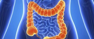
The large intestine (intestinum crassum) is located in the abdominal cavity and in the pelvic cavity follows the small intestine and is the final section of the digestive system. In the large intestine, the processes of digestion of food are completed, fecal masses are formed, which are excreted through the anus. The anatomy of the human large intestine includes the cecum (with appendix), ascending colon, transverse colon, descending colon, sigmoid colon, and rectum ending in the anus.
The length of the large intestine ranges from 1 to 1.65 m, its diameter is 5-8 cm, in the final section - about 4 cm. The large intestine differs from the small intestine in its large transverse dimensions, as well as in the relief of its outer surface. On the outer surface of the colon, three longitudinal strands are visible - ribbons of the colon (taeniae coli), each about 1 cm wide, formed as a result of concentrations of the longitudinal muscle layer in these places.
The mesenteric ribbon (taenia mesocolica) corresponds to the place of attachment of its mesenteries to the colon (transverse colon and sigmoid colon) or the line of attachment of the colon to the posterior abdominal wall (ascending and descending colon).
The omental band (taenia omentalis) runs along the anterior side of the transverse colon, where the greater omentum is attached to it, and continues to other parts of the colon. The free band (taenia libera) is located on the free anterior side of the ascending, descending and sigmoid colon, on the lower side of the transverse colon. At the level of the omental and free bands, finger-shaped protrusions of the serous membrane containing adipose tissue extend from the wall of the colon.
These omental processes (appendices epiploicae) of the human large intestine are 4-5 cm long. Protrusions are formed between the bands of the colon - haustrae coli, which are clearly visible on x-rays. Haustra in the structure of the human large intestine, separated from each other by noticeable grooves, are formed as a result of a discrepancy in the length of the longitudinal ribbons and the sections of the colon between the ribbons.
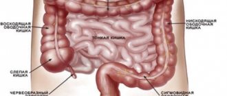
These photos show the structure of the large intestine:
Structure of the human intestine
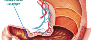
The organ begins and ends with muscle rings: the gastric sphincter and the anus. It consists of several sections, all parts of the intestine perform their task.
Physiological parameters
The general parameters of the organ depend on the individual characteristics of the carrier and his age. On average, the size of an adult ranges from 7.5 to 8.3 m (the length of the small intestine is 6-6.5 m, the large intestine is 1.5-1.8 m).
In women, it is shorter than in males. For the reason that during pregnancy the intestines have to move and it should take up a minimum of space, while maintaining its functionality as much as possible.
In newborns, the size of the organ is from 340 to 460 cm. The period of intensive growth of the child’s intestines and the formation of microflora occurs in the period from 5 months to 5 years. During the transition from breastfeeding to a “general diet” with a gradual increase in portions.
Small intestine and its parts
1. Duodenum (duodenum) - located immediately after the stomach and, compared with other areas, has small dimensions. In turn, it consists of four lobes: superior, descending, inferior and ascending.
It received its name from medieval scientists who, in the absence of modern means, measured it with their fingers and received an indicator of 12 fingers.
In the initial part of the intestine, the primary processes of absorption of nutrients from food and liquids take place.
The main purpose of this department is to stabilize the acid-base balance of the resulting mass, so that the more sensitive structures of the following are not damaged.
A secondary but integral phase of its functioning is the regulation of pancreatitis enzymes and bile.
The duodenum has a direct effect on the functioning of the secretory function of the stomach through reverse interaction, when the sphincter “gatekeeper” opens.
The massive muscular layer of the duodenum helps move the bolus of food further through the intestines.
2. Skinny - located in the upper left region of the abdominal cavity. Like the third part of the small intestine, it belongs to the mesenteric section. It is separated from the duodenum by the duodenal ligament of Treitz.
During dissection, the corpse is empty, which is why it got its name.
Two layers of smooth muscle (longitudinal on the outside and circular on the inside) facilitate the movement of the bolus of food. Motor activity represents different types of contractions: peristaltic and rhythmic.
The walls produce peptide hormones that affect other body systems.
The secretory function is not very active in this zone - intestinal juice is produced in small doses. Microvilli, under the influence of enzymes from the duodenum, participate in parietal digestion.
Interesting: In men, this section is longer.
3. Ileum - located in the right peritoneum. Belongs to the breech section. It borders on the cecum through the bauhinium valve. The hinges operate in two planes: the lower ones are horizontal, and the upper ones are vertical.
Produces up to 2.5 liters of juice. It contains digestive enzymes. Vitamin B12 and the final products of digestion are absorbed here: monosaccharides, amino acids, lipids.
The structural features of the ileum lie in the specific structure of the walls.
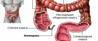
Large intestine and its parts
1. Blind - located in the right iliac fossa. Participates in the processing and absorption of liquid. Its interior is covered with absorbent cells and libercus glands.
A collection of lymphoid tissues is attached to this section - a vermiform appendix, which is called the appendix. Not so long ago, this organ was considered rudimentary - not performing a particularly important function.
But it has now been scientifically proven that the appendix is directly involved in the formation of immunity. People with a removed vermiform appendix are more difficult to tolerate intestinal infections and are more likely to suffer from dysbacteriosis.
A rim consisting of ascending, transverse, descending and sigmoid sections. Does not take special part in digestion, but absorbs water and electrolytes, forming more liquid chyme into thick feces.
2. Direct - The final section of the gastrointestinal tract. It is located downward from the sigmoid and ends with the anus. Forms two bends: sacral and coccygeal.
The structure of the mucous layer is not subject to the negative influence of digestive enzymes and mechanical damage from solid particles of already formed feces.
The length of the rectum is from 14 to 18 cm.
Anus
Conditionally refers to the intestines and has two sections:
- The internal sphincter is a ring-shaped smooth muscle structure that surrounds the anal canal. Approximately 5 mm thick and up to 30 mm long. We are not controlled by human consciousness, therefore it contracts and relaxes reflexively, in an arbitrary manner. During the act of defecation, a certain amount of feces accumulates in the rectum and the mucous membrane of the rectum becomes irritated, causing relaxation of the internal muscle ring.
- External sphincter - consists of striated muscles and surrounds the anal canal. Controlled by consciousness. It is up to 10 cm long and 2.5 cm thick. It has stretch receptors and a rectoanal reflex, which leads to involuntary compression in a protective reaction.
How does digestion occur in the small intestine?
Digestion in the small intestine is performed as follows. The gruel coming from the stomach, pre-treated with saliva and gastric juice, has an acidic reaction. In the small intestine, the presented mass is exposed to alkaline effects. This creates optimal conditions for the processing of nutrients by enzymes. The breakdown of the protein components of food gruel occurs under the influence of the following elements of intestinal juices:
- The enzymes enterokinase, kinaseogen, and trypsin process simple proteins.
- Erepsin breaks down peptides into amino acids.
- Nuclease separates complex molecules of protein origin, known as nucleoproteins, into microelements.
- The enzymes maltase, phosphatase, amylase and lactase break down carbohydrates.
- Lipase processes fats.
After the synthesis of useful substances from food gruel through enzyme treatment, carbohydrate and protein components are absorbed by the villi of the small intestine. Next, the microelements enter through the venous capillaries into the liver tissue. In turn, the fats are sent to the lymphatic system.
Functions of the intestines in the human body
Being part of the gastrointestinal tract, it performs the following processes:
- digestion of food;
- release of microelements and synthesis of vitamins - thanks to the unique microflora;
- formation of immunity (immunoglobulins and antibodies);
- removing toxins and unfavorable compounds that can harm the body.
General health depends on the normal functioning of the intestines.
Sigmoid section (colon sigmoideum)
The continuation of the digestive tube is the long colon. It goes around the loops of the intestinum tenua, which lie in the lower floor of the abdominal cavity. Its beginning is the ascending colon, it is 20 cm long; there are also shorter variants (about 12 cm). It is separated from the caecum by grooves, which always correspond to the frenulum located in the ileocecal angle.
Its continuation is the transverse colon, which can reach 50 cm in length. It is directed slightly obliquely, towards the left hypochondrium. It begins at the level of the tenth costal cartilage. In the middle, this section sags, thereby forming the letter “M” together with other parts of the colon. From the parietal part of the peritoneum to the transverse section there is a mesentery, which covers it on all sides, that is, the intestine is located intraperitoneally.
The place where the transverse part passes into the descending part is the splenic flexure, located immediately below the lower pole of the spleen.
The descending part occupies a marginal location along the posterior wall of the abdomen. Its posterior wall does not have a serosa, and lies in front of the left kidney. At the level of the left iliac crest it becomes the colon sigmoideum. Its average length is up to 23 cm, diameter is about 4 cm, the number of haustrations and their size gradually decreases.
Palpated in the left iliac fossa, forms two loops (proximal and distal). The proximal loop is directed with its apex downward, and the distal loop lies on the psoas major muscle and is directed upward. The colon sigmoideum itself enters the pelvic cavity, and approximately at the level of the third sacral vertebra it gives rise to the rectum.
Common Disorders
Despite the strong functional component, it is quite easy to cause disharmony. Rich microflora is prone to destruction if you neglect the simplest nutritional rules and eat everything.
The slightest disruptions in this part of the gastrointestinal tract cause discomfort, problems with stool, painful syndrome, and other unpleasant factors.
Incorrect bowel function may be accompanied by the following symptoms, which, at first glance, have nothing to do with it:
- headache;
- excessive sweating;
- weakness;
- dermatological diseases;
- stale breath odor.
Even temporary problems with the intestines should not be ignored; they can be a manifestation of more serious pathological processes:
- Constipation is a sedentary lifestyle or poor diet that deprives the body of the required amount of fiber. It may indicate the presence of adhesions in the intestines, a tumor of the uterus or appendages. Often occurs during menopause. Stool retention in combination with flatulence may indicate gynecological peritonitis.
- Neurogenic constipation is a very common phenomenon, as a consequence of psychological blockage. That is, a person clearly feels the need to defecate, but cannot do this due to the fact that he is in someone else’s house, train, or other place that causes emotional stress or discomfort.
Worth knowing: The norm for an adult is stool from 3 times a day to 3 times a week.
Provided there is no emotional discomfort.
- Tenesmus is the urge to defecate. Mostly painless, but can occur even in the complete absence of feces in the rectum. Occurs during dysentery or after radio- and fluoroscopy of the genital organs.
- Diarrhea is frequent loose stools that can be caused by an intestinal infection or intestinal tuberculosis. parametritis.
- Pain during defecation - hemorrhoids, oncology, paraproctitis, and in women it also occurs with inflammation in the area of the peri-vaginal or peri-uterine tissue.
Regular bowel dysfunction is an important reason to visit a therapist, who, judging by the symptoms, will refer you to a specialized specialist.
Histology of the gastrointestinal tract
The walls of the small and large intestine consist of mucous, submucosal, serous and muscular membranes.
The mucosa of the small intestine contains a large number of long villi, while the thick intestine is devoid of them.
The submucosa of the small intestine consists of connective tissue, loose and poorly formed, as a result of which this part of the intestine is very mobile. In the colon, the base is more dense and well defined.
The muscular layer is present in both intestines and consists of two layers: internal (circular) and external (longitudinal). The circular muscles are most developed, especially between the haustra. The combination of circular and longitudinal muscles helps to better mix and promote the contents.
The outer serous part completely reproduces the relief of the outer surface of the intestine.
Diseases and their causes
The intestines are characterized by many diseases, but there are several ways of formation:
- infectious lesion;
- parasite infection;
- unhealthy lifestyle - smoking, lack of exercise, unbalanced diet;
- genetic predisposition;
- long-term use of drugs or their incorrect prescription;
- malfunctions of the immune system.
It’s easy to protect yourself from some points with banal personal hygiene techniques, but no one is protected from others.
The list of pathologies of this organ is so large that there is a separate classification of intestinal diseases:
- Infectious - dysentery, amoebiasis, typhoid fever, cholera, syphilis. All of them arise due to harmful bacteria and amoebas. The simplest illness can turn into serious problems. You shouldn’t even be negligent about dysentery, from which up to 9% of patients die every year.
- Parasitic - scarabiasis, ascariasis, trichinosis, intestinal miasma, trichuriasis, hookworm. They come mainly from food, usually poorly cooked meat or unwashed fruits and vegetables. The symptoms of all parasitic diseases are complex, so cases of erroneous diagnoses are common. May manifest as allergies, fever, anal itching or recurrent abdominal pain. There is a misconception that diseases of this nature are the prerogative of third world countries. The source of intestinal miasma can be the larvae of common house flies.
- Inflammatory - enteritis, Crohn's disease, ulcers, colitis. The causes of some pathologies have not yet been determined, so treatment is predominantly symptomatic.
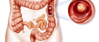
- Neoplasms - rectosigmoid cancer, benign tumors, polyps. Symptoms depend on where exactly the tumor is located. If this is, for example, a disease of the last part - the rectum, then blood, mucus with pus and acute pain accompany each process of defecation and are slightly dulled at rest.
- Atypical - pneumatosis, irritable bowel syndrome, dolichocolon, dolichosigma.
Parameters of different parts of the digestive tube
The small intestine (intestinum tenue) has a length of 1.6 to 4.3 meters. In men it is longer. Its diameter gradually decreases from the proximal to the distal part (from 50 to 30 mm). Intestinum tenue lies intraperitoneally, that is, intraperitoneally, its mesentery is a duplicate of the peritoneum. The leaves of the mesentery cover blood vessels, nerves, lymph nodes and vessels, and fatty tissue.
The length of colon is comparatively smaller - 1.5 meters. Its diameter decreases from beginning to end from 7-14 to 4-6 cm. As described above, it has 6 divisions. Caecum has an outgrowth, a vestigial organ, the appendix, which, according to most scientists, is an important component of the immune system.
Throughout the colon there are anatomical formations - bends. This is the place where one part of it transitions to another. Thus, the transition of the ascending to the transverse colon is called the hepatic flexure, and the splenic flexure is formed by the transverse descending sections.
The intestines are supplied with blood through the mesenteric arteries (superior and inferior). The outflow of venous blood is carried out through the veins of the same name, which make up the portal vein basin.
The intestine is innervated by motor and sensory fibers. The motor branches include the spinal and branches of the vagus nerve, and the sensory fibers include the fibers of the sympathetic and parasympathetic nervous system.
Preventive measures and treatment
Basic rules of personal hygiene save you from most infectious and parasitic diseases.
High-quality heat treatment will make food safe and healthy.
A healthy lifestyle and proper nutrition are undoubtedly important aspects of preventing diseases not only of the intestines, but also of other important systems of your body.
After you see a doctor, you must undergo a complete examination of the abdomen using palpation, a procedural examination and pass the necessary tests. Only on this basis will the doctor be able to make the correct diagnosis.
Description of hardware diagnostic procedures:
- x-ray - checks patency;
- Ultrasound - inflammatory processes, oncological formations are detected, parameters and structure of departments are measured.
- Colonoscopy is the most informative method of early diagnosis, checking the condition of the organ. It is performed with an endoscope with the ability to take tissue samples. Virtual colonoscopy is performed using a computed tomography (MRI) scanner.
- sigmoidoscopy - examination of the mucous membrane.
- irrigoscopy - study of the large intestine and blood supply using x-rays with the introduction of contrast dye.
Most procedures require rinsing or cleansing. The doctor must warn you about this in advance.
For example, ultrasound is performed only on an empty intestine. It is not enough to carry out the act of defecation. After it, undigested accumulations of chyme or feces still remain.
It is necessary to thoroughly cleanse the parts of the organ of everything, including surface waste - perform an enema or take the following medications:
- Fortrans,
- Motilium,
- Loperamide,
- Activated carbon, etc.
It is worth organizing the cleaning in the evening if the event is scheduled for the morning or lunchtime. Do not eat anything before the procedure.
As the most gentle method of basic diagnostics, ultrasound examination is performed for children and adults. During it, it is impossible to harm the mucous membrane or disrupt the tissue structure.
To pass the most informative stool test, you need to know some of the conditions for home collection procedures.
Description of requirements:
- the material must be fresh;
- Before the procedure, the bladder must be empty;
- for women it is important to ensure that menstrual blood does not get in;
- you need to put it into a clean container from there, use a spatula from the kit for collecting stool analysis (which can be purchased at any pharmacy) and transfer it into a jar;
- For bacteriological research, the material must be warm, so the container should be closed immediately.
The main obstacle to effective treatment is a neglectful attitude towards ordinary intestinal problems, which, over time, can result in serious consequences.
Even the most minor problems in the functioning of an organ require a qualified approach. Only in this case will it be possible to really get rid of the problem.
Clinical picture indicating diseases of the colon
When a person has problems with the colon, it is characterized by some signs such as:
- painful sensations. They are aching and spasmodic in nature. Localized in the lower left or right part of the abdomen. The umbilical area is also involved. Relief comes from the act of defecation;
- chronic constipation;
- diarrhea. They bother the patient constantly or occur in periods alternating with constipation;
- bloating. Appear after eating food;
- the appearance of streaks of blood and mucus in the stool.
If such a symptomatic picture occurs, you should urgently visit a doctor.