Treatment of the disease
Treatment of dacryocystitis in adults occurs without complications, with a favorable prognosis. Therapy is prescribed after diagnosis, determination of the stage and causes of the disease.
Treatment methods:
- disinfectant solutions;
- ointments;
- drops;
- tablets;
- injections;
- massotherapy;
- probing.
At the initial stage, massage is used. Then antibacterial and anti-inflammatory medications in the form of ointments or drops.
Swelling of the lacrimal canal is relieved with Sofradex and Chloramphenicol drops. The drugs can reduce inflammation and restore the functioning of the ducts.
In the acute form of the disease, Cefucrosime tablets are prescribed. The product has a beneficial effect on the entire body, the organs of vision are no exception.
Injectable solutions are prescribed to treat bacterial infections. Composition: Sodium Sulfacyl in combination with Levomycetin and Erythromycin.
Corticosteroid drugs and Prednisolone solution, suspension of hydrocortisone and dexamethasone are particularly effective. The quantity and dose of medications is determined by the ophthalmologist.
Treatment
As a rule, if dacryocystitis is without complications, the prognosis for recovery is favorable. Treatment of dacryocystitis, first of all, depends on the form of the disease and the causes of its occurrence.
The treatment process for dacryocystitis is generally divided into two parts:
restoration of patency of the nasolacrimal canal; anti-inflammatory therapy.
When treating dacryocystitis in adults, bougienage and rinsing of the nasolacrimal duct with disinfectant solutions and the use of antibacterial drops and ointments are performed.
Bougienage is the most common, gentle method of restoring the patency of the nasolacrimal canal. During this procedure, the blockage of the nasolacrimal canal is physically removed using a special rigid probe (bougie).
Initially, patients suffering from dacryocystitis are prescribed enhanced antibacterial treatment to avoid infectious complications. This is necessary because with dacryocystitis there is a possibility of a purulent form of encephalitis or a brain abscess.
Dacryocystitis in old age
The acute form of the disease is treated in a hospital setting. As a rule, in this case, intramuscular injections of benzylpenicillin sodium salt (3-4 times a day) or oral administration of tetracycline (4 times a day), sulfadimezine (4 times a day) are prescribed.
If an abscess of the lacrimal sac has formed, it is opened through the skin. Before opening the abscess, systemic vitamin therapy and UHF therapy are performed. After opening, the wound is drained and washed with antiseptic solutions of furatsilin, dioxidin, hydrogen peroxide . chloramphenicol, miramistin, sodium sulfacyl, gentamicin ) and antibacterial ointments ( erythromycin, tetracycline, floxal are instilled into the conjunctival cavity .
In addition to local treatment, systemic antibacterial therapy with broad-spectrum drugs is carried out. For this purpose, cephalosporins, aminoglycosides, and penicillins are used.
In advanced forms of dacryocystitis, when standard drug treatment is ineffective, dacryocystoplasty or endoscopic dacryocystorhinostomy .
Endoscopic dacryocystorhinostomy
Endoscopic dacryocystorhinostomy is a surgical procedure used to treat dacryocystitis in adults. Special modern minimally invasive equipment is used to perform the operation. Dacryocystorhinostomy can only be performed on patients who do not have an allergic reaction to anesthetic drugs. During the operation, a special flexible tube is inserted into the tear duct - an endoscope with a microscopic camera. An endoscope is used to make an incision in the blocked tear duct. The rehabilitation period after surgery is 6-8 days. To avoid inflammation of the cornea, he prescribes a course of antibiotics. The advantage of this operation is that it does not leave visible skin scars on the face or damage to the tear ducts.
Balloon dacryocytoplasty
In most cases, balloon dacryocystoplasty is used. This is a safe operation that can be performed even on children over 1 year old. During the operation, a special thin conductor is inserted into the nasolacrimal canal through the corner of the eye, which is equipped with a microscopic expanding balloon filled with liquid. In a blocked area of the nasolacrimal canal, the balloon expands and opens the duct using pressure and is then removed from the canal. The procedure is performed under local anesthesia. After the operation, a course of antibiotics and eye drops are prescribed to prevent the development of infection.
Complications
Dacryocystitis is a rather dangerous disease, since if left untreated it can cause various complications.
The chronic form of the disease is especially dangerous. In this case, infection of other membranes of the eye is possible. There is a possibility of developing concomitant diseases - blepharitis, conjunctivitis, keratitis . With the further development of chronic dacryocystitis, the cornea is affected and a purulent ulcer is formed. As a result of the occurrence of a corneal ulcer, a cataract may subsequently develop, which can become not only a cosmetic defect, but also reduce the quality of vision.
Further development of the ulcer can also lead to endophthalmitis, which is characterized by inflammation of the internal structures of the eye.
A significant complication can be the development of life-threatening diseases that can lead to disability or death in the patient:
sepsis; orbital phlegmon; thrombophlebitis of the orbital veins; thrombosis of the cavernous sinus; inflammation of the meninges and brain tissue.
Prevention
To prevent dacryocystitis, it is necessary to promptly treat inflammatory diseases of the eyes and ENT organs, as well as avoid eye damage and foreign bodies. With timely diagnosis and treatment of dacryocystitis, complete recovery is possible without serious consequences.
Farsightedness: how to restore vision.
Read this article about why discomfort occurs when wearing lenses.
Do glasses with holes help restore vision: https://eyesdocs.ru/ochki/kompyuternye/s-dyrochkami-perforacionnye.html
Symptoms and what they are
Dacryocystitis is inflammation of the lacrimal sac. Its development is associated with obliteration or stenosis of the lacrimal canals. The natural drainage of tears is disrupted. It accumulates in the cavity cylinder.
Inflammation of the lacrimal duct is observed five times more often in females than in males. The factor is influenced by structural anatomy.
Pathology occurs in persons 25 - 55 years of age. A separate clinical form of the disease is identified in newborns.
Without timely treatment, purulent-septic complications develop. The eyelids, cheeks, nose, and soft tissues of the eye sockets are affected.
Symptomatic manifestations of pathology in adults and children are similar.
Expressed:
- uncontrolled tearing;
- lack of tears;
- swelling of the eyes;
- the appearance of red spots in the eyelids and eye corners.
When the nasal passages fill, lacrimation stops. The release of purulent exudate is observed.
When you press on the area of the lacrimal sac, a pain symptom is noted. The acute form of the disease is characterized by sensitive, sometimes unbearable pain syndromes. Chronic dacryocystitis - absence of pain symptoms.
Causes
Based on the type of dacryocystitis , the root causes of its occurrence also change. It could be:
- Eye injury
- Congenital obstruction of the lacrimal ducts
- Allergy
- Infection with a virus or bacteria
- Complications of nasopharyngeal diseases
- Parasitic infestation
- Incorrect use of cosmetics
- Chemical damage
- Diabetes
- Reduced immune system
Massage
Conservative treatment of dacryocystitis consists of the use of drops, rinsing and massage. Before the start of the event, hands are thoroughly washed and disinfected with antiseptic liquid products. The action will prevent the emergence of a new infection.
Before the massage, the eyes are cleared of pus. For this purpose, a solution of Furacilin is used.
Preparation: equal ratio of the drug to purified water. It is allowed to treat the eyes with chamomile decoction or tea (not strong). The manipulation is carried out from the outer corner of the visual organ to the inner.
Correct massage for dacryocystitis:
- Using your index finger, find a growth (tubercle) in the area of the inner corner of the eye (at the base of the nose).
- The side of the finger is closely adjacent to the growth;
- Press on the tumor. Manipulation will allow you to free yourself from the film covering the lumen of the canal.
- From the tubercle, the finger moves down the bridge of the nose. The pressure is released at the base. Don't tear yourself away from the skin.
- Raise your finger up to the starting position (before increasing).
Perform at least 10 massage movements. When squeezing out purulent fluid, apply Furacilin.
https://medglaza.ru/zabolevaniya/bolezni/dakriotsistit-vzroslyh-lechenie.html
How to properly massage the eyes of a newborn baby
The massage must be carried out in compliance with hygiene and safety requirements so as not to accidentally damage the child’s eyes or cause an infection. Be sure to cut long nails to avoid scratching your baby's skin. Before starting the procedure, wash your hands thoroughly and dry them. It is better to start a massage after feeding, when the baby is in a calm, relaxed state. It is not recommended to do this if he is feeling anxious for any reason or has a fever. First, you should rinse, clearing your eyes of traces of purulent discharge. Weak antiseptic solutions are suitable for this - for example, boric acid, furatsilin, chamomile, etc. A cotton pad is moistened in the solution and traces of purulent fluid are carefully removed by moving from the outer corner of the eye to the inner. Use a separate disk each time.
Next, you should place your index finger on the point in the inner corner of the eye where the two canaliculi meet before entering the lacrimal sac. Ask your doctor to show you this place, and also feel it yourself to better understand where the desired point is. Next, start moving your finger from top to bottom towards the baby’s nose, while applying gentle pressure. It is necessary to repeat such movements 6-10 times. If during the procedure pus is released from the lacrimal sac, it means that the eye massage is carried out correctly. The contents of the ducts are removed using a cotton swab.
It is recommended to eliminate purulent discharge in this way 5-6 times a day. Upon completion of the procedure, you need to rinse the eye with antiseptic solutions, and then administer antibacterial ointments or drops. This could be a solution of Levomycetin 0.25%, Vitabact or other medications. Doctors recommend eye massage in newborns with obstruction of the lacrimal canal for two weeks, and then see a specialist. Usually during this time the process normalizes and the tear begins to flow normally. As a rule, the doctor’s prescription looks like this:
- eye massage - 4-6 times a day, 6-10 repetitions;
- washing the lacrimal canals with an antiseptic solution;
- "Levomycetin" 0.25%, 1 drop 4-5 times a day.
Under no circumstances should you treat your child based on advice from online forums. Incorrect therapy will lead to dacryocystitis taking an acute form, and it will be difficult to cope with it. In general, in 80% of cases, the measures taken act favorably and the film breaks, ensuring normal patency of the tear duct. If this does not happen, surgical treatment is prescribed.
Operation
The course of therapy will depend on the patient’s age criteria, forms and signs of the disease. In addition to therapeutic methods, surgical treatment methods can be used.
The operation is prescribed if:
- there is no effect from the medical procedures performed (massage, drops);
- the abnormal structure of the lacrimal ducts is clearly expressed;
- a secondary chronic form is diagnosed;
- Complications are observed in the visual apparatus or in the area of the sinus space.
Surgery methods:
- Bougienage. One of the simple but effective surgical treatment methods. It is used for minor spread of inflammatory processes. A probe is inserted into the nasolacrimal duct and fluid stagnation is eliminated. After surgery, antibacterial drugs are prescribed to prevent complications.
- Endoscopic dacryocystorhinostomy. A bendable tubular material with a camera and a light source is inserted into the lacrimal sleeve section. An incision is made into the tubule using an endoscope and the pus is removed. Using the presented method, complex forms of pathology are treated.
- Balloon dacryocytoplasty. One of the modern methods of treatment, suitable for children. A thin probe is inserted into the area of the nasolacrimal duct. The device contains a cylinder containing liquid or gas. Pressure causes the blocked duct to expand. As a result: the circulation of the tear fluid composition is normalized. Subsequent therapy: antibacterial drops in combination with antibiotic agents.
To prevent purulent wounds of the cornea, the use of contact lenses and eye patches is prohibited.
Diagnostics
For a specialized specialist (ophthalmologist), making a diagnosis is not a problem, since most of the signs are visible visually. But to clarify the diagnosis, determine the causes and select treatment, additional studies are carried out:
- the patency of the lacrimal ducts is determined - a dye is injected into the eye, which, if patency, enters the nasal cavity;
- probing the nasolacrimal duct to assess the extent of its damage;
- conducting eye biomicroscopy;
- instillation (fluorescein) test;
- Vesta color test;
- passive nasolacrimal test;
- microbiological examination - culture of secretions from the lacrimal canal;
- X-ray examination using special substances injected into the nasolacrimal canal;
- examination of the nose and sinuses;
The doctor may prescribe one or more additional research methods if necessary.
When diagnosing, it is important not only to establish the fact of the disease, but also to determine the causes and select the most effective treatment methods.
Chronic dacryocystitis
The chronic form of dacryocystitis is expressed by increased lacrimation and swelling of the lacrimal sac. The dense formation has a red tint.
Over time, the swelling and redness goes away. An abscess appears, then a fistula.
With a long-term chronic form of the disease, ectasia (stretching) of the lacrimal sac occurs. In this area, the skin becomes thinner and becomes bluish.
Danger: there is a possibility of infection of the cornea and the formation of ulcerative nodes.
The cause is: significant narrowing or obstruction of the tubules. There is no exit to the nasal cavity; fluid accumulates, causing inflammation.
Chronic pathology begins as a result of: sinusitis, prolonged runny nose, bruises of the nose.
Chronic inflammation is treated with surgery. Dacryocystorhinostomy is prescribed. The treatment helps stop the accumulation of fluid and normalize its outflow.
Symptoms of dacryocystitis
With dacryocystitis , most often 1 eye becomes ill. This pathology is accompanied by unpleasant sensations, among which are the following signs of the disease:
- Swelling
- Sharp, cutting pain in the area of the affected eye
- Dizziness
- Involuntary release of tears
- Inflammation of the eyelids
- The appearance of mucus and pus when pressed
- Redness
- General weakness
- Blueness of the skin in the area of the optic duct
- The appearance of a veil before the eyes
- Filling of soft tissues with blood
- Softening of the skin near the lacrimal sac
- Incorrect movement coordination
- Narrowing or complete covering of the palpebral fissure
Disease in infants and children
Dacryocystitis is diagnosed in 5% of infants. In pediatric ophthalmology, it is considered a separate form of the disease. The pathology requires timely treatment in order to subsequently prevent the formation of an inflammatory process, persistent lacrimation, and, in the future, surgical intervention. Complications are the same for both newborns and older children.
Signs
In children, the pathology is unilateral (damage to one eye). In rare cases (3-5%) patients suffer from bilateral lesions.
Symptoms in a child: increased lacrimation, swelling of the lacrimal sac. When pressed, a purulent substance is released.
Symptoms of the acute form of the disease: redness, swelling of the eyelid, enlargement and inflammation of the lacrimal sac. In some cases, swelling covers the upper and lower parts of the eye, and the child has difficulty opening his eyes.
In acute inflammation, the temperature rises and chills begin. Symptomatic signs of dacryocystitis in newborns appear during the first days of life.
Diagnostics
At the first symptoms of dacryocystitis, you will need to be examined by an ophthalmologist.
Diagnostics consists of:
- Palpation. The specialist examines the lacrimal sac by touch and evaluates the nature of the discharge.
- Probe Vesta. A medicine is instilled into the affected eye. The patency and narrowing of the tubules are determined.
- Diagnostic probing. The method determines the extent of the blockage.
When confirming the diagnosis, smears of purulent fluid are taken to determine the microorganisms that have begun to multiply in the lacrimal sac. Then treatment is prescribed.
Therapy
Treatment for infants and newborns takes place at home. Inpatient therapy is not prescribed, since the disease in most cases is associated with physiological causes.
The baby is prescribed a daily massage. These manipulations are quite enough for the baby to fully recover. If massage treatment does not produce the expected results, dacryocystitis begins to progress, inflammation develops, the doctor prescribes probing.
Older children undergo inpatient treatment under the supervision of a doctor. During the maturation of the abscess, UHF and compresses are used.
After opening the purulent focus, the lacrimal sac is cleaned. The course of treatment will depend on the type of pathogen.
For bacterial dacryocystitis: antibiotics (eye drops) or antibiotic-based ointments, tablets and syrups. For viral pathology: the lesion is treated with antiseptic solutions.
Reasons why dacryocystitis may occur
This inflammatory disease occurs when the ducts are blocked or narrowed, which disrupts the normal flow of tear fluid. Here are the factors that can cause this condition.
- Abnormal development or underdevelopment of the lacrimal ducts.
- Stenosis is a congenital narrowing of the tear ducts.
- Infectious diseases of the organs of vision.
- Chronic runny nose.
- Atherosclerosis, etc.
Dacryocystitis of newborns is a common occurrence in ophthalmological practice. This inflammatory disease is diagnosed in 10% of cases of all eye diseases. Let's take a closer look at the causes of dacryocystitis and methods of treating it in newborns.
Congenital type
Dacryocystitis that appears at birth is, by definition, considered congenital. The acquired form of the disease manifests itself at a later age.
Congenital dacryocystitis, what is caused by:
- violation of the patency of the nasolacrimal canal;
- the presence of folds in the mucous lacrimal sac;
- persistence of the fetal membrane (normally its existence is observed at the intrauterine stage).
For comparison, let’s look at what causes the acquired form:
- acute pathologies of the nasopharynx;
- systematic purulent inflammations;
- chronic sinusitis;
- complications after inflammation of the eyeball;
- eye injuries;
- curling eyelashes;
- dust getting into the tear ducts.
The disease occurs in acute and chronic forms. There is a possibility of regular exacerbation.
Symptoms
The clinical manifestations of the disease are distinguished by specific symptoms and signs, which does not cause difficulties in determining the diagnosis.
However, the acute and chronic forms exhibit different clinical pictures.
Of the general symptoms that appear with dacryocystitis of any etiology and form, the following are noted:
- uncontrolled excessive release of tear fluid from the affected eye;
- purulent discharge;
- swelling in the area of the lacrimal sac;
- painful sensations;
- inability to fully open the eye due to swelling and pain;
- the likelihood of an increase in general temperature indicators.
The appearance of at least one of the described symptoms is a reason to visit an ophthalmologist in order to prevent further progression of the disease.
Clinical picture in acute course
Acute dacryocystitis is characterized by particularly severe symptoms:
- expressive hyperemia of the skin in the area of inflamed ducts;
- intense pain in the corner of the affected eye;
- significant swelling of the upper and lower eyelids, which is accompanied by their almost complete closure;
Associated symptoms include headache, chilliness, general weakness, and a slight increase in body temperature.
Symptoms in chronic form
The advanced state of the disease is accompanied by:
- uncontrolled release of fluid;
- the formation of a dense formation in the lacrimal sac - a purulent abscess, as a result of which the skin in this place acquires a bluish tint and becomes thin;
- in the absence of treatment, there is a possibility of fistula formation - internal or external.
Chronic dacryocystitis is dangerous due to its complications, including diseases such as blepharitis, conjunctivitis, phlegmon and even meningitis.
Acute dacryocystitis
Acute dacryocystitis is a rapidly developing inflammation of the lacrimal sac. Purulent fluid is formed as a result of the active activity of pathogenic microbes. An inflammatory process develops.
The main causes of acute forms of pathology include:
- obstruction of the lacrimal canal;
- swelling of tissue structures;
- rhinitis;
- sinusitis;
- polyps;
- adenoids.
Risk factors for the appearance of an inflammatory process include injuries, fractures, and cracks in the nasal cavity.
The presented form of pathology requires contacting a specialist. Treatment is carried out in a hospital and consists of:
- taking vitamins;
- use of antibacterial therapy;
- physiotherapy;
- treatment and drainage of the wound using antiseptic drugs.
After medication and the acute phase of dacryocystitis subsides, surgery is prescribed. Surgery will prevent relapse.
Treatment of dacryocystitis
To treat inflammation of the lacrimal sac, both conservative and surgical treatment are used. If we talk about medications, then antibiotics are prescribed first. They are selected in accordance with the results of bacteriological culture. The most commonly used drugs are cefuroxime, fusidic acid or chloramphenicol. Alternatives to these drugs are tetracycline, doxycycline, ofloxacin or levofloxacin. Drug treatment is accompanied by local therapy using a Solux lamp or exposure to a high-frequency electromagnetic field (UHF). If an abscess forms, it is opened and washed.
Primary dacryocystitis can be treated through surgery if medications and therapy do not help or abnormalities in the development of the lacrimal ducts are too pronounced. The secondary form of the disease can only be treated surgically. For the purpose of recovery, external and endosial operations are performed. In the second case, this is surgery through the nose. Its advantages are that postoperative scars are less traumatic and invisible. As a result of the manipulations, the passage between the lacrimal sac and the nasal cavity increases. In 98% of cases, a chronic disease can be completely eliminated.
If we talk about newborns, they are treated with massage of the affected area, eye washing and probing. The operation is performed only after three months. Here is a possible treatment option for newborns:
And here is what they say about the probing procedure and the consequences of dacryocystitis if it is not treated in time:
Drops for treatment
For eye diseases, complex therapy is used. It consists of the use of medications, massage, folk recipes and surgical procedures. The choice of tactics depends on the degree of damage and the stage of the inflammatory process.
After examination and diagnosis, drops are prescribed. Their action is aimed at eliminating pathogenic bacteria and microorganisms.
Drops that effectively combat dacryocystitis:
- Gentamicin. Bactericidal antibiotic agent. Designed to eliminate bacterial eye infections.
- Tobrex. An antibiotic with high antibacterial activity. Effectively destroys pathogens that belong to the group of aminoglycosides.
- Vitabact. The active ingredient of the drug is piloxidine. The drops have antiseptic properties and relieve inflammation.
- Vigamox. Effectively eliminates the development of pathogenic microbes. Actively fights gram-negative, gram-positive, anaerobic bacteria. Has anti-inflammatory properties.
- Oftaquix. Antimicrobial agent from the group of fluoroquinolones. Treats superficial bacterial infections. Suitable for use from 1 year of age. Used for preventive purposes. After eye surgery.
The medications are taken on the recommendation of a doctor.
Dacryocystitis is an ophthalmological disease. If symptoms are detected, it is recommended to contact a medical facility for assistance. Treatment is prescribed by a specialist, after diagnosis, individually for each patient.
Characteristic symptoms
At an early stage of development, dacryocystitis, as a rule, is asymptomatic and does not manifest itself in any way. But during development, the patient may experience swelling in the area of the lacrimal sac and a feeling of fullness. In addition to these symptoms, discomfort and pain and increased lacrimation may occur. Upon palpation, purulent contents or clear liquid may be released from the lacrimal sac.
Symptoms of the disease
As the pathology develops, the skin around the patient's eyes becomes inflamed and red. This is due to constant lacrimation. To avoid serious consequences, you should immediately consult a doctor for a diagnostic examination.
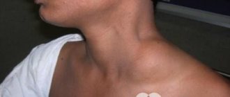
Dacryocystitis in adults - photo
Causes
The main reason for the development of dacryocystitis is a violation of the obstruction of the nasolacrimal duct as a result of narrowing or complete closure. Such pathological deviations lead to problems with fluid circulation. As a result, pathogenic microorganisms develop in the cavity of the blocked lacrimal canal due to stagnation of secretions.
Viral diseases, such as sinusitis, rhinitis or respiratory infections, can provoke swelling of the tissues of the nasolacrimal duct.
But there are other reasons for the development of dacryocystitis:
- effects of low or high temperature on the body;
- contact with hazardous chemicals that negatively affect the functioning of the visual system;
- the body's reaction to various allergens; decreased immunity;
- elevated blood sugar levels;
- violation of metabolic processes;
- foreign objects entering the eye;
- benign neoplasms in the nasal cavity;
- mechanical damage to the lacrimal canals;
- nose fracture
All these factors can lead to the development of dacryocystitis in children and adults. In order to promptly detect pathology and begin treatment, you need to know how it manifests itself.
Causes of the disease
For newborns and infants, common factors in the development of the disease are:
- too narrow nasolacrimal duct;
- partial fusion of the nasolacrimal canal or preservation of the membrane in it;
- plugs that have not resolved after intrauterine development;
- complete overgrowth of the duct (extremely rare).
In adults, the most common causes of darkyocystitis are:
- diseases of the nasopharynx, such as sinusitis, sinusitis, nasal polyposis, allergic or catarrhal rhinitis;
- nasal injuries that lead to rupture of the canal or blockage due to pressure on it from traumatic edema;
- eyelid injuries leading to blockage of the secretion of the lacrimal glands;
- foreign bodies (sand, dust, cosmetic residues) getting into the duct;
- viral or bacterial infections can be both the cause of blockage and its consequence;
- infectious skin lesions around the eyes;
- hypothermia, leading to thickening of the tear secretion and blockage of the canal;
- prolonged exposure to high temperatures and dryness, which leads to drying out and blockage of the canal.
Constant exposure to certain risk factors can lead to chronic dacryocystitis. Possible influence of external factors, such as working in hazardous conditions. The structural features of the lacrimal ducts can also cause partial blockage and the occurrence of chronic dacryocystitis.
Risk group
Dacryocystitis is a fairly common disease that can occur in any person. However, for some groups of the population the risk is higher.
These include people:
- prone to allergic reactions, especially during “seasonal allergies”;
- with a chronic decrease in immunity, including in the autumn-winter period;
- patients with diabetes mellitus;
- having chronic nasal diseases or ophthalmological diseases;
- working in hazardous or dusty industries;
- women who abuse decorative cosmetics.
Folk remedies
Treatment of dacryocystitis at home is possible only after examination by an ophthalmologist and his permission. Folk recipes use eye drops, compresses, and lotions to wash the tear duct in adults. Alternative medicine is strictly contraindicated for children.
What to do if the eye canal becomes inflamed:
- Use aloe juice diluted 50 to 50 with boiled water for eye drops or lotions.
- An aqueous solution of honey is a natural antiseptic; it is used in the eyes for inflammation.
- Eyebright juice, thyme tincture, and calendula are used for compresses.
- Wash your face with a decoction of chamomile, sage, and birch leaves and wash out the tear ducts.
- Black tea bags are applied as warm compresses.
Dacryocystitis in adults
Often this eye pathology occurs in middle-aged and elderly people, and women are diagnosed seven times more often than men. Unlike children, in whom the disease is usually congenital, in adults dacryocystitis occurs for a number of reasons:
- viral or bacterial eye pathologies that contribute to the development of edema that compresses the tear ducts;
- chronic inflammation of the tear ducts - dacryoadenitis;
- violation of the integrity of the tear ducts due to injuries;
- ingress of particles of cosmetic products due to untimely removal of makeup;
- polyps in the nasal cavity;
- consequences of severe bleeding - blood clots form and increased secretion of exudate begins, which can contribute to the formation of dacryolites.
Opening an abscess
This procedure is carried out through the skin. The manipulation is preceded by UHF therapy and vitamin therapy.
An abscess is opened in this way:
- Under local anesthesia, the lacrimal sac is opened through the skin.
- The wound is drained;
- Next, the opened area is treated with furatsilin, hydrogen peroxide or dioxidine;
- To prevent further development of infection and inflammation, drops with an antibacterial effect are instilled into the conjunctiva. This may be “Gentamicin”, “Sodium Sulfacyl”, “Miramistin”, “Levomycetin”;
- You can also put ointments with an antibacterial effect into the conjunctiva - Floxal, tetracycline or erythromycin ointments.
In addition to local treatment, general antibacterial therapy is required using antibiotics with a wide spectrum of effects. Penicillins, aminoglycosides or cephalosporins are used for it.
If the disease is advanced, these measures may not bring the desired result, and then more radical methods are resorted to.
Methods of treating pathology
Depending on the stage of the disease, the course of therapy may vary. At an early stage, there is no need to use medications, since therapeutic massage or nasal rinsing can help cope with the symptoms. But in other cases, stronger treatment is required, including the use of antiseptic and antibacterial drugs, available in the form of eye drops. No less effective traditional medicine can be used as a supplement.
Treatment of the disease
Pharmacy drugs
The use of potent drugs will help alleviate and eliminate the symptoms of dacryocystitis. The most common ones include:
- "Doxycycline"
- "Tetracycline";
- "Chloramphenicol";
- "Cefuroxime";
- "Fusidic acid".
Drops for dacryocystitis
On a note! If the pathology is accompanied by an abscess, then to enhance the effect of the drugs used, doctors often prescribe physiotherapeutic procedures. Since the drugs described above are potent, they can only be taken as prescribed by your doctor.
Massotherapy
As noted earlier, at an early stage of the development of pathology, it is enough to perform therapeutic massage, especially since doctors recommend doing only it for the first time. Regular massage helps remove purulent contents from the cavity of the lacrimal sac.
Effective treatments
You can perform therapeutic massage on your own; there is no need to seek help from a doctor every time. The peculiarity of this massage is to apply gentle pressure on the corner of the affected eye. In this case, the movements should smoothly transition from the outer corner to the inner one. Despite the fact that during the procedure purulent contents will be released from the lacrimal sac, a certain part of it, even a small one, will still remain inside the affected tubules. Therapeutic massage is recommended to be performed twice a day - morning and evening . According to medical research, this time is considered the most optimal. The duration of the procedure should be at least 5 minutes. Some doctors recommend combining massage with the use of pharmaceutical drugs or folk remedies. This allows you to enhance the effect of therapy.
Newborn massage
Folk remedies
The use of traditional medicine is justified only at the early stage of treatment of dacryocystitis. Even though they all consist exclusively of natural ingredients, improper use of folk remedies can negatively affect your health. Therefore, before starting therapy, you should consult your doctor.
Table. Recipes for folk remedies for dacryocystitis.
| Product name, photo | How to use |
| Euphrasia decoction | An excellent medicinal plant used in the treatment of dacryocystitis and other eye diseases. To prepare the decoction, add 200 ml of boiling water to 2 tbsp. l. crushed plant and leave for 30 minutes. Use the prepared decoction to wash your eyes 3 times a day. |
| Herbal collection | To prepare the herbal mixture, you need to mix 10 g of sage leaves, eucalyptus, peppermint, dill seeds and chamomile flowers in one bowl. Pour 300 ml of boiling water over 1 tbsp. l. prepared collection and leave for 10-15 minutes. Strain the cooled product through cheesecloth to get rid of plant residues, and use it to wash your eyes or as a lotion. The duration of the therapeutic course is 7-10 days. |
| Kalanchoe juice | Grind in a blender or mince several washed Kalanchoe leaves and squeeze the juice out of the resulting pulp. Since the latter is very concentrated in its pure form, it must be diluted before use. Saline solution is used for dilution. Instill the prepared product into the nasal passage 2-3 times a day. |
| Chamomile decoction | Using a decoction prepared from chamomile, you can get rid of the symptoms of a mild form of dacryocystitis. Pour boiling water over 1-2 tbsp. l. plants and leave for 20 minutes. Then soak a cotton swab in the prepared broth and apply to the affected area. Make sure the compresses are warm, not hot. |
| Tea bag lotions | Brew 2 tea bags (no matter what kind) and apply them to your eyes for about 10-15 minutes. The bags should not be hot, so after brewing you need to wait until they cool down. |
Alternative treatment for dacryocystitis also involves the use of cold compresses. The main objective of this method is to relieve the inflammatory process and eliminate eyelid twitching. To do this, you need to moisten a small piece of cloth (a regular handkerchief will do) in cool water and apply it to the sore spot. After about 10 minutes, the compress can be removed. Repeat the procedure 2-3 times a day. Regular use of cold compresses will also help get rid of bags under your eyes (if you have any).
Cold compress
Surgery
If traditional treatment is ineffective, doctors are forced to resort to surgery. It is worth noting that advanced forms of dacryocystitis can only be cured surgically . Restoring the normal functioning of the nasolacrimal ducts is possible in two ways - endoscopic dacryocystorhinostomy (creation of a new passage between the patient’s lacrimal sac and the nasal cavity) and balloon dacryocytoplasty (the doctor inserts a special device into the lacrimal duct).
Transcanalicular laser endoscopic dacryocystorhinostomy
The choice of method depends on many factors, for example, the patient’s health and age, and the presence of concomitant diseases. You also need to take into account the financial component, because the cost of a particular procedure can also vary depending on the severity of the disease and the clinic in which the operation is performed. The duration of the procedure is short, but it is followed by a long recovery period, during which the patient must comply with all doctor’s instructions.
Complications and prognosis
As a rule, with timely treatment of dacryocystitis, the prognosis is quite favorable. If ulcers appear on the cornea, this can cause the formation of a cataract and further progressive deterioration of vision in the affected eye.
If dacryocystitis in an adult is not treated, the infection from the lacrimal sac will spread further: to the cornea of the eye, other organs of vision, and sometimes even the internal organs of the eye ulcerate.
Dacryocystitis due to the accumulation of purulent contents is very dangerous for the eyeball. In stagnant, infected tear fluid, pathogenic microflora rapidly develops, including staphylococci and pneumococci. Tuberculosis and chlamydia microflora develop less frequently.
The course of the disease can be complicated by the appearance of such pathologies as:
- orbital phlegmon;
- sinus thrombosis;
- thrombophlebitis of the orbital veins;
- meningitis;
- sepsis.
As you can see, the consequences of the disease are very serious and can lead not only to a person’s disability, but also to death. So, we learned what a disease like dacryocystitis in adults is, and we also found out how this disease can be cured.
The consequences of the disease are quite frightening, so in this case you should not delay visiting a doctor. Adequate measures taken in time will help quickly stop inflammation and can prevent loss of vision and blood poisoning.
Complications of dacryocystitis
Complications of dacryocystitis occur due to incorrect therapy or as a result of a complex course of the disease. They consist in the widespread spread of infection in the tissues that are located next to the affected eye. This:
- Belmo
- Phlegmon
- Sepsis
- Ectasia of the lacrimal sac
- Endophthalmitis and eye subatrophy
- Blepharitis
- Thrombophlebitis of the ophthalmic veins
- Conjunctivitis
- Sinus thrombosis
- Keratitis
- Damage to brain tissue
Ways to combat the disease
Dacryocystitis can be easily treated for children of any age if they contact an ophthalmologist in a timely manner. At the initial stage, therapy is carried out using conservative methods in the form of eye drops, physiotherapy and specially designed massage, which we will talk about below.
If the disease is advanced and accompanied by complications, surgical intervention cannot be avoided. Often a probing procedure is used to pierce the plug. If this therapy does not bring a positive effect, more serious surgical treatments will be required.
Methods excluding surgical surgery
Conservative methods of treating dacryocystitis include gentle remedies in the form of eye drops, preventive massage and physiotherapy. The treatment plan is prescribed by an ophthalmologist, since self-medication can lead to the development of phlegmon of the lacrimal sac, which will inevitably lead to surgical intervention.
Drug therapy
Antibacterial eye drops are prescribed to the baby to relieve inflammation and speed up the healing process. They help fight the infection that has arisen. The following medications are prescribed to children:
- Vigamox,
- Gentamicin,
- Levomecitin.
| It is important to apply drops to the baby's eyes with clean hands, pre-treated with Chlorhexidine. |
The baby's eye is wiped with Furacilin solution, after which the baby must be swaddled tightly in order to avoid unexpected injuries. The pipette with which the instillation will take place must be boiled. Gently holding and opening the baby's eyelids, you need to drop one drop into each eye, and blot the remaining product with a sterile swab.
The procedure is repeated 2-4 times a day. If the symptoms of the disease are severe, your doctor may prescribe antibiotics for internal administration.
Massage equipment
Properly performed massage of the tear duct can break through the plug. In 30-40% of cases, this technique allows you to save a baby from dacryocystitis without resorting to surgery. The ophthalmologist explains the procedure to the mother, since independence in this matter can provoke complications of the disease.
Initially, you should thoroughly wash and treat your hands with Miramistin or Chlorhexidine solution. The baby's eyes must first be wiped with Furacilin solution, removing excess mucus and pus. Watch the force of pressure on the child's sinuses. Since the baby's bones have not yet fully strengthened, there is a possibility of damage to the cartilage tissue.
The best effect is achieved when the procedure is carried out during or after the newborn’s crying, due to the fact that the natural process promotes the film’s spontaneous rupture.
The massage must be performed using the following technique:
- Place your fingers on the bridge of your nose,
- Lightly pressing on the cavity, gradually palpate from the eye to the beginning of the bridge of the nose in a direction from top to bottom,
- Remove the resulting suppuration with a cotton swab treated with Furacilin solution.
Massage should be performed 5-7 times a day. After the session, the baby’s eyes must be washed with Furacilin or herbal decoction. Then apply antibacterial drops to your eyelids.
Learn more about the massage technique in the video.
https://youtu.be/6JcfpsEnlfw
Probing
If conservative treatment methods do not produce a positive effect, the baby is prescribed a probing procedure. The operation is performed under local anesthesia and is indicated for infants aged 2 months and older. The method involves mechanical rupture of the resulting plug using a special tool (probe) resembling a metal wire.
Probing is indicated in the following cases:
- Profuse lacrimation,
- An inflammatory process that develops into a chronic form,
- Lack of positive effect after massage sessions and eye drops,
- Abnormal development of tear ducts.
Upon completion of the procedure, the baby is prescribed rinsing of the nasolacrimal duct for 1-3 months to avoid relapse of the disease. In addition, the ophthalmologist prescribes antibacterial drops, a vitamin complex and immune-supporting drugs.
Dacryocystorhinostomy
If the disease is at an advanced stage or there is no positive effect from the above methods of getting rid of dacryocystitis, children are prescribed surgical intervention in the form of intubation of the lacrimal ducts. The surgical method is called dacryocystorhinostomy. The procedure is designed to restore the process of fluid passage through the nasolacrimal duct.
| The formation of an abscess involves surgically opening the abscess and then prescribing antibiotics. |
The surgical intervention is performed under general anesthesia and requires a one-month recovery period. During this time, it is necessary to protect the baby from hypothermia, excessive physical exertion and rubbing the eyes with hands.
Return to contents
Dacryocystitis: characteristics
Dacryocystitis is an inflammatory process of the nasolacrimal duct located between the nose and the inner corner of the eye. The disease spreads due to the lack of natural outflow of tears. The disease manifests itself as a result of the non-rupture of the film that protects the baby’s airways during the mother’s pregnancy from amniotic fluid.
With proper development, this veil should break through with the child’s first cry or in the first weeks of his life. In another option, the nasolacrimal canal becomes clogged, fluid accumulates inside the eye sac and inflammation begins.
The consequence of an introduced infection is the proliferation of bacteria. The baby's periorbital area acquires a reddish tint and swells, which causes discomfort to the child.
| Today, the disease is treated with simple methods, subject to timely treatment; the treatment regimen is different and is prescribed taking into account the age of the child. |
Doctor Komarovsky
The famous children's doctor E. O. Komarovsky believes that the most effective and safe way to treat pathology in newborns is the correctly applied massage technique.
Massage should be performed daily and as often as possible. In this case, all movements should be as careful as possible so as not to injure the already damaged lacrimal canaliculi.
The movements should take place along the tubule, which contributes to the rapid removal of the plug. In most cases, massage combined with drug treatment gives positive results after just 2 weeks of regular use. If this does not happen, alas, you cannot do without surgery.
Diagnosis of the disease
If an inflammatory process is detected in the eye area, you need to make an appointment with an ophthalmologist. The specialist will evaluate the functioning of the lacrimal canals, the condition of the lacrimal openings and conjunctiva, and study the volume and composition of the discharge. If symptoms are not expressed, you may need to undergo examination by an allergist, otolaryngologist and pediatrician.
To objectively assess lacrimal drainage, the newborn's nose is cleared and then the lacrimal sac is compressed. After this, collargol is instilled into the conjunctival cavity, and the specialist evaluates how quickly the dye disappears and whether the cotton swabs inserted into the nasal passages are stained.
To clarify the degree of duct obstruction, dacryocystography is done. the infection is detected by laboratory testing of a smear taken from the conjunctiva and scraping from the eyelids.
Acute and chronic form
Keep in mind! In adult patients, dacryocystitis can develop in acute or chronic forms.
The two types of disease can be distinguished by symptoms, and in the acute form the following symptoms can be observed in the patient;
- increased body temperature;
- swelling in the area of the lacrimal sacs ;
- narrowing of the palpebral fissure;
- possible swelling of the eyelids ;
- pain in the orbit of the eye;
- manifestation of general symptoms of intoxication of the body.
The tumor , which can be easily felt in the area of the lacrimal sac, may initially be dense to the touch , but after a few days it begins to soften and the swelling subsides.
During this period, an abscess forms, which can spontaneously open, and due to the outflow of pus from it, swelling decreases.
In the chronic form, the patient does not experience pain , but at the same time he experiences strong constant lacrimation , and the swelling in the area of the lacrimal sac turns into a tumor, upon pressing on which pus begins to flow out of the lacrimal canaliculi.
Forecasts
In most cases, the prognosis is favorable. The presence of an inflammatory process, purulent discharge from the eyes of a newborn is a characteristic symptom that can be noticed visually.
These signs are considered a signal that the baby needs to be shown to a doctor.
Treatment started on time in most cases leads to complete healing. If therapy is delayed, the development of unfavorable and very dangerous complications that threaten the health and life of the child (meningitis, blindness, sepsis) is possible.
Is it dangerous?
If the eyes are very itchy due to dacryocystitis, and timely therapy is not carried out, the pathology becomes more complicated and dangerous complications arise. If deviated, the infectious focus spreads to other healthy tissues, causing blepharitis, conjunctivitis and keratitis. Dacryocystitis can pose a threat not only to the health, but also to the life of an adult, causing the following disorders:
- blood poisoning;
- orbital phlegmon;
- blood clot formation;
- inflammatory reaction in the meninges and tissues.
Probing for dacryocystitis
Probing is considered a surgical intervention prescribed for blockage of the tear duct. This procedure helps prevent the development of complications and eliminates the cause of the disease.
https://youtu.be/Q8CYHcalegM
The operation is performed as follows:
- Alcaine or other desensitizing eye drops are usually used as a local anesthetic. The effect occurs after a quarter of an hour.
- A thin Sichel probe is placed into the nasolacrimal canal for a few seconds, slightly stretching the tissues and preparing them for more serious intervention.
- After this, a Bowman probe is lowered into the duct, piercing the plug that blocks the path of secretions.
- The canal is disinfected with saline solution.
In order to ensure the effectiveness of the procedure, a Vesta test is performed.
To do this, a colored liquid is dropped into the baby's eyes; fluoreiscin or collargol is usually used for this purpose. At the same time, a cotton swab is placed in one of the nasal passages for 5-7 minutes, which can only become colored if the duct is free.
A contraindication for the operation is a deviated nasal septum and the development of phlegmon of the lacrimal sac. But in some cases, specialists still decide to prescribe the procedure even if there are contraindications, if the expected benefit justifies the possible risks.
You need to prepare for sounding:
- The diagnosis must be confirmed by examination by specialists and laboratory blood tests.
- To prevent the newborn from belching during the procedure, he is not given food for several hours.
- During the operation, the baby is swaddled tightly. Otherwise, careless movement can cause a dislocated neck, nosebleeds, ruptured tear ducts and the spread of infection.
During the rehabilitation period, the child needs special care. To do this, he is prescribed antibacterial eye drops. A light massage of the lacrimal openings and washing the eyes with herbal decoctions are also useful to wash off the crusts that have formed.
Differences between dacryocystitis and conjunctivitis
Dacryocystitis in newborns is quite easily confused with an inflammatory disease of the mucous membrane - conjunctivitis. Only upon careful examination can you see the difference. Both pathologies have similar clinical signs. Symptoms of dacryocystitis in newborns include severe lacrimation, the corner of the eye turns red, and pus is released when pressed.
Conjunctivitis is characterized by the following manifestations:
- redness of the entire eye;
- swelling;
- photophobia;
- cutting pain;
- slight lacrimation;
- is bacterial, less often allergic in nature.
In any case, parents need to visit a pediatric ophthalmologist in order to get tested and begin treatment in a timely manner.
Sometimes inexperienced parents try to treat their baby’s eyes with drops containing antibiotics, but they, as a rule, do not help or allow them to get rid of the concomitant infection without relieving all the symptoms. In order not to harm the child, in no case should you self-medicate, but you should consult a doctor as soon as possible.
Prevention
To prevent the development of dacryocystitis, it is recommended to promptly consult a doctor if an inflammatory focus appears in the ENT organs and other structures located near the visual organs. If a foreign body gets in, you need to carefully remove it, avoiding infection. It is possible to prevent pathology in adults with the help of a balanced diet and an active lifestyle. If a person is involved in a hazardous sport, then protective equipment is used to prevent eye injury.
https://youtu.be/ZlM0V1kDL0M
Drug therapy
Treatment of dacryocystitis in newborns should be aimed at relieving inflammation and fighting infection:
- Collargol . Antibacterial drug in powder form for dilution with water and rinsing the conjunctiva. Reduces mucus secretion, eliminates the inflammatory process. The composition of the medicine includes silver - 70% and albumin - 30%.
- Vitabact . The active ingredient of the drops is dihydrochloride, which disinfects the mucous membrane of the eyes.
- Levomycetin eye drops . They have an antiviral effect and are indicated for severe forms of pathology in children over 1 year of age.
- Phloxal . Thanks to the antibacterial component ofloxan, the drops effectively fight pathogens.
For new mothers, putting drops into the eyes of a newborn can be difficult.
Video with instructions on how to properly instill drops in a newborn baby:
https://youtu.be/_joCiS_u-Vw
In order to cope with this action as quickly as possible, you need to follow this algorithm:
- Wash your hands with soap.
- Place the newborn on a flat surface.
- Prepare a couple of cotton balls.
- Using your thumb, gently pull back the edge of the child’s lower eyelid and squeeze out one drop from a pipette or bottle with a dispenser, which should fall on the conjunctiva. If a little more of the drug accidentally spills out, the excess will end up on the skin of the face and should be blotted off with cotton wool.
- The used tampon should be immediately put aside to avoid using it by mistake a second time. The tip of the bottle or pipette should not touch the eye so that the sterility of the containers is not compromised.
- Similarly, drops are instilled into the second eye.
- The active ingredient in medications may cause a burning sensation that will disappear on its own within a few seconds. You should not allow your child to rub their eyes immediately after the procedure. If the baby reacts too violently, he can be swaddled.
- When finished, you need to clean your hands with antibacterial soap to prevent infection in your eyes.
To wipe the child's eyelids, use a weak solution of manganese or furatsilin. Alternative to potassium permanganate – 1 tsp. 2% boric acid combined with a glass of boiled water.
Acute form of the disease
Dacryocystitis in newborns, treatment of exacerbation of which should ensure effective fight against infection, poses a danger to the health of the baby. With severe symptoms of pathology, the child’s condition rapidly deteriorates.
https://youtu.be/s8B-pGnXPxY
The following signs are noted:
- redness of the epidermis;
- swelling starting from the eye area and gradually spreading to the entire face;
- redness of the whites of the eyes;
- soreness of swollen tissues;
- narrowing of the eyes;
- gluing eyelashes;
- numerous crusts on the skin of the eyelids and in the corners of the eyes;
- inflammation of the skin resembling erysipelas;
- high body temperature;
- lethargy;
- intoxication of the body.
The resulting swelling becomes less dense after 1-2 days and acquires a yellow tint. An abscess is formed that can open on its own.
Treatment of dacryocystitis of the eye
Massage of the tear duct must be completed by rinsing the eyes
To treat dacryocystitis, use daily acupressure of the tear duct, which should be carried out after feeding several times a day. The technique for carrying it out is quite simple and does not require any special skills, and the goal is to remove the plug from the canal. For those who want to know how to do a massage with dacryocystitis, we inform you: they massage with the very tips of their fingers, it’s a pity for the child, but for the effectiveness of the massage, the pressure during it the conduction should be perceptible, the movements should be directed from bottom to top.
The correct implementation of the massage is indicated by an increase in purulent discharge from the canal. To prevent the development of complications, the use of antibiotics and antibacterial drugs may be prescribed. Such manipulations help improve the flow of tears in the canal and push through the plug that blocks it. Massage can be effective only in the first months of a child’s life, then other methods of treating dacryocystitis should be prescribed.
Whatever massage technique you like for dacryocystitis, you always need to finish the tear duct massage session by rinsing the eyes. It is carried out to remove selected content. At home, you can use freshly brewed chamomile herb or tea leaves, you can also use Furacilin solution. Before the rinsing procedure, it is necessary to check the temperature of the liquid; it should be room temperature. Washing is done using a pipette; a solution is collected into it, which is instilled into the conjunctival sac. After washing, you need to remove all the liquid and washed pus.
If after you have performed an eye massage for dacryocystitis, a lot of purulent discharge appears, in addition to rinsing, it is also necessary to apply drops to the affected eye. For this purpose, antibacterial agents are used as prescribed by a doctor.
Very often, such procedures are sufficient to overcome dacryocystitis; the effectiveness of treatment is indicated by such signs as the absence of purulent discharge, cessation of disorders tear discharge.
Conservative treatment, if dacryocystitis of the eye is detected, is carried out only in the first two months of the child’s life. If it does not have the desired effect, you should contact your doctor again: in these cases, a procedure for probing the tear ducts should be prescribed. This is a simple procedure that is performed in children's ophthalmology departments on an outpatient basis, that is, there are no indications for hospitalization after it is performed.
This intervention is carried out using local anesthesia. Probing is carried out by introducing a probe into the nasolacrimal canal through the natural ocular passage. The purpose of the procedure is to push through the gelatin plug and resume the normal flow of tears. After completion of the procedure, treatment with a septic tank is required. Since pain relief is carried out, the child does not feel anything. The effectiveness of probing in the treatment of dacryocystitis reaches over 90%, which indicates the feasibility of its implementation. To consolidate the results obtained, the child is prescribed antibacterial eye drops.
Preventive measures
Unfortunately, it is impossible to completely protect a baby from the development of such an unpleasant disease as dacryocystitis, but minimizing the risk of its development is within the power of every parent.
To do this, it is necessary to pay due attention to the hygiene of the baby's eyes; it is recommended to wash the eyes several times a day (especially in the morning) after the baby has slept (or, as necessary, if there is intense discharge from the eyes).
Dacryocystitis is a disease that develops due to congenital (in most cases) or acquired causes. The pathology has characteristic manifestations and requires urgent treatment.
Therapy can be conservative (using medications, massage techniques) or operative (probing method). The earlier treatment is started, the more favorable the prognosis will be. In the absence of competent therapy, the development of more serious pathologies is possible.
5 / 5 ( 8 votes)
Consequences of late contact with a specialist
Dacryocystitis in medicine has a favorable prognosis and can be successfully treated. If you neglect to contact an ophthalmologist, the disease can lead to complications:
- Inflammation of the tissues of the eyelids,
- Infection of the lacrimal sac
- Consolidation in the area of the inner corner of the eye, indicating a cystic formation,
- Loss of vision,
- Sepsis,
- Meningitis.
| Failure to contact an ophthalmologist in a timely manner can cause a child to become disabled and even lead to death. |
Classification of dacryocystitis
Dacryocystitis in newborns, the treatment of which depends on the form of the pathology, is divided into congenital and acquired forms of the disease. The first type is associated with genetic abnormalities, and the second is associated with the influence of external or internal factors.
Congenital dacryocystitis is usually diagnosed at the age of 1 to 2 weeks.
The acquired disease is classified according to the type of factor that caused it:
- parasitic;
- traumatic;
- bacterial;
- viral;
- allergic.
Depending on the course of the disease, dacryocystitis can be chronic or acute.
Chronic disease occurs when tissues in the immediate vicinity of the lacrimal sac become inflamed. The pathology can become a complication of sinusitis, ethmoid labyrinthitis or rhinitis. Patients with syphilis and tuberculosis are also at risk.
The acute form of the disease has severe symptoms and may be accompanied by frequent relapses. Exacerbation is often a consequence of the lack of effective treatment for chronic dacryocystitis.