Lymphadenopathy is a pathological condition characterized by enlarged lymph nodes and is one of the leading symptoms of many diseases.
Approximately 1% of patients with persistent lymphadenopathy are diagnosed with malignancy during medical examination.
Lymph nodes are peripheral organs of the lymphatic system. They play the role of a kind of biological filter that cleanses the lymph entering them from the limbs and internal organs. There are about 600 lymph nodes in the human body. However, only the inguinal, axillary and submandibular lymph nodes, i.e. those that are located superficially, can be palpated.
Lymphadenopotia - mechanism of occurrence
Even a slight increase in lymph nodes in the human body is an alarming signal, since this indicates the occurrence of a pathological process inside the body. Lymph nodes are unique indicators. Pathogenic agents entering them cause a response, increased production of lymphocytes. Because of this, the lymph node increases in size. As the pathology progresses, inflammation may develop in it. The lesion may involve one lymph node or several at once. Therefore, even hidden pathology in the body becomes obvious.
How does abdominal lymphadenopathy develop?
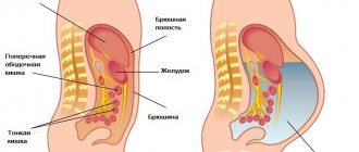
The mesenteries in the human body are responsible for supporting the small intestine and attaching it to the posterior wall of the intestine. Lymphadenopathy of the lymph nodes located in this area is also called mesenteric. The mechanism of development of pathology in adults:
- In various diseases, pathogenic microorganisms enter the lymph nodes.
- The nodes increase in size.
- As the pathology progresses in adults, inflammation occurs, the lymph nodes become painful, the skin over them turns red, and hyperemia occurs.
- Pain in the navel area in women - causes and associated symptoms
- The lymph node under the arm is inflamed: what to do and how to treat it
- What is the danger of contamination of the human lymphatic system - structure, functions and diseases
In most cases, abdominal lymphadenitis is a complication of other diseases. The following agents can cause inflammation:
- Bacteria. Causes bacterial inflammation of the lymph nodes in adults. In this case, mesadenitis is provoked by streptococci, staphylococci, E. coli, and mycobacteria.
- Viruses. Lymphadenitis of a viral nature can be caused by adenoviruses, Epstein-Barr virus, and enterovirus.
- Fungi. They provoke fungal lymphadenitis.
- Parasites. Inflammation may be a consequence of helminthic infestation.
Lymphadenitis causes damage to both individual lymph nodes of the abdominal cavity and their entire group. Depending on the type of infectious agent that caused the inflammation, mesadenitis occurs:
- Specific. It develops when the body is exposed to Koch's bacillus (Mycobacterium tuberculosis) or Yersinia.
- Non-specific. Associated with damage to the body by viruses or bacteria that migrated from other foci of infection. Such mesadenitis is divided into purulent and simple.
Principles of treatment
Treatment of lymph nodes depends on the underlying disease. Enlarged lymph nodes cannot be treated, since the size of the organs of the lymphatic system returns to normal after the cause of lymphadenopathy is eliminated and immunity is increased. For inflammation of the lymph nodes, antibacterial drugs are prescribed. The choice of medication is made after a series of examinations to identify the causative agent of inflammation. As a rule, broad-spectrum antibiotics are used, for example, macrolides, fluoroquinolones or combination drugs based on penicillin. Along with antibacterial therapy, agents are prescribed to strengthen the immune system.
Related Articles
What are lymph nodes: location, functions, norm and pathology
Lymph nodes in the groin in men: location features, causes of inflammation
How to treat lymph nodes at home: effective drugs and folk remedies
Abdominal lymphadenopathy: what it is, how it manifests itself, types and causes, diagnosis and treatment
Lymphadenopathy: classification, signs and treatment
Treatment of the disease
The axillary immune system reacts to diseases of neighboring organs. Adenopathy of these lymph nodes is treated based on the cause that led to its occurrence. If the pathology is infectious in nature, then the doctor recommends antiviral drugs or antibiotics. Malignant tumors are eliminated through radiotherapy, chemotherapy, and surgery.
Physiotherapeutic methods are used at the discretion of the specialist:
- Laser therapy, in which the body is influenced by light waves, as a result of which pain is relieved and the intensity of the inflammatory process is reduced.
- Galvanization, which involves applying a small electric current to soft tissue. The procedure is aimed at relieving pain and restoring tissue and nerve fibers.
In exceptional cases (usually with a purulent process), surgical intervention is performed, after which antibiotics are prescribed for prophylactic purposes.
To strengthen the immune system, echinacea preparations, various herbal decoctions and infusions are recommended. Traditional methods are popular.
Reasons for the development of pathology
It is impossible to accurately and unambiguously determine the causes of enlarged retroperitoneal and mesenteric lymph nodes. But this happens to a greater extent because infections penetrate into the lymph nodes through the intestines or lymph.
This lesion can be caused by absolutely any infection under “favorable” conditions:
- adenovirus or enterovirus infection;
- streptococcus and staphylococcus;
- Epstein-Bar virus (herpetic virus type 4);
- mycobacteria;
- Koch's wand;
- syphilis;
- parasites and protozoa;
- microbes that cause brucellosis.
The viruses, infections and bacteria listed above are pathogenic in nature and can affect any of the 500 lymph nodes located in the abdominal area.
Causes of enlarged lymph nodes
Knowing what pulmonary lymphadenopathy is, you should understand why it occurs. In general, the reasons can be divided into two large groups: general and specific. Common causes of swollen lymph nodes:
- infections (viruses and bacteria);
- fungal diseases;
- parasitic infestations;
- tuberculosis;
- systemic connective tissue diseases;
- long-term drug therapy.
The listed factors can cause an increase in any group of lymph nodes, including intrathoracic ones.
Pathology can also be caused by a number of specific reasons. They are any pathological processes affecting the organs of the chest.
Note! Local enlargement of lymph nodes is always associated with infection, inflammation or a tumor process occurring in nearby organs.
Enlarged intrathoracic lymph nodes may be associated with:
- pulmonary tuberculosis;
- sarcoidosis;
- metastases;
- Hodgkin's lymphoma.
Only a doctor can accurately determine the cause of enlarged lymph nodes after examining the patient. It is recommended to visit a doctor as soon as possible after the first alarming symptoms are detected.
Tuberculosis
Untreated tuberculosis can lead to chronic lymphadenopathy
This disease in ICD-10 is designated by code A15. The cause of the development of the disease is the penetration of Koch's bacillus into the body. The disease is highly contagious and is transmitted by airborne droplets. At the initial stage, tuberculosis may not have specific symptoms, which greatly complicates timely diagnosis. In general, the characteristic symptom of this disease is a productive cough that persists for more than 4 weeks.
Other symptoms of the disease:
- severe fatigue;
- general weakness;
- night sweats;
- loss of appetite;
- weight loss;
- increased body temperature;
- chills.
The disease is dangerous not only due to its high degree of contagiousness, but also to the risk of spreading throughout the body. The most common complication of tuberculosis is infection of the lymphatic system. In this case, enlargement and inflammation of the intrathoracic lymph nodes can be either one of the first symptoms of the disease or a complication of advanced tuberculosis. The disease must be treated in a timely manner. Self-medication for tuberculosis is unacceptable, as it may lead to the development of a resistant form of the disease.
Sarcoidosis
An inflammatory disease that affects the tissues of the respiratory system is sarcoidosis. According to ICD-10, the disease is designated by code D86. With this pathology, changes occur in the tissues of the respiratory system, including the lungs, with the formation of nodules (granulomas). The peculiarity of this pathology is a long asymptomatic course. As a rule, the disease is detected by chance, during a routine examination with ultrasound, CT of the respiratory system or fluorography.
At the first stage of the disease, the intrathoracic lymph nodes become enlarged. In this case, malaise, cough, and difficulty swallowing appear. This is an alarming symptom that should force the patient to see a doctor as soon as possible. As a rule, enlarged lymph nodes allow sarcoidosis to be diagnosed at an early stage, which greatly facilitates treatment.
With sarcoidosis, the patient may feel quite well, while examination of the lungs may reveal a significant number of granulomas in the organ tissue. The pathology must be treated, otherwise there is a risk of developing respiratory failure.
Hodgkin's lymphoma
Night sweats are one of the symptoms of damage to the intrathoracic lymph nodes
Lymphogranulomatosis, or Hodgkin's lymphoma, is accompanied by severe pulmonary lymphopathy. This is a malignant disease in which the pathological process spreads to lymphoid tissue. The first symptom of the pathology is enlarged lymph nodes while maintaining fairly good general health. Typically, this disease primarily affects the cervical lymph nodes, but in every fifth case, lymphogranulomatosis begins with an enlargement of the mediastinal nodes.
When the intrathoracic lymph nodes are affected, the following symptoms are observed:
- labored breathing;
- cough;
- night sweats;
- hoarse breathing.
However, in the vast majority of cases, there are no significant symptoms until the late stage of the disease. Timely detected Hodgkin lymphoma is successfully treated in 83% of cases, allowing a five-year remission to be achieved, which is considered to be a complete cure.
According to ICD-10, the disease is designated by code C81.
Metastases
Enlargement of the thoracic lymph nodes is observed with the spread of metastases against the background of stage 3-4 cancer. In approximately 15% of internal organ cancers, metastases spread to the lymphatic system. First of all, lymph nodes that are located close to the site of cancer are affected. In case of damage to the intrathoracic lymph nodes by metastases, the cause of lymphadenopathy can be lung cancer, thyroid cancer, or tracheal cancer.
The presence of metastases is an unfavorable symptom. The process of metastasis begins in the later stages of cancer and can be fatal.
In addition, enlarged lymph nodes may be the first symptom of cancer before metastases begin to spread. In this case, it is caused by a violation of the movement of lymph due to a pathological process in nearby organs. In such cases, seeing a doctor because of enlarged lymph nodes often helps save the patient’s life, since cancer can be detected at an early stage, when the pathology responds well to treatment.
Etiology
It is possible to find out the exact cause of lymphadenopathy only after conducting appropriate studies. The most common causes of enlarged lymph nodes may be the following:
- viral diseases;
- lymph node infection;
- connective tissue injuries and diseases;
- serum sickness (effect of medications);
- fungus;
- infectious diseases that suppress the immune system.
The child most often develops abdominal lymphadenopathy. The reason for this is bacterial and viral infection of the body. Lymphadenopathy in children requires immediate examination by a physician, as symptoms may indicate a severe infectious disease.
Causes of armpit lymphadenopathy
The most common causes of disorders of the lymph nodes in the axillary region are:
- infectious diseases of various origins;
- parasitic diseases;
- oncological pathologies;
- blockage of the sweat glands (often occurs when using low-quality deodorant or failing to comply with personal hygiene rules);
- skin damage in the chest area, near the arms and shoulders;
- psoriasis, eczema and other skin diseases;
- hepatitis;
- rheumatoid arthritis;
- melanoma;
- serum sickness due to the use of medications (in particular antibiotics);
- allergy;
- cat scratch disease.
In women, the causes of lymphadenopathy of the axillary lymph nodes include breast cancer. The most common triggering factor for illness in both sexes is infectious and parasitic diseases. Inflammation of the lymph nodes also occurs with mastopathy.
Classification
Depending on how many lymph nodes are enlarged, the pathology is classified into three types:
- local;
- regional;
- generalized.
Local damage affects one lymph node. Regional enlargement of nodes affects several nodes located next to each other. As for the generalized development of pathology, this is the most severe case, since at least three groups of lymph nodes are involved, which are located in different parts of the body.
About 70% of cases of enlarged lymph nodes occur in the local form of the pathology. Generalized inflammation in the nodes indicates serious problems in the functioning of the immune system.
Another type of classification subdivides lymphadenopathy according to the period of limitation:
- acute;
- chronic;
- recurrent.
Mesadenitis can take any of these three forms of the disease. But it is worth noting that in the chronic form, purulent inflammation is already observed in the lymph nodes, which spreads throughout the body.
Some experts use a classification of pathology according to the degree of hyperplasia, but this is a rather controversial division, since lymph nodes from different areas of the body vary in size even in normal condition.
Symptoms
There are general symptoms of lymphadenopathy, characteristic of any location:
- Increasing node size.
- Painful sensations when pressed (can serve as a sign of tissue necrosis, in particular caseous necrosis, or the beginning of an inflammatory process).
- Changing the node structure. A dense and enlarged node indicates the presence of a metastasized tumor, a soft one indicates the penetration of an infection into the body.
- Simultaneous inflammation of several nodes in one area is a sign of a carcinogenic process or another serious disease, tuberculosis, for example.
In addition to the above signs, there are specific symptoms for specific areas of inflammation.
Hilar lymphadenopathy is characterized by:
- difficulty breathing,
- temperature rise, sometimes significant,
- unpleasant sensations when swallowing,
- cough,
- discomfort and pain in the chest area.
Lymphadenopathy of the intrathoracic lymph nodes often indicates the presence and maturation of a tumor or its transition to the stage of metastasis. The risk group includes middle-aged patients; in younger patients, hilar lymphadenopathy is observed much less frequently.
Inflammation of the chest and axillary lymph nodes is no less dangerous. PAP in these areas can serve as both a sign of the presence of infection and cancer. Statistics show that lymphadenopathy in the axillary region is one of the first signs of a metastatic tumor in the mammary gland. In addition to changes in the lymph node, additional signs are noted. The nature of their appearance depends on the cause of the pathology.
The accompanying symptoms are considered:
- protrusion of rashes and skin lesions,
- constantly elevated temperature,
- sweating, especially worse at night,
- chills and fever,
- enlarged liver and spleen,
- Unreasonable weight loss, sometimes very significant.
When a doctor diagnoses “breast lymphadenopathy”, the first questions he hears are: “what is it, is it already cancer or not yet and... is it possible to do without surgery.” The doctor decides whether surgical intervention is necessary after examining the patient. And it is quite possible that the final diagnosis will not be so scary at all.
Breast lymphadenopathy can be a consequence of mastitis, a benign formation, or a carcinogenic process. The first thing you can notice even during self-examination is the location of the tumor:
- A change in the nodes in the upper part of the chest will most likely be a sign of a benign neoplasm. But there is always a real danger of degeneration of a benign process into an oncological disease.
- A change, or worse, inflammation of a node in the lower part of the mammary gland is almost always a sign of cancer.
- Most often the process is two-way. Unilateral axillary lymphadenopathy on the left or right can be caused by a purulent wound or furunculosis of the upper limbs or shoulder area. Bilateral enlargement of the node is a sign of sarcoidosis, secondary syphilis or tularemia.
- Lymphadenopathy of the mammary gland with mastitis is characterized by severe pain in the area of pressure. When the root cause is eliminated, the lymph nodes return to normal.
Symptoms
Mesadenitis is a pathology, the existence of which people most often do not suspect. Abdominal lymph nodes are located inside the peritoneum, and their enlargement can only be diagnosed using ultrasound.
The mesentery is a fold of membrane that attaches the intestine to the abdominal wall and holds it in place. Mesenteric lymphadenitis is inflammation of the lymph nodes in the mesentery
Symptoms of the disease appear suddenly and are more typical only in acute cases. Patients suffer from pain in the abdominal area, in some cases it is impossible to indicate the exact location of the discomfort. If enlarged lymph nodes occur in the lower abdomen on the right, patients often confuse the pathology with appendicitis, since the pain and all the symptoms are very similar:
- Increased body temperature.
- Nausea and vomiting.
- Lack of appetite and stomach pain.
- Diarrhea or constipation.
- Tachycardia.
- Enlarged spleen and liver.
- Dry mouth, dehydration.
If symptoms are ignored for a long time, the patient may receive unpleasant symptoms in the form of peritonitis, intestinal obstruction and other serious diseases. This happens because the lymph nodes begin to fester without proper treatment.
The chronic form of the course is less noticeable to humans, the clinical picture is blurred and rarely causes concern. There is practically no pain syndrome, pain is felt only during physical activity.
The pathology often affects children. According to statistics, girls get sick less often than boys. Age range from 6 to 13 years. The symptoms are no different from adult mesadenitis. When palpating the abdomen, you can find that it is tense, this is due to the formation of lymphoid follicles. It is necessary to consult a specialist to avoid complications and begin the necessary treatment on time.
Symptoms of pathology
The clinical picture of the disease depends on the nature of the pathological process. In the acute form, inflammation of the lymph nodes develops suddenly and has pronounced symptoms. The chronic version is distinguished by a more blurred clinical picture over a long period of time. Main symptoms of mesadenitis
| Nature of the disease | Main symptoms |
| Spicy |
|
| Chronic |
|
Signs of complications
If lymphadenopathy of the retroperitoneal space in adults is not eliminated in time, complications may develop. Common consequences of inflammation of the lymph nodes:
| Possible complications | What signs are accompanied by |
| Peritonitis |
|
| Sepsis |
|
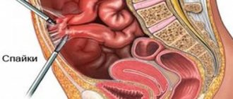
| Adhesive disease of the abdominal organs |
|
| Generalization of the process with the development of extensive inflammation of the body’s lymph nodes |
|
Diagnostics
Since inflammation does not have specific symptoms, diagnosing mesadenitis is difficult. It is important to distinguish the disease from acute appendicitis, gastric or duodenal ulcers, renal colic, adnexitis, and ovarian apoplexy. To confirm mesadenitis, the following studies are carried out:
- diagnostic laparoscopy;
- general analysis and blood culture for sterility;
- MRI and ultrasound of the abdominal cavity.
Diagnosis of pathology
The lymph nodes of the abdominal cavity are subject to disruption under the influence of various factors, so the diagnosis must be comprehensive, aimed at identifying the exact cause of the pathology. The following is carried out for the patient:
- Examination by a surgeon. Palpation of the abdomen reveals uneven formations of high density, concentrated in different places. Positive symptoms of Klein, McFadden, Shtenberg are determined.
- Ultrasound of the lymph nodes of the abdominal cavity and retroperitoneal space. According to the study, enlarged and dense lymph nodes and increased acoustic density in the mesentery area are detected.
Examination data of the pancreas, spleen and gallbladder are compared with ultrasound of the abdominal lymph nodes. This makes it possible to exclude pathologies with identical symptoms (for example, acute pancretitis).
- MRI allows you to identify the location, diameter and number of affected formations, visualize changes in the digestive tract and other abdominal organs.
- Laboratory research:
- the CBC shows an increase in leukocytes and an increase in ESR;
- blood culture for sterility makes it possible to identify a specific pathogen located in the circulatory system and triggering the development of pathology;
- tuberculin test or diaskintest (in case of suspected tuberculous origin of mesadenitis);
- serological methods of blood analysis to detect a pathogen or the presence of antibodies to it (including the causative agent of viral hepatitis);
- blood biochemistry, based on the results of which it is possible to determine abnormalities in the functioning of the liver, kidneys and pancreas;
- general urine test (to assess the functioning of the urinary system).
- Diagnostic laparoscopy. It is advisable to carry out when there is insufficient information obtained from other sources. During the procedure, it is possible to visualize the affected immune components, their number and location. Diagnostic laparoscopy allows you to examine the abdominal organs to exclude concomitant diseases. To draw conclusions, lymph node material is collected intraoperatively for histological examination.
- X-ray may be needed for the purpose of differential diagnosis (excluding peritonitis).
Complications of mesenteric lymphadenitis
Mesadenitis requires treatment; if the pathology starts, it is incredibly dangerous for a person. If you do not contact a specialist for a long time, there is a risk of developing an abscess or peritonitis due to lymphatic suppuration.
In extremely severe cases, generalized lymphadenitis affects the entire human lymphatic system, resulting in enlarged and inflamed nodes throughout the body. These complications are especially common in patients suffering from tuberculosis; in other cases, this is a rather rare occurrence.
Lymph nodes of the abdominal cavity: location features, causes of enlargement and inflammation
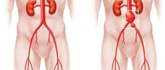
The lymph nodes of the abdominal cavity are a large group of lymph nodes that provide lymph flow to the organs of this area. For a number of different reasons, these nodes can become enlarged and inflamed. Due to their deep location, the lymph nodes in the abdominal cavity are not palpable, so a pathological process can be suspected by indirect symptoms. It is important to know the localization and function of the lymph nodes in the abdomen in order to promptly pay attention to the body’s alarm signals and consult a doctor.
Prevention of inflammation of the lymph nodes in the abdomen
To avoid damage to the lymph nodes in the peritoneum, as a preventive measure it is worth diagnosing and treating the following diseases in a timely manner (especially if they are chronic):
- tonsillitis;
- bacterial kidney damage;
- bronchitis;
- inflammation of the walls of the gallbladder;
- bacterial overgrowth syndrome.
The patient’s life expectancy, as a rule, is not reduced, because the disease does not pose a mortal danger. It is important to begin prompt treatment of the inflammatory process. High-quality treatment will not only quickly return the patient to the usual rhythm of life, but will also save life if the patient is diagnosed with tuberculosis.
Unfortunately, it is very difficult to detect the development of pathology in the early stages; patients are often hospitalized with severe attacks of pain, when the likelihood of suppuration increases. Doctors do not recommend neglecting the first signs of the disease, but rather contact a medical facility as soon as possible. Only timely examination and treatment will avoid complications.
Causes, treatment of enlargement and inflammation of the subclavian lymph nodes
Previous post
Enlarged lymph node on one side under the jaw
Next entry
Prevention and prognosis for enlarged lymph nodes
To prevent the development of lymph node hyperplasia, it is important to adhere to a healthy lifestyle. A person is advised to stop smoking, drinking alcohol and drugs. It is very important to subject your body to systematic physical activity, move a lot, and walk in the fresh air. The list of preventive measures for lymphadenitis includes the following recommendations:
- if any health problems are detected - exacerbation of gastritis, increased body temperature, general weakness - you should immediately consult a doctor;
- it is necessary to avoid contact with stray animals; after interacting with pets, you must wash your hands thoroughly;
- Promiscuity should be avoided and barrier methods of contraception should be used;
- at least once a year, all men and women need to undergo general tests and undergo a preventive examination of the body for early detection of possible pathologies.
The prognosis for lymphadenopathy depends on the etiology of its development, the general condition of the patient and timely treatment. With the local form of the disease, it is often possible to fully recover without negative consequences for the body. With generalized lymphadenopathy, the outcome is less favorable.
Establishing diagnosis
If you notice the above symptoms, you must immediately contact a specialist and undergo an examination. An accurate diagnosis requires a comprehensive laboratory instrumental examination of the patient. First of all, it is necessary to exclude inflammation of the appendix, since pain in the lower abdomen suggests this diagnosis.
The doctor initially collects an anamnesis of the disease. In order to identify the causative agent of the pathology, questions are asked about previous injuries, blood transfusions, transplantations, place of work, recent travel, etc. During the examination, the specialist checks the condition of the mucous membranes and palpates the abdomen to determine the presence or absence of mesenteric follicles.
It is necessary to do a number of laboratory tests:
- General blood and urine analysis.
- Biochemical blood test.
- General stool analysis.
- Feces for occult blood.
- Serological tests.
- Wasserman test.
An accurate and unambiguous diagnosis can only be made after the results of an ultrasound or x-ray. Only after the cause of the disease has been established is drug therapy prescribed. If you take measures only to eliminate symptoms, then relapses will occur after some time.
Prevalence and risk groups
Persons with weakened immunity and inflammation of the gastrointestinal tract are susceptible to inflammation of the lymph nodes in the abdominal cavity. In childhood, the symptom occurs more often than in adults, which is associated with insufficient formation of the immune system.
Malignant tumor processes with metastasis are one of the factors for enlargement of lymphatic organs. This happens due to the spread of metastases from the abdominal cavity.
People suffering from Epstein-Barr virus are potentially susceptible to enlarged nodes. This occurs due to infection with infectious mononucleosis. The pathogen is also found in the detection of nasopharyngeal cancer and Burnitt's lymphoma.
Diseases such as HIV and hepatitis can provoke changes in the size of nodes. After infection with Giardia and some types of helminths, this pathology also develops.
Principles of nutrition for inflammatory bowel disease
Diet No. 4 according to Pevzner involves excluding from the diet any chemically or mechanically irritating foods. Nutrients that can cause fermentation or rotting processes in the intestines with increased gas formation are also limited. All dishes should be warm; hot and cold food will disrupt intestinal motility and irritate the gastrointestinal mucosa.
You should not eat fried, pickled, salted or smoked foods. Food should be steamed, stewed, boiled, baked. The products must be well ground in a meat grinder or blender. Vegetables can be rubbed through a sieve. Rough foods containing a lot of plant fiber should also be excluded.
Advice: an important principle of therapeutic nutrition is fractionation. It is better to eat more often so as not to overload the intestines. At the same time, you should not overeat; portions should be small - 150-200 g.
What dishes can you cook?
The diet for intestinal inflammation can include the following dishes and products:
- steamed meatballs, dumplings or cutlets from lean fish or meat (veal, beef, chicken, hake, pink salmon, rabbit);
- meat or fish soufflé;
- pureed vegetable and cereal soups in low-fat (second) meat, fish or vegetable broth;
- eggs, soft-boiled or steamed as an omelet;
- fresh non-sour cottage cheese, cottage cheese casserole;
- porridge from crushed cereals in water, vegetable broth or diluted milk (preferably oatmeal, buckwheat, semolina and rice cereals);
- yesterday's wheat bread or crackers made from it;
- unsweetened savory cookies.
What is the violation?
In young children, lymphadenopathy of the cervical lymph nodes is more noticeable due to the lack of fat in the neck
Nodes in the lymphatic system are important elements of the immune system. They perform a barrier function, acting as a kind of obstacle to pathogenic agents. If a person is healthy, it is almost impossible to palpate the nodes in the neck. Lymphadenopathy of the neck is manifested by a noticeable increase in nodes, and they are easily palpated even by a non-specialist.
Cervical lymphadenopathy is a common symptom of various diseases, from common acute respiratory viral infections to autoimmune pathologies. At the same time, the symptom is not always dangerous and indicates a serious illness, but it cannot be ignored, especially if no other signs of any pathology are found.
According to ICD-10, this violation is designated by code R59. Lymphadenopathy of the cervical nodes is equally common in both adults and children. Moreover, in children the pathology is more noticeable due to the lack of fat in the neck.
Surgical intervention
In most cases, adenopathy is treated with medications. Performing surgical intervention is advisable only when the contents of the lymph nodes are represented by purulent fluid.
In such situations, the formation is opened. It is usually carried out using a standard technique - open surgery. After gaining access, the surgeon removes the purulent contents, installs a drainage system through which the discharge will drain, and applies sutures.
Methods for diagnosing pathology
Enlarged lymph nodes are often confused with the development of appendicitis, since in the acute phase it is accompanied by vivid symptoms - pain, vomiting and fever. When the first signs of such health appear, the patient should call an ambulance, since taking medications can complicate diagnosis.
During the examination, the doctor must feel the patient’s abdomen: on the right, left, below and around the navel. Anamnesis is collected, specifying previous events and additional symptoms, including the presence of infections and poisonings. Laboratory tests are prescribed that will help identify the pathogen and make a diagnosis - to clarify the disease that caused the enlargement of the nodes:
- general urine test to determine the condition of the genitourinary system;
- stool analysis - to determine the absence of parasites and internal bleeding;
- do a mantu test to exclude tuberculosis;
- study a general and biochemical blood test to determine viral pathogens.
Tests help rule out most diseases that can cause enlarged lymph nodes.
An instrumental examination is prescribed - ultrasound. It helps determine the condition of nodes and internal organs. Additionally, X-rays and tomography are used - this equipment helps determine whether the patient has serious conditions such as tumors or intestinal damage.
After numerous examinations, a puncture of the inflamed nodes may be prescribed, which gives an accurate idea of the presence of tumor processes and sources of inflammation.
Without a full examination, which includes laboratory and instrumental tests, it is impossible to accurately establish a diagnosis. Only after receiving medical data does the doctor determine the disease causing the enlargement of the nodes. It is the disease that needs to be treated, not the symptom.
And little about secrets
- Do you often experience discomfort in the heart area?
- Suddenly you have the opportunity to feel weak and tired...
- There is nothing to say about shortness of breath after the slightest physical exertion...
- And you have been taking a bunch of medications for a long time, going on a diet and watching your weight...
But judging by the fact that you are reading these lines, victory is not on your side. It is on this basis that we recommend reading the story of Olga Markovich, who found an effective remedy for cardiovascular diseases. Read later>>>
If you think you have Lymphadenopathy
and the symptoms characteristic of this disease, then a general practitioner can help you.
In addition, we offer you to use our online disease diagnostic service. which, based on the entered signs, selects possible diseases.
Diseases with similar symptoms:
A disease characterized by acute, chronic and recurrent inflammation of the pleura is called tuberculous pleurisy. This disease has the peculiarity of manifesting itself through infection of the body with tuberculosis viruses. Quite often, pleurisy appears if a person has a tendency to pulmonary tuberculosis.
Lymphoma (matching features: 8 out of 20)
Lymphoma is not one specific disease. This is a whole group of hematological diseases that seriously affect the lymphatic tissue. Since this type of tissue is found virtually throughout the human body, malignant pathology can appear in any area. In addition, internal organs are likely to be damaged.
Histoplasmosis (matching features: 8 out of 20)
Histoplasmosis is a disease that develops due to the penetration of a specific fungal infection into the human body. With this pathological course, internal organs are affected. The pathology is scary because it can develop in people of different age categories. In addition, in the medical literature you can find such names of the disease as Ohio Plain disease, Darling disease, reticuloendotheliosis.
Thyroid cancer is a malignant pathology in which a compaction (nodule) is formed that affects the thyroid gland and develops on the basis of its follicular epithelium or parafollicular epithelium. Thyroid cancer, the symptoms of which are predominantly detected in women aged 40 to 60 years, is diagnosed on average in 1.5% of cases when considering any type of malignant tumor formation in a particular area of localization.
Pleurisy (matching symptoms: 7 out of 20)
Pleurisy is a general name for diseases in which inflammation of the serous membrane near the lungs - the pleura - occurs. The disease in most cases begins against the background of pre-existing diseases and may be accompanied by the formation of effusion on the surface of the membrane (exudative pleurisy) or fibrin (dry pleurisy). This nuisance is considered one of the most common pulmonary pathologies (300–320 cases per 100 thousand population), and the prognosis for treatment depends entirely on the severity of the primary disease and the stage of inflammation.
Types of violation
With local lymphadenopathy, 1-2 lymph nodes are affected
Having understood what axillary lymphadenopathy is, you should pay attention to the fact that the pathology is divided into several types. Based on localization, local, regional and generalized forms of the disorder are distinguished.
Local is called axillary lymphadenopathy, affecting 1-2 nodes in one area (left or right).
Regional is a form of pathology in which several nodes enlarge in one area. This may be lymphadenopathy of several nodes on one side, or symmetrical enlargement of the lymph nodes in the armpits.
Axillary lymphadenopathy can be unilateral or bilateral.
A generalized form is a disorder in which nodes in different regional zones (at least three) enlarge, for example, in the axillary, cervical and inguinal regions.
According to the nature of the pathology, there are:
- acute – appears for the first time against the background of any diseases, accompanied by vivid symptoms;
- chronic - manifested by a moderate increase in nodes, without other obvious symptoms;
- recurrent - appears again in the same group of lymph nodes after treatment of the cause of lymphadenopathy.
Women often encounter recurrent axillary lymphadenopathy, since an increase in nodes in this area is often due to changes in hormonal levels.
Traditional methods
The use of unconventional methods does not eliminate the need to seek qualified medical help. The use of traditional methods is permissible, but only after obtaining the approval of the attending physician. This is because in some cases natural remedies can significantly worsen the course of the disease.
The most effective recipes:
- Grind freshly picked dandelion grass. Moisten a cotton swab in the released juice and apply it directly to the affected lymph node. Lotions can be done up to 3 times a day.
- Mix nettle and yarrow in equal proportions. Combine these components and fill them with 500 ml of water. Place the container on the fire. Boil for 20 minutes. Let the broth cool. Take the product three times a day before meals.
- Mix 500 g of goose or badger fat with norichnik (medicinal plant). The latter must be taken 6 tbsp. l. Treat the affected areas with the resulting ointment.
Symptoms characteristic of the disease
The symptoms of this disease are very different and they depend on the cause of the inflammatory process. For example, the most characteristic is the appearance of “bumps” on the affected lymph node, the palpation of which brings unpleasant sensations, even severe pain. The lymph nodes of the muscle area, groin or neck become inflamed. In this case, you can visually detect redness of the skin.
The second type of lymph nodes is visceral. If they are affected, it is much more difficult to detect the inflammatory process, since the lymph nodes of the porta hepatis or mesenteric nodes are located in hard-to-reach places. And here it is no longer possible to do without diagnostic methods within the walls of the laboratory.
Recovery period
When the exacerbation subsides, the diet for intestinal inflammation allows you to include soups with boiled cereals and finely chopped vegetables in the menu. Fish, meat and poultry can be cooked in sliced form. You can add whole milk and cream to pureed vegetables. Porridges should be well boiled, this also applies to pasta. The following vegetables and fruits are allowed:
- green pea,
- broccoli,
- baked apples and pears;
- sweet berries (raspberries, strawberries - no more than 100 g/day);
- fruit juices diluted half with boiled water.
During recovery, the diet for intestinal inflammation is expanded to include fermented milk drinks (low-fat), mild varieties of cheese. For sweets, you can eat marmalade, marshmallows, and jams at this time. During the recovery period, daily energy consumption is 3000 kcal, fats - 100 g, proteins - 100-110 g, carbohydrates - 400 g, liquid - 1.5 liters, table salt - 10 g. The fractional diet is maintained until complete recovery (at least 4 meals per day). Then it is allowed to include beets, ripe tomatoes, sweet apples, peeled, into the diet).
Important! It should be remembered that these nutritional rules for intestinal inflammation must be followed for quite a long time until remission or complete recovery is achieved.
Cervical lymphadenopathy: causes
Cervical lymphadenopathy: causes
The causes of inflammation of the nodes that collect lymph in the neck can be infectious or non-infectious. Most often this is:
- fungal, parasitic, bacterial or viral infections of tissues and organs of the neck;
- measles, cytomegaly, infectious mononucleosis;
- AIDS and HIV;
- toxoplasmosis;
- blood diseases from the field of oncology;
- sarcoidosis
As we can see, cervical lymphadenopathy has a variety of causes, therefore, regardless of the symptoms and degree of discomfort, if you have the slightest suspicion, you should immediately contact your physician. This will help quickly localize the disease and prevent a more serious illness.
Conservative methods of therapy
The scope of treatment directly depends on the patient’s age, the form of the disease, and the severity of the patient’s condition. The choice of treatment tactics for lymph node adenopathy is made after identifying the underlying pathology that served as a provoking factor.
If the accumulations and surrounding tissues are affected by infection, taking antibacterial drugs is indicated. Before the degree of sensitivity of pathogens to certain substances is revealed, doctors prescribe antibiotics belonging to the group of cephalosporins and fluoroquinolones. As a rule, experts recommend taking Medaxone and Levofloxacin. The duration of treatment depends on the individual health characteristics of the patient and the severity of the disease.
Pathogens spread very quickly throughout the body through the lymph nodes. In this regard, local therapy for the disease should be treated with caution. All medications must be prescribed by a doctor; only he can assess the appropriateness of their use. In almost all cases, experts recommend using Vishnevsky ointment. The product must be applied directly to the affected lymph node.
If during the biopsy it was determined that the pathological process is malignant, the patient is prescribed a course of chemotherapy. The outcome of the disease in this case directly depends on the severity of the underlying disease.
As an additional treatment, doctors prescribe immunostimulating agents. They are designed to strengthen the body's defenses during the fight against the disease. The drug “Glutoxim” has shown high effectiveness in practice.
Diagnostic features
To determine the causes of the development of pathology and make an accurate diagnosis after identifying enlarged lymph nodes in the celiac cavity, doctors begin collecting anamnesis. The patient is asked about his lifestyle, medical procedures performed, possible contacts with animals, and the presence of casual sexual relations. It is necessary to inspect the sites of inflammation, determine the size of the lymph nodes, the presence of pain and other symptoms.
To identify the origin of pathology and the degree of development of negative processes in the body, hardware and laboratory diagnostics are carried out:
- biochemical, serological and general blood tests;
- urine test;
- blood tests for syphilis and HIV infection;
- Ultrasound, MRI, CT;
- radiography.
In clinically severe cases, a lymph node biopsy, bone marrow biopsy, osteoscintigraphy and other studies are performed at the discretion of the doctor. If the cause of the pathology is unclear, laparoscopy is recommended. Only after an accurate diagnosis is made, a decision is made on the choice of treatment tactics.
Methods for determining violation
The retroperitoneal space cannot be examined, as well as visually assess the condition, however, examination of the abdominal wall, palpation and percussion are the first clinical methods used during consultation with a specialist. Pay attention to the color of the skin, the presence of depressions or protrusions, determine infiltrates, neoplasms of the abdominal wall.
The patient is placed on the couch and a cushion is placed under the lower back. As a result, the abdominal and retroperitoneal organs protrude forward, allowing palpation. Pain that appears when pressing or tapping on the abdominal wall may indicate a purulent-inflammatory process, neoplasms (including cystic).
X-ray examinations are also used:
- X-ray of the intestines and stomach;
- urography - study of the functioning of the urinary system with the introduction of a contrast agent;
- pancreatography - assessment of the condition of the pancreas with the introduction of a contrast agent;
- pneumoperitoneum - injection of gas into the abdominal cavity with further x-ray examination;
- aortography - examination of the patency of the abdominal aorta;
- angiography of aortic branches;
- cavography - assessment of the condition of the vena cava;
- lymphography.
Instrumental research methods include ultrasound, CT and MRI of the retroperitoneal space. They are carried out in a hospital or outpatient setting.
The primary method for detecting pathology is palpation of the abdomen. Anamnesis is being collected. Based on the information received, the following laboratory tests are prescribed:
- UAC
- OAM;
- blood biochemistry;
- coprogram.
| Survey | Peculiarities | Indications for use |
| Ultrasound of retroperitoneal lymph nodes | Allows you to identify the diameter of formations, their location relative to other organs and other characteristics |
|
| Contrast radiography | A special composition is injected into the intestines and an image is taken | Differential diagnosis of lymphadenitis from intussusception and other diseases |
| CT and MRI | Allows you to obtain a detailed image of the peritoneum | Detection of neoplasms or distant metastases |
If the retroperitoneal lymph nodes are not visualized, this means that they are in normal condition. An increase in formations indicates infections of nearby organs or cancer.
What is lymphadenopathy?
Lymphadenopathy is a condition in which the size of lymphatic vessels of various origins increases.
In 75% of cases with this disease, only one lymph node is affected by inflammation.
Most often, lymphadenopathy occurs in the axillary or postauricular area.
Sometimes ordinary inflammation of the lymph nodes is confused with a disease called lymphadenitis. They are similar, but have one difference. With lymphadenitis, in places where lymphatic vessels are enlarged, accumulations of pus form, which is more dangerous to human health.
Diagnostic measures
You should always remember that enlarged lymph nodes in the neck are only a symptom of a disease. It can be very difficult to detect the source or cause of this phenomenon even in specialized medical institutions, and without instrumental and laboratory tests it is simply impossible.
A sample diagnostic list includes:
- General blood analysis. They look for changes in cellular composition, ESR.
- Biochemical research makes it possible to detect protein metabolism disorders and proteins specific to certain diseases.
- Analysis for antigens to viruses and malignant cells.
- X-ray or computed tomography.
- A puncture biopsy of the affected node provides the most valuable diagnostic data.
Puncture of an enlarged lymph node allows you to take a piece of tissue for histological examination. Often, only such a study allows one to distinguish metastasis from hemoblastosis.
However, even these studies may not make it possible to quickly make an accurate diagnosis. Then additional methods are used, which must be selected by the attending physician.
Retroperitoneal injuries
The most common hematoma is the result of mechanical trauma. Immediately after damage, it can reach enormous sizes, which makes it difficult to differentiate the diagnosis. A specialist may confuse a hematoma with damage to a hollow organ. The injury is accompanied by hemorrhagic shock due to massive blood loss.
The brightness of the manifestations decreases faster than in the case of damage to internal organs. Laparoscopy allows you to determine the condition. Pneumoperitoneum shows displacement of the retroperitoneal organs and blurring of their contours. Ultrasound and computed tomography are also used.
LYMPHADENOPATHY OF THE MEDIASTINUM
Found (22 posts)
AIDS October 16, 2012 / Maria
... 5 mm to 29 mm, enlarged lymph nodes of the mesogastrium and mediastinum
, headaches, sharp deterioration of vision, very much covered... what direction should I be examined, what may be associated with
lymphadenopathy
.
The infectious disease doctor throws up his hands and advises taking tests before... open (23 more messages)Last 5:
endocrinologist August 30, 2012 / Victoria... / Chernigov
...cells without pathological changes, in the middle parts of the mediastinum
a rounded formation with clear contours is visualized...: Sonographic picture of a left-sided nodular goiter.
Lymphadenopathy
. Consultation with a Surgeon is recommended. The surgeon is probably... open
oncologist July 8, 2012 / Vitaly / 0
..., no pathological formations were found in the mediastinum. Mediastinal lymph nodes
and roots of the lungs: upper paratracheal up to 19mm, ... process of the lower lobe of the left lung (peripheral form),
lymphadenopathy
.
Is it possible to do something or cure... open
oncologist March 13, 2012 / Elena / 0
... hydrothorax (pleurisy), minimal hydropericardium. Intrathoracic lymphadenopathy
.
Atherosclerosis of the aorta and coronary arteries. Previously, 11 years old... deformed, its front plate is crushed. mediastinal
shadow is shifted to the right. In the right lung there are focal infiltrative ... open
oncologist December 1, 2011 / Natalya / Abakan
...: I have LGM 2B with damage to the supraclavicular nodes and mediastinal
.
The diagnosis was made a year ago. there was severe night sweats, ... CT scan of the chest 10/12/2011 mediastinal lymphadenopathy
with positive x-ray dynamics. l/u with a diameter of up to 2.2 cm. open
oncologist November 10, 2011 / Christina... / 0
... conclusion k-1 signs of a space-occupying formation of the left lung (susp.cr pulm.centralis. sin) with atelectasis of the c1 lung.
Mediastinal
lymphadenopathy . Osteolytic lesions with a soft component of the 10th rib on the right. 10- …open
oncologist October 22, 2010 / VLADIMIR... / FEODOSIA
CT signs are most characteristic of a space-occupying lesion in the right lung. Lymphadenopathy
.
Mediastinal
lipomatosis . Atherosclerosis of the aorta and coronary vessels. Ascites. decipher please! open
oncologist October 22, 2010 / VLADIMIR... / FEODOSIA
CT signs are most characteristic of a space-occupying lesion in the right lung. Lymphadenopathy
.
Mediastinal
lipomatosis . Atherosclerosis of the aorta and coronary vessels. Ascites. decipher please! open
oncologist July 14, 2010 / Andrey / Osinniki
Hello, please tell me what kind of disease I was diagnosed with; Mediastinal lipomatosis
.
Lymphadenopathy of the mediastinum
and roots of the lungs. And how long will I live if I don’t get personal? open
oncologist June 22, 2009 / Olga
...) and sent me for a computed tomography of the chest to oncology. In oncology I was diagnosed with Lymphadenopathy
l / at the anterior and central
mediastinum
. We discovered a permanently enlarged lymph node in the neck - they did an ultrasound, I … open
Walls of the retroperitoneum
The anterior wall is the peritoneum of the posterior wall of the abdominal cavity together with the visceral layers of the pancreas and colon.
The upper wall runs from the costal and lumbar part of the diaphragm to the coronary ligament of the liver on the right and the phrenic-splenic ligament on the left.
The posterior and lateral walls are represented by the spinal column and nearby muscles, covered with intra-abdominal fascia.
The lower wall is a conventional border through the boundary line separating the pelvis and the retroperitoneal space.
Treatment of lymphadenomopathy
To lymph nodes it is strictly prohibited:
- Apply cold
- Warm up
- Rub.
Do not self-medicate, because incorrect actions will only worsen the situation. Contact a specialist.
The doctor should find out the medical history, while specifying:
- Fact of injuries
- The presence of inflammatory diseases of organs,
- Contact with sick people
- Contact with animals
- Eating raw foods: meat, fish,
- Have there been trips abroad, etc.
Carefully examine all lymphatic organs, which includes:
- Examination of the tonsils,
- Palpation of all groups of lymph nodes, indicating their location, shape, pain, consistency, etc.,
- Palpation,
- Percussion of the liver and spleen.
Conduct an examination of the organs from which lymph flows into the corresponding group of lymph nodes. Conduct standard diagnostic tests (general and biochemical blood tests, etc.)
If there is no further enlargement of the lymph nodes, it is necessary to monitor the patient for two to four weeks.
If bacterial flora is present in the drainage area of the lymph nodes, antibiotic therapy is prescribed taking into account data on sensitivity to it.
In the presence of dense, painless lymph nodes measuring more than 2×2 cm, or localized in the supraclavicular region, as well as over 40 years of age, a biopsy of the lymph nodes with cytological and histological examination is indicated.
It is strictly forbidden to prescribe hormonal drugs for lymphadenopathy of unknown etiology.
The classic treatment for lymphadenopathy of bacterial etiology is:
- Broad-spectrum antibacterial drugs (for example, sulfonamides, etc.) are used to suppress bacterial microflora in the lymph nodes and the primary site of infection.
- Surgical tactics are opening and drainage of the primary purulent focus and purulent lymphadenitis.
- Increasing the body's resistance and reducing the influence of intoxication.
For chlamydial and mycoplasma infections:
- Initially, an immunogram is assessed,
- Then complex therapy is carried out, which includes the use of macrolide antibiotics, or tetracyclines, or difluoroquinolones in an age-specific daily dosage for 1-3 weeks in one or two courses,
- In the 2nd half. course, licopid is prescribed in an age-specific dosage for 10 days
- As well as drugs that contain immunomodulators (recombinant alpha-two interferon, interleukin-two) for 5-20 days.
In conclusion, I would like to say that the treatment of lymphadenopathy is aimed at eliminating the underlying disease.
Symptoms and diagnosis
Mediastinal lymphadenopathy behaves asymptomatically when a particular disease is at the initial stage of development. Subsequently, when the lymph nodes put pressure on other anatomical formations, the pathology manifests itself:
- chest pain;
- cough, difficulty breathing (shortness of breath);
- hoarseness in the head;
- problems with swallowing;
- swelling in the face, neck, shoulders;
- loss of body weight;
- elevated temperature;
- sweating;
- enlarged spleen and liver.
Since lymphadenopathy is not the root cause of the patient’s suffering, but only a symptom of more serious diseases , the patient should undergo a comprehensive examination.
Diagnostics will consist of:
- radiography;
- computed tomography of the chest;
- ultrasound examination;
- blood sampling;
- biopsies.
Magnetic resonance imaging
A CT scan of the retroperitoneum is performed to determine pathologies or identify abnormal structure of internal organs. For convenient implementation and a clearer result, the injection of a contrast agent is used. The procedure is indicated for injuries to the abdomen or lumbar region, suspected neoplasm, damage to the lymphatic system of this area, urolithiasis, polycystic kidney disease, prolapse or the presence of inflammatory diseases.
CT scanning of the abdominal cavity and retroperitoneal space requires preparation for the procedure. For several days, foods that cause increased gas formation are eliminated from the diet. If constipation is present, laxatives and a cleansing enema are prescribed.
The patient is placed on a surface that is located in the tomograph tunnel. The device has a special ring that rotates around the body of the subject. Medical staff are located outside the office and observe what is happening through a glass wall. Communication is supported using two-way communication. Based on the results of the examination, the specialist chooses the method of necessary treatment.
If ultrasound and CT are not informative or if it is necessary to collect more accurate data, the doctor will prescribe an MRI of the retroperitoneum. What this method reveals depends on the chosen area of study. MRI can determine the presence of the following conditions:
- pathological enlargement of organs;
- retroperitoneal tumor;
- the presence of hemorrhages and cysts;
- conditions with increased pressure in the portal vein system;
- pathology of the lymphatic system;
- urolithiasis disease;
- circulatory disorders;
- presence of metastases.
Neuroblastoma
The formation has a high degree of malignancy. It affects the sympathetic part of the nervous system and develops mainly in children. The early appearance is explained by the fact that neuroblastoma develops from embryonic cells, that is, the tumor is of embryonic origin.
The typical location is one of the adrenal glands, the spinal column. Like any tumor, retroperitoneal neuroblastoma has several stages, which makes it possible to determine the necessary treatment and make a prognosis of the disease.
- Stage I is characterized by clear localization of the tumor without involvement of the lymph nodes.
- Stage II, type A - the location does not have clear boundaries, the neoplasm is partially removed. Lymph nodes are not involved in the process.
- Stage II, type B - the formation is unilaterally localized. Metastases are determined in the part of the body where the tumor is located.
- Stage III is characterized by the spread of neuroblastoma to the second half of the body, metastasis to local lymph nodes.
- Stage IV tumor is accompanied by distant metastases - to the liver, lungs, and intestines.
The clinical picture depends on the location of neuroblastoma. If it is in the abdomen, it easily reveals itself upon palpation, causes digestive disorders, lameness and bone pain appear in the presence of metastases. Paralysis and paresis may develop.
Possible complications of the pathological condition
Untimely treatment, uncontrolled course of the condition for a long time, can provoke serious complications:
- Purulent softening, dissolution of the affected node, development of abscesses, fistula tracts;
- Bleeding due to germination of the vascular wall;
- Blockage of the veins of the affected area;
- Impaired lymph flow in the affected area, resulting in the development of lymphostasis;
- Blood poisoning.
Timely adequate therapy will help get rid of the pathological condition and prevent possible consequences and serious complications.
Diagnosis and treatment of the syndrome
Since PAP is a manifestation of a certain disease, the attending physician tries to determine the root cause of the problem.
First of all, non-traditional methods of therapy pose a danger to the patient, since such drugs do not prevent the disease that caused PAP from progressing and affecting more and more new areas.
The choice of treatment method for lymphadenopathy depends on many factors in each specific case, including the individual characteristics of the patient’s body, the expected disease and information obtained after undergoing a comprehensive comprehensive examination.
Anatomical features
The range of organs is quite diverse. This includes the urinary system, the digestive, cardiovascular, and endocrine systems. Organs of the retroperitoneal space:
- kidneys;
- ureters;
- pancreas;
- adrenal glands;
- abdominal aorta;
- colon (its ascending and descending parts);
- part of the duodenum;
- vessels, nerves.
Fascial plates, which are located in the retroperitoneal space, divide it into several parts. Along the outer edge of the kidney are the prerenal and retrorenal fascia, formed from the retroperitoneal fascia. The prerenal centrally connects with the fascial layers of the inferior vena cava and the abdominal aorta. The retrorenal fascia is “invaded” into the intra-abdominal fascia at the site of covering the diaphragmatic crus and the psoas major muscle.
Perirenal tissue passes through part of the ureter and is located between the prerenal and retrorenal fascia. Between the posterior surfaces of the colon and the retroperitoneal fascia there is peri-colic tissue (retrocolic fascia).
Tumors
Neoplasms can arise from dissimilar tissues:
- fatty tissue - lipoma, lipoblastoma;
- muscular system - fibroids, myosarcoma;
- lymphatic vessels - lymphangioma, lymphosarcoma;
- blood vessels - hemangioma, angiosarcoma;
- nerves - retroperitoneal neuroblastoma;
- fascia.
Tumors can be malignant or benign, and multiple or single. Clinical manifestations become noticeable when the neoplasm begins to displace neighboring organs due to its growth, disrupting their functionality. Patients complain of discomfort and pain in the abdomen, back, and lower back. Sometimes a neoplasm is detected by chance during a routine examination.
A large tumor of the retroperitoneal space causes a feeling of heaviness, venous or arterial stagnation of blood due to compression of the vessels. It manifests itself as swelling of the legs, dilation of the veins of the pelvis, and abdominal wall.
Benign tumors change the patient’s condition little, only in the case of particularly large tumors.
Diseases
The development of an inflammatory process becomes a common pathology. Depending on the location of inflammation, the following conditions are distinguished:
- inflammation of the retroperitoneal tissue;
- paracolitis - a pathological process occurs behind the descending or ascending colon in the tissue located in the retroperitoneal space;
- paranephritis - inflammation of the perinephric tissue.
Symptoms begin with manifestations of an intoxicating nature: chills, hyperthermia, weakness, exhaustion, an increase in the number of leukocytes and erythrocyte sedimentation rate. Palpation determines the presence of painful areas, protrusion of the abdominal wall, muscle tension.
One of the manifestations of purulent inflammation is the formation of an abscess, a common symptom of which is the appearance of flexion contracture in the hip joint on the side of the affected area.
Purulent processes in which the abdominal and retroperitoneal organs are involved are severe due to their complications:
- peritonitis;
- phlegmon in the mediastinum;
- osteomyelitis of the pelvis and ribs;
- paraproctitis;
- intestinal fistulas;
- pus flows into the gluteal region, onto the thigh.
Lymphadenopathy in children
Currently, in pediatric practice, cases of lymphadenopathy in various age categories of patients have become significantly more frequent, and previously changes in the lymph nodes were largely of an inflammatory nature, and in the last decade, damage to the lymph nodes by paraneoplastic processes has not been uncommon, which is largely explained by the unfavorable environmental situation.
It is necessary to distinguish between such concepts as “lymphadenitis”, which is nothing more than an inflammatory-changed lymph node, and “lymphadenopathy”, which is an intermediate diagnosis until the cause of the enlarged lymph node is reliably established (scarlet fever, infectious mononucleosis, lymphogranulomatosis).
It should be borne in mind that until a child reaches the age of 12 years, the lymphatic system is considered immature, although the beginning of its formation occurs in the early intrauterine period. This functional immaturity of the structures of the lymphatic system explains the high frequency of lymphadenopathy that is observed among patients of the pediatric age category.
When examining a newborn child, the detection of any palpable lymph nodes indicates their enlargement, since in this age period the lymph nodes are usually not accessible to palpation. The first year of a child’s life is considered critical, since at this age there is a reactive enlargement of large lymph nodes in the neck, occipital and groin areas. In adulthood, the criterion for the normal functioning of the lymphatic system, which is observed in most healthy children after three years, is considered to be palpation of no more than three groups of superficial lymph nodes.
If we consider the structure of etiopathogenetic forms of lymphadenopathy, which are observed with greater or lesser frequency in pediatrics, then the leading positions are occupied by immune reactive lymphadenopathy that occurs in various infectious diseases. Lymphadenopathy resulting from existing chronic hyperplastic diseases (immunodeficiency) and systemic connective tissue pathologies are observed equally often in childhood.
Fortunately, lymphadenopathy of a specific tumor nature makes up no more than 10% of the total morbidity, however, timely early diagnosis of this type of change is important for the prognosis of the patient’s recovery. It is extremely rare that lymphadenopathy in children is observed with a severe allergic reaction and helminthic infestation.
The development of signs of cervical lymphadenopathy is more typical for children with a lymphatic-hypoplastic type of constitution, and changes in the lymph nodes of the cervical group are always accompanied by the development of inflammatory changes in the oral cavity, as well as an enlargement of the thymus and spleen. The reactive type of lymphadenopathy of the cervical groups of lymph nodes is often a manifestation of inflammatory changes in the gums in children during teething. In order to determine the suspected chronic source of infection from which inflammatory agents entered a particular regional lymph node, it is necessary to take into account the direction of normal flow of lymphatic fluid from a particular anatomical region.
In a situation where a child, after one month of life, has persistent generalized lymphadenopathy, combined with fever, dermatitis, widespread oral candidiasis and chronic diarrhea, it should be assumed that the patient has AIDS.
Damage to the lymph nodes by paraneoplastic processes can occur as a primary tumor of the lymphatic system or in the form of the development of secondary metastasis. A malignant form of the tumor process localized in the lymphatic system, which prevails in childhood, is lymphosarcoma, which primarily affects the mediastinal and mesenteric lymph nodes.