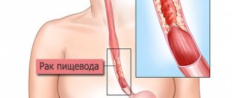
Esophageal atresia is a severe congenital anomaly in which the lumen of the organ becomes closed at a distance of about 10 cm from the oral cavity.
The lower segment is often connected to the trachea. The defect is diagnosed from birth, and the newborn develops specific signs during the first feeding. Code according to ICD 10 (esophageal atresia ICD 10) – esophageal atresia without fistula (Q39.0), with tracheoesophageal fistula (Q39.1).
Esophageal defect in newborns is the most common variant of this type of anomaly among the gastrointestinal tract. As a result of this deviation, the child cannot eat and without radical treatment, death occurs from exhaustion or food entering the respiratory system.
Course of the pathology
Esophageal atresia in newborns is a disease characterized by the inferiority of the child’s digestive system. The baby’s esophagus, which connects to the trachea, when normally it should go to the stomach, is not fully formed. There are several types of this pathology:
- the process of the esophagus has a fistula only in the upper part;
- the presence of a fistula in the lower part of the esophagus, extending into the trachea;
- the presence of fistulas on both sides of the esophagus.
Each type of pathology is equally dangerous for the life of a newborn, since during the first feeding food may enter the lungs, which leads to complex pneumonia. Therefore, a baby with a similar defect in the digestive system needs proper care and surgical intervention.
https://www.youtube.com/watch?v=yf5WaYpYB6I
The reasons that influenced the formation of such a defect in the fetus are considered:
- bad habits of the mother (smoking, alcohol, drugs, drinking energy drinks);
- bad heredity;
- infections during pregnancy;
- poor nutrition, unfavorable lifestyle of the expectant mother.
Watch a video about esophageal problems in newborns.
It is possible to identify esophageal atresia in newborns and determine the consequences of the pathology in the maternity hospital. More often it can be diagnosed in the first few hours of a baby’s life.
When trying to feed a child, the body rejects food. Severe vomiting with foamy secretions begins, the baby refuses to eat, it becomes difficult for him to breathe, and attacks of suffocation occur. If you have not yet offered your baby the breast, a clear indicator of the presence of a pathological process will be wheezing coming from the respiratory tract, a strong cough begins, and a possible increase in temperature.
After some time, the baby is diagnosed with dehydration.
Symptoms of the defect appear almost immediately after birth, even before the first feeding. They are quite typical, which makes it possible to quickly organize treatment. The newborn has a constant discharge of viscous foamy mucus from the mouth and nose (symptom of “false hypersalivation”), attacks of suffocation with cyanosis of the face and body due to aspiration (mucus entering the trachea).
Improvement occurs temporarily with the removal of mucus, then everything repeats. When the first breastfeeding begins, the fluid is immediately regurgitated, and in the presence of a tracheoesophageal fistula, it enters the respiratory tract, stimulating coughing, respiratory distress, and cyanosis. The reverse movement of fluid (regurgitation), in contrast to true vomiting, appears immediately after two sips.
Within a few hours, a newborn with esophageal atresia develops respiratory failure. It is aggravated by the entry of stomach contents into the trachea.
Respiratory disorder causes consequences in the body in the form of respiratory acidosis (deviation of the acid-base balance towards acidification), increased content of red blood cells and hematocrit.
The condition is rapidly deteriorating. Complications of respiratory failure are:
- aspiration pneumonia;
- exhaustion;
- dehydration.
In case of atresia, the doctor is unable to move the catheter further than 10–12 cm from the gums because it rests on the blind end
Examination of the baby makes it possible to identify:
- retraction or bulging in the upper abdomen;
- rapid breathing with disturbed rhythm;
- temporary and then complete cyanosis of the body;
- moist rales in the lungs.
These disorders intensify when trying to feed. Meconium is passed in the stool after birth on the first day, then defecation does not occur.
Symptoms, course
Signs of esophageal atresia appear immediately after birth. The child experiences regurgitation when feeding in small doses, false hypersalivation. Hypersalivation is an increased secretion of saliva of low viscosity, the secretion of foamy liquid with mucus from the mouth and nose. Newborns with esophageal atresia in the majority of cases are born premature and low birth weight.
Norms for the size of the fetal stomach on ultrasound and possible deviations
The fetal stomach at 16-20 weeks can have variable sizes. They are defined in millimeters. The normal sizes of the fetal stomach by week are presented in the table.
| Gestational age | Abdominal circumference |
| 16 weeks | 88-116 |
| 17 weeks | 93-131 |
| 18 weeks | 104-144 |
| 19 weeks | 114-154 |
| 20 weeks | 124-164 |
The study may reveal the following deviations:
- large or small stomach in the fetus on ultrasound;
- absence of an organ;
- presence of pseudo-content;
- slit-like stomach;
- gastric atresia in the fetus.
Description
Esophageal atresia is a severe malformation. With pathology, the lower (proximal) section of the esophagus is completely closed, and the upper (distal) is connected to the trachea through the fistula canal. An anomaly occurs during intrauterine development in the first three months of pregnancy. Atresia is formed due to cellular degeneration.
The frequency of recorded cases is 1 in 5000 newborns. If esophageal atresia is not detected in a timely manner and without surgical intervention, death occurs. The disease is often accompanied by defects in other organs and systems, such as the heart, gastrointestinal tract, genitourinary and musculoskeletal systems, and pathologies of the craniofacial part.
Combination with other vices
The pathology in question, according to statistics, occurs in 0.1-0.4% of infants. In most cases, it is combined with defects of other systems and organs. The most common diseases are:
- Radial bone dysplasia.
- Developmental defect of the limbs.
- Pyloric stenosis.
- Tracheoesophageal fistula.
- Choanal atresia.
- Spinal malformation.
- Agenesis of the gallbladder.
- Abnormalities in kidney development.
It should also be noted that in 5% of all cases of atresia, a chromosomal pathology is diagnosed - Down, Edwards or Patau syndrome. Another 40% of babies are born premature or with intrauterine growth retardation.
Treatment of esophageal atresia in infants
In order to avoid consequences that could lead to death of the newborn, it is necessary to urgently make a decision regarding treatment of the disease. First of all, it is important to correctly diagnose the pathology. To do this, an examination is performed using x-ray therapy, as well as using a special probe, which is passed into the baby’s nasal cavity and the degree of maturity of the esophagus is checked.
Each stage of the examination is performed in duplicate for a better understanding of the problem. Based on the diagnosis, an operation is prescribed, which involves prolongation of the esophageal process and its adhesion to the stomach.
After this, the baby is transferred to the intensive care unit, where his condition is monitored. Subsequently, specialists monitor the baby during the first six years of life, and if there are no consequences of surgery, they give a certificate that the child is healthy.
Diagnostic measures
It is possible to detect that a child has a defectively formed esophagus even during the period of intrauterine development during ultrasound diagnostics. You can assume that the fetus has atresia based on polyhydramnios. This is explained by the fact that the fetus does not swallow water, as it should do during its normal development. In addition, there is another sign - the stomach is small or completely absent.
After the birth of a child, atresia can be identified by the characteristic signs of the disease - bluish skin, foam from the mouth, shortness of breath. If such symptoms appear, the first thing the doctor does is check the patency of the esophageal tube using a special probe inserted through the nose. If atresia occurs, the probe will not reach the stomach cavity.
The diagnosis is confirmed through instrumental diagnostics, which includes the following methods:
- radiography;
- bronchoscopy;
- esophagoscopy.
X-ray of esophageal atresia
Additionally, consultation with an endocrinologist and surgeon may be required.
Treatment tactics
The only way to save a child's life is to perform surgery. The operation must be performed within the first 36 hours after birth. Conservative treatment methods will not bring any results.
Preparatory measures are carried out in the maternity hospital in the first hours of the patient’s life. Immediately after diagnosing the anomaly, pathological fluid is sucked out from the oral and nasal cavities every fifteen minutes.
Children are given antibacterial treatment. Feeding the child through the oral cavity is excluded. The baby is inhaled with moist air.
In order to improve the speedy emptying of the gastric sac and to avoid gastric juice from entering the respiratory tract through the fistula, the child must be positioned lying on his stomach with his head raised and the right side of his body lowered.
With increasing signs of respiratory failure, the baby is transferred to artificial ventilation.
In the absence of associated anomalies and provided there is sufficient body weight, the operation is performed in one stage. Surgery involves separating the tracheal and esophageal tubes and suturing the upper and lower lobes of the esophageal tube.
The baby should be under constant supervision of medical staff
Carrying out the operation
Surgery can be performed in different ways. The choice of technique largely depends on the form of the anomaly, as well as the general condition of the baby. If a distal tracheoesophageal fistula is present, a thoracotomy is performed to open the chest.
In the case of combined malformations, the operation most often begins with the application of a double gastrostomy, that is, the introduction through incisions into the stomach and duodenum of two rubber tubes, the outer ends of which are fixed to the skin.
After the operation, the baby is in the intensive care unit. Electrolytes, antibiotics, and glucose solution are administered parenterally. A long stay of the endotracheal tube in the trachea is undesirable, so specialists try to transfer the baby to his own breathing as soon as possible.
In the first week after surgery, it is strictly forbidden to straighten the baby’s neck. This is fraught with divergence of seams. After seven days, you can start feeding the baby with formulas with the addition of prokinetics.
With isolated atresia in the absence of associated malformations after surgery, cure occurs in one hundred percent of cases. Such children must be observed by a pediatrician and surgeon.
Parents should monitor the baby’s lifestyle and nutrition, and also maintain immunity.
Feeding after surgery is carried out through a tube or gastrostomy tube
Treatment of the disease is only surgical. Conservative therapy will have no effect. Preoperative preparation involves aspiration of contents from the oral and nasal cavities and oxygen supply. Next, the introduction of antibacterial drugs and hemostatics is required. This is necessary so that complications do not begin to progress in the child during and after surgical intervention.
An operation for esophageal atresia is performed to separate the esophageal tube and trachea, and subsequently form a full-fledged esophagus. A week after the intervention, radiography is performed, which is necessary to assess the condition of the connection. If everything is fine and there are no pathologies, then the child will now be able to feed himself through the oral cavity (orally).
Diagnostics
The diagnosis can be tentatively made during the period of intrauterine development, when an ultrasound examination shows polyhydramnios and the stomach is not visualized in the fetus. After the birth of a child with combined easily diagnosed anomalies of the limbs or spine, probing is first performed to exclude or confirm atresia. The diagnosis is made when the probe passes no more than 10 cm and rests.
Prenatal examination:
- obstruction of the esophagus in newborns, decreased turnover of amniotic fluid, which gives an indirect sign of atresia - polyhydramnios ;
- reduction of the stomach or inability to see it;
- developmental delay at different stages of embryogenesis;
- visualization of the proximal esophagus as it periodically fills and empties.
At the stage of prenatal diagnosis, one can only suspect atresia; most of the signs are characteristic of other developmental anomalies.
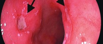
The newborn undergoes esophagoscopy to visualize the organ from the inside. The trachea is examined using an endoscope. The child is monitored by a surgeon and an endocrinologist. At the same time, a general assessment of the condition of the internal organs is carried out in order to decide on the method of treatment.
Newborn examination:
- blood analysis;
- electrocardiography;
- echocardiography;
- Ultrasound;
- MRI and neurosonography .
Confirmation of atresia is an indication for urgent surgery.
Surgery
When diagnosed with esophageal atresia, treatment is only surgical. The main option for the operation will be isolation of the fistula and ligation with the application of esophagoesophageal anastomosis . An umbrella is passed through the anastomosis and fixed well. A drainage is left in the mediastinum, which is connected to the aspiration system.
If the tension of the organ segments is high, anastomosis causes difficulties, and sutures may cut through. Their tension provokes tissue death due to insufficient blood circulation below the level of the anastomosis. a diastasis of more than 1.5 centimeters between segments is detected during the operation
First, the esophageal-tracheal fistula of the lower section is eliminated and then the upper segment is brought out to the neck. This approach to treatment prevents a common consequence of radical treatment in the form of aspiration pneumonia, which allows saving the life of the newborn.
After the main operation, an artificial esophagus is created, which is formed from the intestine. Feeding the child between two operations is carried out using an artificial external fistula.
For pain relief, endotracheal anesthesia is performed; many surgeons believe that mask anesthesia or local anesthesia is unacceptable in the surgical treatment of atresia.
Preparation for surgical treatment:
- the newborn is given a semi-sitting position to prevent reflux;
- glucose, saline, blood plasma, vitamin B are administered intravenously;
- the newborn is placed in an oxygen tent.
After the operation, the child is transferred to the surgical or children's department and placed in an individual box. For some time after the operation, which is determined individually, the child does not take any food by mouth. Feeding is carried out by drip infusion of glucose and Ringer's . An enema is prescribed less often. From about 3 or 4 days, catheter feeding begins.
Some experts believe that in order to prevent frequent postoperative complications, it is necessary to apply a gastrostomy tube for feeding until the wound has completely healed. But this approach to treatment often leads to various complications.
Preparation for surgery
Preparatory activities are carried out in the hospital:
- the baby is placed on a raised platform;
- ensures constant pumping of excess mucus from the digestive tract;
- feeding stops completely;
- antibiotics, hemostatic agents, and infusion solutions are prescribed to maintain the stability of the condition;
- with the development of hypoxia, oxygen inhalations are performed;
- in case of severe respiratory dysfunction, artificial ventilation of the lungs is carried out through a tracheal tube. This procedure is not possible due to the increased risk of complications if there is a fistula. In this case, a ventilation tube is inserted below the fistula canal or one lung is ventilated. In particularly severe cases, the fistula is surgically ligated with or without gastrostomy on an emergency basis.
The operation is prescribed after confirmation of the diagnosis and if the baby weighs more than 2 kg. Otherwise, the child’s condition is stabilized and studies are conducted to determine the presence of additional pathologies. While surgery is delayed, a temporary gastrostomy tube is placed on the stomach.
Preparation for surgery begins from the moment the diagnosis is made in the maternity hospital. The child is continuously given humidified oxygen (through the Bobrov apparatus), cardiac medications, antibiotics and vitamin K are started. The mucus released in large quantities is carefully sucked out every 10-15 minutes through a soft rubber catheter inserted into the nasopharynx. Oral feeding is absolutely contraindicated.
The patient is transported to the surgical department according to the rules provided for newborns, with a mandatory continuous supply of oxygen to the child. Children are transported from remote areas and regions by plane; The child tolerates the flight relatively satisfactorily.
Further preparation for the operation continues in the surgical department, striving mainly to eliminate the phenomena of pneumonia. The duration of preparation depends on the age and general condition of the child, as well as on the nature of pathological changes in the lungs.
Children admitted within the first 12 hours after birth do not need long-term preoperative preparation (1 1/2-2 hours is sufficient). During this time, the newborn is placed in a heated incubator, humidified oxygen is constantly given, and mucus is sucked out from the mouth and nasopharynx every 10-15 minutes. Antibiotics, cardiac drugs and vitamin K are ADMINISTRATED. Previously, 45 minutes before the operation, drug preparation for anesthesia is carried out.
Children admitted later after birth with symptoms of aspiration pneumonia are prepared for surgery within 12 hours to 3 days. The child is placed in an elevated position in a heated incubator with a constant supply of humidified oxygen. Every 10-15 minutes, the mucus is sucked out (an individual nursing station is required!).
Parenteral nutrition is carried out at the rate of 30 ml of liquid per day per 1 kg of child’s weight. This includes intravenous administration of 30 ml of blood plasma, 40% glucose solution 2 times 3 ml and 10% calcium chloride solution 2 times 1 ml. The child receives the remaining amount of liquid (5% glucose solution) intravenously (in three doses).
If during the first 6 hours preoperative preparation does not give noticeable success, it is necessary to assume the presence of a fistulous tract between the upper segment and the trachea, in which mucus inevitably enters the respiratory tract. Continuing preoperative preparation in such cases will be useless, and it is necessary to proceed to surgical intervention.
Some authors recommend special schemes for the main options for preoperative preparation, depending on the child’s condition, the presence of complications and associated developmental defects. We present the diagram by D. E. Bablyak (1974).
The first option is used for full-term newborns admitted on the 1st-2nd day of life, the second and third - for newborns with aspiration pneumonia, with prematurity and the presence of combined malformations, the fourth and fifth options - for newborns with a severe form of aspiration pneumonia, with serious combined pathology, severe prematurity, as well as in newborns with large diastasis between segments of the esophagus.
Manifestation of the disease
Symptoms of atresia appear immediately after birth. The very first and most characteristic of these is considered to be copious foamy discharge from the child’s mouth and nose. At the first feeding, uncurdled milk is regurgitated; if parenteral (intravenous) nutrition is not provided in a timely manner, the newborn becomes exhausted and dehydrated.
And if the esophagus communicates with the trachea, the baby begins to cough and choke heavily, and respiratory failure may develop. After the airways are cleared, his condition improves somewhat until the next feeding. When gastric juice enters the lungs, a serious complication develops - aspiration pneumonia.
Esophageal atresia comes in two forms:
- Fistula. It is characterized by the presence of a communication between the trachea and the esophagus (tracheoesophageal fistula).
- Isolated. With it, the trachea and esophagus do not communicate with each other.
Much more common are fistulous forms of esophageal atresia, which, in turn, come in several types:
- fistulous atresia of the trachea and both ends of the esophagus;
- fistulous atresia of the trachea and proximal segment of the esophagus;
- fistulous atresia of the trachea and distal segment of the esophagus.
Sometimes a child has a tracheoesophageal fistula, but without esophageal atresia.
The most severe form of esophageal atresia is aplasia, that is, its complete absence.
The most typical location of esophageal atresia is the tracheal bifurcation area. The section of the esophagus that is not connected to the trachea by a fistulous tract ends blindly. The isolated form of the disease is characterized by the presence of two blind-ending sections of the esophagus, which either overlap each other or touch each other.
Symptoms
Immediately after the baby is born, symptoms of atresia appear. This makes it easier to quickly diagnose the defect and make a decision to correct it. These symptoms include:
- false hypersalivation - continuous profuse viscous mucous, bubbling or foaming discharge from the baby’s mouth and nose;
- due to the reflux of mucus into the respiratory organs through the trachea, coughing, moist rales, shortness of breath, and asphyxia appear;
- bluish skin due to suffocation;
- with a distal fistula, the child has bloating due to air entering the stomach;
- if the lower tracheoesophageal fistula is absent, then the baby’s stomach sinks;
- immediately after the start of feeding, the child regurgitates milk, vomiting, coughing, and choking begin;
- due to the inability to eat normally, frequent, heavy regurgitation, the newborn’s body gradually becomes dehydrated, exhaustion sets in, and if urgent measures are not taken, the child dies.
Often esophageal atresia is combined with Down syndrome, bone dysplasia, heart disease, absence of the gallbladder, and anal atresia.
Rehabilitation
Treatment tactics for a child in the postoperative period are selected according to certain criteria:
- degree of prematurity;
- level of respiratory dysfunction;
- type of combined pathologies.
In the absence of complications and the above parameters, extubation is allowed, that is, removal of the breathing tube from the child’s trachea. If there is severe respiratory instability, the baby is connected to a mechanical ventilation device, which is turned off only when the ability to breathe independently is restored.
In the postoperative period, the baby’s neck is securely fixed for 3-7 days in order to minimize the risk of tension on the anastomosis and suture divergence. To relieve pain, opioid analgesics are prescribed through an IV. On days 3-5 it is possible to switch to injections. Antibacterial therapy is performed with metronidazole in the amount of 15 mg/(kg*day). In the future, the appointment depends on the condition of the baby.
Further lifestyle
If the operation is performed on time and the rehabilitation period is completed successfully, the child will live a full life.
But in the future he will have to limit himself to eating junk food, which is an undesirable irritant for the entire digestive system. This is everything spicy, smoked, salted, fatty, heavily fried, as well as fast food, canned food, etc.
It is better to adhere to a healthy lifestyle and a balanced diet, consuming enough vitamins and macro-/microelements. And, of course, undergo examination at least once a year.
Feeding
In the postoperative period, the child is fed by infusion. The volume must be sufficient to maintain the functioning of the organs and systems of the body. On the first day after surgery, a glucose solution with electrolytes is prescribed. In the absence of a negative reaction from the baby, parenteral nutrition is administered through a crater connected to the duodenum.
If esophago-esophagoanastomosis is installed, parenteral nutrition is allowed for 5-7 days. For this, a probe inserted into the stomach is used. You can feed orally from day 10, if there are no complications. It is important to know that reverse reflux is possible, that is, the reflux of contents from the stomach into the esophagus. To avoid this, it is recommended to start feeding with medicinal mixtures Humana AR, Nutrilon Antireflux, Friso.
Anatomical variants of atresia
Esophageal atresia in newborns is possible both without connection to the trachea and in the most common form - with a tracheoesophageal fistula (80–90% of cases).
The dimensions of the fistula tract may vary
The fistula is located at the level of the last cervical vertebra, upper thoracic and below. When a fistula tract is formed from above, the end of the esophageal tube remains at the level of the thoracic vertebrae (II–III), the lower part is connected along the posterior or lateral surface to the trachea (bronchus).
The lowest option is the most common. Usually the upper segment of the esophagus is wider and larger than the lower. The length of the esophageal tube is 8–12 cm short of the mouth opening.
The blind end shows hypertrophy on tissue histology, while the other shows wall thinning. A tracheoesophageal fistula is an unusual canal lined internally with epithelium or granulation tissue. The formation of a double fistula tract is possible.
Varieties
There are several types of this anomaly:
- isolated form. In this case, there is no formation of a fistula canal that could communicate the esophageal tube with the trachea;
- the fistula tract and tracheoesophageal fistula are formed simultaneously;
- atresia with tracheoesophageal fistula, where the upper section of the organ ends blindly, and its lower section is connected to the trachea. This type of pathology is most often diagnosed;
- formation of a tracheoesophageal fistula in an isolated form;
- atresia, in which two tracheoesophageal fistulas are formed simultaneously.
Types of esophageal atresia
Consequences
Children after surgery to eliminate esophageal atresia are registered. This is necessary due to the risk of developing in the first months:
- swallowing dysfunction;
- obstruction or failure of the anastomotic area;
- infection of the junction.
After a long period of time, the following consequences of surgical intervention may occur:
- muscle weakness, against the background of which reverse reflux will develop with the release of food from the stomach into the digestive tract;
- narrowing of the passage, which will lead to difficulty swallowing food.
If a newborn develops one of the consequences listed above, an urgent esophagoscopy is prescribed, carried out to examine the inner surface of the esophagus with optical equipment.
Prevention
Newborn babies do not play any role in the prevention of atresia. You can prevent it only by taking care of the health of the expectant mother. Moreover, it is necessary to begin the prevention of possible severe malformations at the stage of pregnancy planning. To do this, future parents need:
- lead a healthy, active lifestyle;
- The expectant mother needs to give up bad habits even before pregnancy;
- in the first trimester, take your health especially seriously, avoiding taking strong medications, contact with toxic chemicals, and x-ray exposure;
- perhaps change your place of residence to an environmentally friendly one, away from large industrial enterprises and busy highways.
An extremely quickly performed operation to eliminate esophageal atresia in newborns gives them the right to a full life. After all, after a course of rehabilitation and repeated plastic surgery, the child is no different from his peers, if there are no concomitant pathologies. But healthy, proper nutrition that is gentle on the gastrointestinal tract should become the norm for a growing person in later life.
We recommend: Why do children develop gastroesophageal reflux and how to treat it?
Possible complications
After the end of the rehabilitation period, the newborn is discharged from the hospital, but until the end of the first year of life is under the supervision of a doctor at a dispensary. Complications during this period may include narrowing of the esophagus in the area of the anastomosis and difficulty swallowing. In these cases, esophagoscopy is prescribed.
The following are also possible: suture failure, bleeding, anemia, repeated pneumonia, mediastinitis. Subsequently, cardia insufficiency and reflux may appear - the reflux of stomach contents into the esophagus due to impaired motility of the latter. To eliminate it, drugs that improve motor skills are prescribed - prokinetics.
Forecast
The prognosis for esophageal atresia depends on the type of pathology and the impact of conditions aggravating the treatment process. In the isolated form, the survival rate is about 90-100%, in the case of severe congenital defects and varying degrees of prematurity - 30-50%.
For about a year after treatment, the baby is under the supervision of pediatric doctors: a surgeon and a gastroenterologist.
With timely diagnosis and surgical intervention, the survival rate is 40-90%. This depends on the severity of the pathology, the number and type of concomitant pathologies.
Reasons for development
The trachea and esophagus begin to form simultaneously, directly from the cranial end of the primary embryonic intestine, and during early embryogenesis they are completely connected to each other. The process of separation of these organs begins from the 4th week of pregnancy. The digestive organ should be completely separated by the 12th week.
- late pregnancy (mother's age is more than 40 years);
- direct impact on the body of the expectant mother of chemical or toxic substances;
- use of drugs prohibited during pregnancy;
- exposure to x-ray radiation in the early stages of pregnancy;
- taking drugs;
- smoking and alcohol abuse.
It should also be taken into account that the main risk group includes older primiparous women, even if they have no other negative causes that could provoke this fetal malformation.
The causes of esophageal atresia are not yet fully understood. The formation of the defect is associated with various developmental disorders in the fetus. Atresia occurs if the growth rate of the esophageal tube is disrupted during the period from the fifth to the twelfth week of intrauterine development.
The main factors that can disrupt the formation of the esophagus in the prenatal period include the following:
- maternal drug use;
- smoking;
- unfavorable environmental situation in the area where the pregnant woman lives;
- exposure of a woman’s body to various chemical compounds;
- exposure to x-rays on the body in the first trimester;
- use of medications that negatively affect the fetus;
- age of the fair sex. The older a woman is, the higher the likelihood that she will have a child with various defects.
Provoking factors
It is important to note that everything here is ambiguous. It happens that children with this defect are born to absolutely healthy parents. But still, there are a number of provoking factors that can contribute to the development of pathology, namely:
- Pregnant woman smoking.
- Drug use.
- Early exposure to radiation.
- Alcohol abuse.
- Impact of chemicals on the female body.
- Use of medications prohibited during pregnancy.
- Age over 35 years.
The listed factors can also lead to the development of other pathologies. That is why you need to give up all bad habits a few months before the planned conception, start taking folic acid, adhere to proper nutrition and a healthy lifestyle.
Rehabilitation period
The ventilatory breathing support and tube are removed as soon as the child can breathe independently. In the first week after surgery, it is not recommended to straighten the neck, since there is a possibility of sutures coming apart due to tension in the esophageal anastomosis.
When suctioning mucus from the trachea, the catheter is inserted no deeper than the length of the endotracheal tube, so as not to touch the sutured fistula. Drainage from the mediastinum is removed on days 5-7, provided there is no unhealthy discharge.
Postoperative pain is controlled with opioid analgesics in combination with paracetamol and analgin.
When a gastrostomy tube is placed, you can feed the baby 2 days after the operation. If an esophago-esophagoanastomosis was performed, nutrition is administered through a tube only for 5-7 days. You need to start with antireflux mixtures (Humana AR, Nutrizon).
After the operation, the child is in the intensive care unit. Intensive therapy continues. A solution of glucose and electrolytes is injected drip into a vein. Broad-spectrum antibiotics, Metronidazole, are used.
Feeding is carried out through a tube or gastrostomy when intestinal motility is restored
Doctors try to transfer the baby to his own breathing as soon as possible, since a long stay of the endotracheal tube in the trachea can cause a state of tracheomalacia and edema, in which case an urgent tracheostomy will be required.
In the first 7 days after surgery, the child’s neck should not be straightened. This movement puts tension on the esophageal anastomosis and can cause suture dehiscence. On days 6–7, it is necessary to check the anastomosis using an X-ray method with the introduction of Yodlipol. The patency of the esophageal tube and the absence of leaks are checked.
If plastic surgery is not planned, then the child can begin to be fed through the mouth. Nutrition begins with special mixtures (Humana AR, Frisov, Antireflux, Nutrizon), prokinetic drugs (Domperidone) are added to ensure peristalsis of the stomach and intestines.
At 2–3 weeks, esophagogastroscopy is performed, the anastomotic zone of the nearest parts of the stomach is visually assessed. If a narrowing is detected (occurs in 30–40% of patients), bougienage is added to treatment. Thick rubber bougies (sizes 22–24) are inserted into the esophagus to develop a narrowed canal.
Treatment
Urgent surgical intervention after a detailed instrumental examination and a decision on methods for eliminating the malformation is the main type of treatment for atresia. For this purpose, the newborn is transferred to the surgical department and undergoes preoperative preparation, which consists of:
- fixing the baby in a special position;
- aspiration (clearing by sucking out mucus) of the airways every 30 minutes;
- tube feeding;
- in case of oxygen starvation - inhalation;
- laryngoscopy with tracheal catheterization;
- prescribing antibiotics, special solutions to stabilize and improve the condition.
If the child weighs more than 2 kilograms and the diagnosis has been clarified, proceed directly to the operation. In the case of a complex, lengthy operation, it begins with the installation of a gastrostomy in the stomach and duodenum in order to reduce pressure and facilitate the feeding process.
The tubes are brought out, secured to the skin of the baby’s abdomen. In this case, the operation begins only after the child’s condition improves for 2-4 days. It involves replacing the esophagus with a large intestine - this is esophagoplasty. The second stage of such surgical intervention is carried out when the baby reaches the age of 3 months to 3 years.
If the child is full-term, there are no birth injuries, no serious malformations, and only atresia with a tracheoesophageal fistula is present, then a thoracotomy is performed - opening the chest followed by surgical separation of the trachea and esophagus. The next stage of the operation is the restoration of the esophagus by direct anastomosis - suturing two sections of the esophagus together, if the distance between them is 1.5-2 cm, no more. With significant diastasis, a more complex manipulation is performed - cervical esophagostomy, as with the non-fistula form of atresia.
After the operation, the child continues to be fed through a tube. After 7 days, using fluoroscopy with a contrast agent, the condition of the suture on the esophagus and the patency of the tube are determined. If there are no streaks of contrast, then the baby begins to feed through the mouth, but with caution, and with special mixtures. After another 2-3 weeks, endoscopy is performed to examine the inner surface of the esophagus and the condition of the fusion site. If there is a narrowing of the esophagus, which occurs in 30-40% of cases after surgery, bougienage is prescribed with rubber tubes number 22-24 to increase the lumen.
The newborn is discharged only after the start of steady weight gain. If there are no complications, then this occurs 5-6 weeks after surgery.