Author of the article
Vashkevich Irina Vladimirovna
in detail
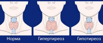
Changes in the condition of the cervical epithelium can become alarming signals, which the gynecologist first of all pays attention to when examining patients of different ages. There are various reasons for such changes, one of which is dysplasia, which occurs mainly against the background of papilloma virus infection. The danger of this disease lies in the tendency of the affected cells to undergo cancerous degeneration.
Official medicine considers human papillomavirus (HPV) to be the main cause of cervical dysplasia. Among all the varieties of this virus, the most dangerous types for women are types 16 and 18. They most often cause cancerous lesions of the cervix, which were preceded by the stage of dysplasia. The difficulty of diagnosis and the asymptomatic course of the stages of dysplasia emphasize the danger of the disease. After dysplasia, cervical cancer is the second leading cause of death among women, after breast cancer. Such statistics require the closest attention to the signs of the disease detected during gynecological examinations. Regular visits to the gynecologist will allow you to identify the disease in the early stages and prevent cancer.
What is cervical dysplasia
Dysplasia is a change in the cells of the squamous epithelium of the cervix and the appearance of so-called atypical cells. Such cells have an enlarged, shapeless nucleus or many nuclei, are actively dividing, and their correct location in the epithelium and ability to perform the function inherent in nature are disrupted. It is giant nuclei and active division that the cytologist looks for when he examines your smear for atypical cells (PAP test).
| Normal squamous epithelial cell and atypical cells with enlarged nuclei. |
The causes of cell atypia can be divided into bacterial and viral.
Bacterial dysplasia of the first degree is essentially a chronic inflammation of the cervix (cervicitis, colpitis). Cellular changes in this case are caused by an aggressive environment. This form of dysplasia does not require oncological alertness. After proper anti-inflammatory treatment, the size of the nuclei and the shape of the cells return to normal.
And it’s a completely different matter if the cell becomes infected with the human papillomavirus (HPV) of high oncogenic risk.
The virus in the epithelium can exist in two forms - in a “sleeping” and active state. At the moment of weakening of the immune system, HPV enters an aggressive stage; the genetic material of the virus is integrated into the DNA of the cell and triggers the development of the disease.
Typically, the body repairs or destroys such defective cells with altered DNA. But the papilloma virus “turns off” the alarm, and the broken cell loses the opportunity to inform the “repair teams” about its illness. Our immune system continues to perceive it as healthy, so it does not interfere with division and transformation.
The virus embedded in DNA actively reproduces itself, stimulates the cell to actively divide, and damage accumulates with each division.
Thus, the disease progresses to moderate and severe dysplasia, which are already considered precancerous diseases of the cervix and require treatment by a gynecological oncologist. The same mechanism underlies the development of cervical cancer.
Treatment of grade 3 dysplasia
The second (moderate or moderate) and third (severe) degrees of dysplasia have the same indications for treatment. To avoid further development of the disease, which can lead to oncology, the affected areas of the cervix must be surgically removed and antiviral and immune therapy must be carried out in parallel.
Surgery has 4 methods for removing or localizing the affected area:
- conization of the cervix (produced using the most progressive loop method);
- laser exposure;
- radio wave exposure;
- cryodestruction (freezing of affected areas, leading to their disintegration).
The best results are obtained using surgical treatment and drug therapy. When infected with carcinogenic types of HPV, patients are prescribed immunomodulatory, antiviral or universal drugs such as Galavit, which have a complex effect.
In difficult cases, for example, when less invasive treatment methods do not provide a positive effect, a patient with severe dysplasia may be prescribed cervical amputation.
What is cervical LSIL and CIN 1
Historically, grade 1 dysplasia has had many names. One of the reasons is the wealth of epithets in the Russian language. It's all the same disease:
- mild cervical dysplasia,
- dysplasia 0 degree,
- low grade epithelial dysplasia,
- mild dysplasia of squamous epithelium,
- initial stage of dysplasia,
- mild dysplasia,
- dysplasia type 1,
- slight dysplasia, etc.
The second reason is that the diagnosis of dysplasia 1 has changed its official name many times. Until 1988, the international name for dysplasia was Cervical Intraepithelial Neoplasia - grade 1 cervical intraepithelial neoplasia of the cervix. Abbreviated as CIN I, or CIN 1 of the cervix.
But since the term “neoplasia” means “new formation,” that is, a tumor, the name was adjusted. The modern international name for this disease is LSIL - low grade squamous intraepithelial lesion - low grade intraepithelial lesion . The term “lesion” more accurately reflects the nature of the cell changes.
In contrast to LSIL, severe dysplasia with extensive epithelial lesions is called HSIL - high grade squamous intraepithelial lesion. The HSIL group includes moderate dysplasia and carcinoma in situ (carcinoma in situ).
You need to remember that now the disease is called LSIL. And only so.
But since gynecologists still use a wide variety of formulations, we allow ourselves the same.
Types of disease
In the international list of diseases, cervical dysplasia is listed under the abbreviation CIN, which is translated into Russian as cervical interepithelial neoplasia. Depending on the stage of change in cervical epithelial cells, three degrees of dysplasia are distinguished:
- 1 (weak) degree is noted with superficial damage to the epithelium;
- 2 (moderate or moderate) degree is characterized by more extensive changes in the epithelium, reaching half of its depth;
- 3 (severe) degree has signs of damage to 2/3 of the thickness of the epithelial layer and pronounced morphological manifestations of cell atypicality.
The risk of transition from the first degree to the second and third is estimated by statistics as very low (at the level of 1%). At least 75% of patients with moderately developed dysplasia recover completely within 5 years after treatment. The statistics of the transition of the third degree of dysplasia to a cancerous state is estimated based on various sources at levels from 12% to 32%.
The degrees of the disease are characterized not only by the depth of damage to the epithelial layer. Along with the deepening into the structure of the mucous membrane, the morphology of the cells also changes, in particular, their nucleus increases. In the severe stage, the nuclei become hyperchromatic, their volume close to the volume of the cell itself. But cellular changes do not extend beyond the basement membrane. They affect only the mucous membrane, but cover almost the entire area of the outer part of the cervix. Damage to the basement membrane in epithelial dysplasia is interpreted as stage zero cancer. Epithelial cells are already acquiring carcinogenic morphological characteristics, and the disappearance of the membrane opens the way for them to further invade the organ tissue.
Correct diagnosis of grade 1 dysplasia: how to keep the disease under control
To detect dysplasia on time, you need to visit a gynecologist at least once a year, and from the age of 21, additional research is required.
Here are instructions on what to do and why.
STEP 1. Examination by a gynecologist. It's called a mirror inspection.
Often they are limited to only this stage, and then in the medical record, instead of the suspicious suspicion of LSIL, a diagnosis of “cervical erosion” may appear. Indeed, in this case, even a trained specialist cannot always “by eye” distinguish one from the other.
The main thing in this case is not to start active treatment of erosion, as it can accelerate the process of transition from mild cervical dysplasia to a severe form. It is necessary to undergo additional studies to exclude the presence of atypical cells and HPV.
STEP 2. Cervical smear for cytological examination.
The second name for this analysis is PAP test.
The analysis for identifying/excluding atypical cells goes through several stages, and it is extremely important to follow the technology of the analysis at each of them.
A cytological examination begins not in the laboratory, but in your gynecologist’s office, and its first stage is the correct collection of material.
A smear from the cervix is taken with a special brush - such as in the illustration. It is very important! Only this brush allows you to take cells for analysis from the entire circumference of the cervix and from the cervical canal. This significantly reduces the likelihood that any modified section will be missed.
Previously, spatulas were used for this purpose. Today they are no longer relevant.
| This is what the best brush for taking a smear from the cervix for atypical cells looks like. |
| This brush allows you to take cells for analysis from the entire circumference of the cervix and from the cervical canal. |
In the second stage, the smear is applied to glass and sent to the laboratory to a cytologist, who will stain it and determine the presence or absence of atypical cells. In case of dysplasia, 1 cytologist will write in the conclusion “the cytogram corresponds to LSIL”, or “squamous epithelium with LSIL phenomena”.
It will be better if you do not take a classic Pap test, but a smear from the cervical canal using liquid cytology . It is considered the most reliable. In this case, the contents of the brush are not applied to the glass. The entire brush head is immersed in a container with a special solution and sent to the laboratory.
For greater reliability, I recommend conducting a cytological examination using the LIQUID CYTOLOGY method.
| When performing liquid-based cytology, the entire brush tip is sent to the laboratory in a special solution. |
According to the letter of the Ministry of Health of the Russian Federation dated November 2, 2019 N 15-4/10/2-7676 On the direction of clinical recommendations (treatment protocol) “Benign and precancerous diseases of the cervix from the perspective of cancer prevention”, if there are no atypical cells in the smear, it is recommended to take it Once every 3 years.
Through the prism of my experience, I cannot agree with this and recommend doing a cytological smear once a year.
For various reasons, the reliability of cytological analysis for mild dysplasia ranges from 60 to 90%. In my practice, there are many girls 20-30 years old with in situ cancer, and women under 40 years old with invasive cervical cancer, who regularly undergo cytological examinations once every three years. If cytology had been taken once a year, this development of events could have been avoided - “intercepted” and treated the disease at a safe stage of dysplasia.
Many of my colleagues also note the fact that only a few months pass between newly diagnosed mild dysplasia (LSIL) and CIN III (HSIL).
STEP 3. Extended colposcopy.
This is an examination of the cervix under a microscope, during which the cervix is stained with special solutions.
It is important to understand that the diagnosis of “grade I dysplasia” is not made either on the basis of examination or on the basis of colposcopy. But colposcopy gives the gynecologist a lot of related information and allows you to make a more accurate diagnosis. For example, it shows flat condylomas and suspicious areas on the cervix, which, even before the appearance of visual manifestations of dysplasia, can indicate the dangerous viral nature of the disease.
To see problem areas, the cervix is stained sequentially with vinegar and Lugol's solution. After treatment with a solution of acetic acid, whitish areas (the so-called acetic-white epithelium) appear on the epithelium of the vaginal part of the cervix. Areas treated with acetic acid are stained with Lugol. Problem areas are also painted unevenly – the so-called “iodine negative zones” are visible. These are the first signs of dysplasia caused by HPV.
| Whitish areas on the cervix after treatment with vinegar solution (left) and uneven coloring of the cervix after treatment with Lugol's solution (right). A healthy cervix is evenly colored. |
Diseases not associated with HPV manifest themselves differently: the cervix acquires an excessively bright shade or is covered with a large number of “bubbles” (cysts).
Treatment of the cervical epithelium with acetic acid and Lugol's solution is an absolutely harmless procedure.
Typically, colposcopy is recommended only after identifying atypical cells in a cytological smear (see STEP 2) or after identifying high-risk HPV (STEP 4). But practice shows that often a cytological smear shows the absence of atypia, even if it is present. And only colposcopy allows you to suspect something is wrong.
Therefore, I strongly recommend having an extended colposcopy annually.
STEP 4. Determination of viral load.
If atypical cells are detected in a cytological smear, it is necessary to continue the examination and confirm the viral cause of the cell changes - take a liquid Digene test for the quantitative determination of viral particles of HPV infection.
If you are over 30 years old, this test must be taken once a year, regardless of the result of the cytological examination.
The fact is that HPV is present in some quantity in almost all of us. And only exceeding the norm allows us to talk about the activity of the virus and evaluate it.
Therefore, I cannot agree with the recommendation of the same Letter of the Ministry of Health of the Russian Federation dated November 2, 2017 No. 15-4/10/2-7676 on the need for high-quality tests for HPV.
A qualitative test for determining high oncogenic risk HPV is not informative . It shows only the presence or absence of the virus in the epithelium. But the fact of its presence does not convey any significant information - HPV is present in 80-90% of people, and does not cause disease in the vast majority. Therefore, I do not see any obvious need to take a PCR smear.
The Digene test is a quantitative research method and is much more informative. It determines the so-called viral load. Only if the result of the Digene test is negative (when the viral load is less than 0.8 units), we can say that initial dysplasia is caused by an infection without the participation of the papilloma virus and it does not require special antitumor treatment. It will be enough to cure the inflammation. Only in case of combination with ectopia or pseudo-erosion of the cervix, it may be necessary to “cauterize” with a laser or radio wave (Surgitron, Fotek).
If, along with previously identified atypical cells, quantitative analysis shows an excess of the normal threshold for HPV of high oncogenic risk, we can reasonably say that the dysplasia condition is of a viral nature. Typically, with CIN1, quantitative analysis shows higher HPV values than with any other type of dysplasia. Contrary to the literature, this condition is not self-limiting and sooner or later progresses to HSIL and invasive cancer.
The body’s immunity is strong enough to contain the activity of the virus and the “degeneration” of dysplasia into a tumor for a long time. But my observations over the past 10 years show that dysplasia in some cases goes from a mild stage to a severe and even invasive cancer in less than 12 months.
Therefore, if there is atypia and high levels of papilloma virus in the cells, I strongly advise my patients to treat grade 1 dysplasia using the non-surgical method of photodynamic therapy.
You shouldn’t be complacent with the “fearless” diagnosis of “low-grade cervical dysplasia” and waste time waiting for it to go away on its own.
There is one more indication. It occasionally happens that in the presence of atypical cells, the Digene test does not detect papillomavirus. The fact is that viruses transform, new serotypes of the papilloma virus constantly appear, unknown to existing test systems.
In this case, colposcopy reveals flat condyloma on the external or internal genitalia or leukoplakia on the vaginal vault. Both of these diseases are clearly viral in nature. They talk about the presence of the virus in the body and its high activity. Accordingly, the presence of atypical cells against their background should raise oncological suspicion. In this case, the absence of papillomavirus in the analysis is not a reason to refuse photodynamic treatment of mild cervical dysplasia.
STEP 5. Immunohistochemical determination of p16 protein expression
This is an analysis for which material is taken from the same container as for the Digene test.
p16 is a protein that appears in aging cells and tells the immune system to send the cell for disposal. Typically, tumor cells try to suppress the expression of p16 so that they can begin to divide uncontrollably. But contrary to logic, in cells that have undergone the transforming effect of HPV, there is an increase in the expression of this protein, which serves as a sign of the virus entering the active phase and the integration of the DNA of a high-oncogenic risk virus into the cell genome. Since this process is irreversible, analysis for p16 is a marker for the initiation of carcinogenesis.
In the presence of atypical cells or a suspicious picture of extended colposcopy, I consider this analysis mandatory. It is the determining test in choosing the tactics and method of treating dysplasia.
Determination of p16 protein is NOT carried out if the cytogram is normal and there are no atypical cells.
A positive test for p16 against the background of CIN I - II indicates that the cells have already undergone transformation by the virus. This change in cells is irreversible - the active virus makes the cell invisible to the radar of the immune system.
That is why the positive result of this analysis indicates the urgent need for immediate treatment of even mild dysplasia. The disease will only progress further. Observational tactics against the background of p16 expression are detrimental, and the “wait and watch” recommendation can lead to in situ cancer. It is necessary to undergo treatment.
The presence of atypical cells and detected p16 protein expression is an OBJECTIVE INDICATION for treatment with photodynamic therapy.
STEP 6. Biopsy.
A biopsy is the removal of a small piece of cervical tissue for histological examination. This analysis is more informative than a cytological study. But since the procedure for collecting material is considered traumatic, the need for a biopsy is determined individually by the attending physician. It is usually required if severe dysplasia is suspected.
Diagnostics
The most accessible and accurate method for diagnosing dysplasia and early stages of cervical cancer is a cytological study of a cell preparation (scraping of epithelial cells of the cervix). This method is also known as a PAP smear, named after the Greek physician Papanicolaou. The procedure for taking a smear is painless. The gynecologist uses a sterile instrument to scrape cells from the outer mucosa of the cervix. The taken material is sent to the laboratory, where it is examined under a microscope. During a microscopic examination, specialists determine the condition of the cells based on morphological characteristics. Healthy cells form a uniform epithelial structure, and their nuclei are small. Enlarged nuclei and scattered structure indicate the presence of the disease. The degree of dysplasia is determined by the morphology of the cells and the depth of epithelial damage.
The initial stage of dysplasia is invisible when examining the outer part of the cervix. Erosive manifestations are observed when damaged epithelial cells form lesions. They may appear as redness with a whitish coating. But the final diagnosis is made only after a biopsy.
A PAP smear allows you to detect the presence of atypical cells in the scraping. If such cells are identified, then a biopsy procedure is prescribed to clarify the diagnosis and determine the degree of dysplasia. The colposcope is inserted into the vagina. The optical system helps to view areas at high magnification. A tissue sample is taken from the most changed areas, usually from three points.
Of the two hundred HPV subtypes, only a few are classified as carcinogenic. To determine the strain, a vaginal smear is prescribed. Laboratory testing using the PCR (polymerase chain reaction) method allows you to detect and identify the DNA of human papilloma viruses. This is the most informative method for determining the causative agent of dysplasia.
Mild grade 1 cervical dysplasia can very quickly turn into severe dysplasia
Why is it necessary to treat HPV-associated dysplasia of the 1st degree, if it is not clear whether it will turn into severe dysplasia or not? We know from the literature that self-healing from HPV is possible even for serotypes with high oncogenic risk. But according to our observation, not everything is so optimistic.
If the diagnosis does not include the five mandatory steps that I wrote about above, then based only on examination and a negative cytological conclusion, an incorrect diagnosis is often made.
I tried to highlight the main reasons why a doctor may miss a serious disease or underestimate the risks.
| Cell transformation begins in the deep basal layer, and atypical cells in the initial stages of dysplasia may not be included in the cytological smear. |
Reason 1. My experience shows that often “dysplasia 1” is not always a correct diagnosis. The virus affects the deepest layers of the cervical epithelium - the basal cells. Since atypical cells begin their development at depth and gradually “rise,” it is often impossible to assess the stage of the process with the eye, even when using a colposcope. And the cytological brush “collects” material only from the upper layers of the epithelium.
Therefore, against the background of good colposcopy and cytology, it often happens that after taking a biopsy - a piece of tissue that allows you to evaluate the problem at its entire depth, from the epithelial to the basal layer - the diagnosis worsens to precancerous HSIL.
Reason 2. Previously, mild dysplasia was considered a background process. Only grades 2 and 3 were classified as true precancer. It was believed that the transition from one stage to another takes about 3-5 years.
In fact, it is impossible to predict the rate of progression of dysplasia. My observations and those of my colleagues show that viral lesions today pass through all stages much faster, and the process of tumor growth can begin within 12 months after the diagnosis of dysplasia 1.
In order not to be unfounded, I suggest you read the message from the woman.ru forum:
“In the summer in July, we were diagnosed with “grade 1-2 dysplasia”, took courses of antiviral drugs and suppositories, in December we had DEC (diathermoelectrocoagulation) and sent the material for a biopsy. The result was “severe dysplasia with transition to squamous cell carcinoma of the cervix.” I’m terrified, in shock... I haven’t slept (at all!) for 4 days now. How in 5 months from grade 1 to severe?...
What to do?! Is there a cure for this? We are collecting everything for the oncology clinic, we have been doing all the paid tests, ultrasound, FGDS, etc. for 4 days now.
I have two children, I’m very afraid of not seeing them graduate from school... I sit and roar.”
I think that it’s still not worth checking the rate of progression of viral dysplasia on yourself.
Reason 3. I have already said that dysplasia originates in the deep basal layer of cells. When treating nulliparous women with the currently popular method of gentle excision, the surgeon tries to remove a minimum amount of tissue. And if after the operation, virus-infected cells remain in this layer, they are highly likely to cause a recurrence of dysplasia with its transition to a more severe stage.
Reason 4. Any infection - no matter viral or bacterial - has a long period of latent course. This period is called persistence, when the virus is present in the cell in a “dormant” state. It is impossible to predict the moment of weakening of the immune system, which will serve as an impetus for the awakening of the virus and the development of the disease. It could be a common cold or stress. Even strong positive emotions reduce immunity. Then persistent HPV of high oncogenic risk is integrated into the cell’s DNA and begins its transformation.
It is possible that at the time of examination by a doctor, the virus has already passed into an active form, but the damage has not yet accumulated a critical mass, they are almost invisible to the eye or do not arouse suspicion.
Reason 5. Different strains of papillomavirus have different degrees of aggression, which also cannot be predicted from the results of an examination or cytological analysis. And where one strain will persist for years, the second will transform the cell in a few months, first into an atypical one, and then into a cancerous one.
Without clarifying studies, it is impossible to give any guarantee that your dysplasia will behave calmly, and that in a year the cytologist will not write anything worse than “LSIL” in his conclusion.
Reason 6. If dysplasia 1 is caused by the human papillomavirus of high oncogenic risk, and the virus is integrated into the DNA of the cell, in my practice I have not observed a single case of self-healing - dysplasia always progresses.
Viruses of high oncogenic risk include serotypes 16, 18, 31, 33, 35, 39, 45, 51, 52 and 56. The most dangerous are serotypes 16 and 18 of the virus; they cause severe dysplasia and cervical cancer in 80% of cases.
I look at viral dysplasia systemically, regardless of its degree.
Considering the fact that independent regression of mild dysplasia cannot be excluded, in the absence of colposcopic signs of HPV infection, I am inclined to observe.
If CIN I is accompanied by obvious viral signs of human papillomavirus infection (flat condyloma, vinegar-white epithelium, p16 protein expression), then I definitely recommend treatment with a non-surgical PDT method.
Because with such a combination of aggravating factors, we may simply not have time to think.
Diagnostic methods
For timely diagnosis of moderate dysplasia, a woman who is sexually active or over 30 years of age should be examined by a gynecologist once a year for safety reasons. In addition to a preventive examination, it is recommended to conduct cytology - a laboratory test.
The main medical methods for detecting the disease are:
- gynecological examination by a doctor of the patient’s external and internal genital organs with mirrors, taking samples;
- collection and further laboratory examination of a smear (cytology), colposcopic examination. The probability of the result being accurate is approximately 60%;
- taking a piece of the uterine epithelium for a biopsy. The most accurate test that allows you to simultaneously detect cancer cells.
During the examination, the doctor can visually identify dysplasia areas; they differ from healthy tissue in appearance - they are lighter in color. Also, when conducting trial studies according to Schiller (treatment of the surface of the cervix with Lugol's solution), it will be seen that the affected areas will not completely change their color. They have a more whitish tint.
Cytology gives the most accurate result, especially with a large degree of damage, that is, at stages 2 and 3.
A cervical biopsy is performed to remove a tissue sample from the cervix and examine the sample in detail.
Colposcopy is an examination using a special instrument. During which changes in the epithelial mucosa and the entire uterine cavity are visible in more detail. This makes it possible to more clearly identify areas affected by dysplasia.
The doctor is able to draw more accurate conclusions than when examining using mirrors. But the most accurate method, which, along with examining the degree of dysplasia, can also determine the presence of cancer cells, is the examination of epithelial tissue as part of a biopsy. This laboratory study gives 100% correspondence of the results to the actual state of affairs.
Review of treatment from Elizaveta, Yaroslavl
Dear Maxim Stanislavovich, leaving this review, first I want to tell you how my story began. So, I came for a preventative appointment with a gynecologist, the doctor noticed changes in the uterus, after learning the test results, it became clear that I have HPV and first-degree dysplasia... The doctor recommended that I be examined again six months later, BUT I, not understanding the seriousness Given this situation, a year later I came to see a paid gynecologist. I took tests and the diagnosis was cr in situ. The doctor offered two options, either coanization or the innovative PDT method, but in order not to injure the young body (I’m 23 years old), she insisted on PDT. After that, I started scouring the Internet in search of the best doctor, and then I found you, with vast experience, and most importantly, with a large number of positive reviews from cured patients. Without a doubt, I chose you, after that I contacted the administrator Oksana, and literally within two days I was given a date for PDT, which is very convenient, because I live in Yaroslavl. Arriving in Moscow at the clinic, I felt the highest level of service.
That's why I wrote this story. To express words of gratitude and so that girls with the same problem, without hesitation, come to you, because you are a qualified and competent doctor who is focused on results.
Causes
The most common cause of dysplasia is the human papillomavirus. In 2008, German scientist Harald zur Hausen received the international Gairdner Prize for studying the degree of danger of this virus for the female body.
When infected with papillomavirus, the body begins to fight it. If the immune system is strong, then the tissues affected by the virus are restored and antibodies are produced, but this does not always happen. The 16th and 18th strains of the virus are characterized by high oncogenic activity, and therefore most often become the cause of the development of complications leading to dysplasia.
From the moment of infection until the appearance of the first signs of the disease, described below, several months and even years pass.
Factors that provoke the accelerated development of dysplasia are:
- promiscuity (change of more than 3-4 partners per year);
- having sexual intercourse before the age of 14;
- infectious diseases transmitted through direct sexual contact (chlamydia, herpes simplex virus, cytomegalovirus);
- frequent consecutive pregnancies and associated hormonal imbalances;
- medical abortions;
- constant use of oral contraceptives;
- failure to maintain personal hygiene during sexual intercourse;
- cervical injuries;
- advanced forms of erosion;
- weakened immune system;
- smoking, alcoholism.
If cervical dysplasia is detected at stages 1 or 2, then timely treatment can slow down the development of the disease and promote further recovery.
What reasons increase the risk of developing dysplasia?
A natural question arises: why, after infection with the papilloma virus - and according to various sources, up to 95% of the world's population are its carriers - only a few of them will encounter the disease? What factors enable the virus to emerge from its “dormant” state and begin active division?
The immune system can restrain the development of the virus in the cell for a long time. But against the backdrop of weakened immunity, the virus becomes active. Here are the main risk factors:
1. Stress. Emotional experiences reduce immunity, and even strong positive emotions are stressful for the body.
2. Pseudo-erosion (ectopia) of the cervix is a physiological feature of the body, which nevertheless creates favorable conditions for infection with a large amount of virus. Young women with unestablished stable relationships with the opposite sex are most susceptible to this risk.
3. Inflammatory diseases (colpitis, cervicitis). The endometrium of the vagina and cervix performs the function of the skin - it protects against infections. Inflammatory diseases not only reduce its barrier ability, but they are a favorable condition for the progression of the virus. Therefore, women with frequent inflammation are at risk for developing dysplasia.
4. Smoking. Nicotine itself is one of the most powerful carcinogens, and its concentration in the mucus of the cervical canal is often much higher than in the blood. The second factor in the carcinogenic effect of cigarettes is tar. They radically change the properties of vaginal mucus and cervical plug, weaken local immunity and make the epithelium extremely vulnerable to HPV and other infections.
5. Heredity. Genetic predisposition to the development of atypical processes, which can often occur even without the participation of HPV. You need to be especially attentive to yourself if your direct relatives are diagnosed with cervical dysplasia or cervical cancer.
6. HPV of high oncogenic risk. These serotypes increase the risk of developing dysplasia because they are better adapted than other viruses to protect against suppression by the immune system.
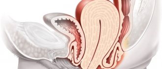
Dysplasia and pregnancy
Cervical dysplasia and pregnancy are compatible in all respects:
- pregnancy does not accelerate the development of dysplasia;
- dysplasia does not have a negative effect on the course of pregnancy;
- atypical cells do not lead to disturbances in fetal development;
- dysplasia does not impair placental function.
Some hormonal changes that occur physiologically during pregnancy can change the appearance of the cervix. Some areas look like dysplasia, but in fact it is pseudo-erosion or some other disease.
Mild dysplasia detected during childbirth does not require treatment. The woman is only prescribed a repeat examination and monitoring of the condition of the tissues 12 months after childbirth. With an average degree of pathology, the dynamics of improvements begin to be monitored several weeks after birth, so as not to miss a sharp progression. If stage 3 is detected or a malignant tumor is confirmed, the patient is managed together with an oncologist, who helps make decisions regarding childbirth and further observation.
How to treat grade 1 cervical dysplasia to avoid relapse
Is it possible to cure grade 1 dysplasia? It is possible, and it can be done without surgery.
Unlike conization, photodynamic therapy is a non-traumatic therapeutic method. It targets dysplasia in several directions at once:
1. destroys atypical cells,
2. destroys HPV-affected cells in the mucous membrane of the cervix and cervical canal,
3. destroys concomitant infection (cervicitis).
PDT completely destroys cells affected by the human papillomavirus, which serves as a reliable prevention of cervical precancer. Leading Oncology Institute of Russia, Moscow Research Institute named after. Herzen recommends treating HPV infection with PDT as a preventive measure for recurrent dysplasia.
Using PDT for treatment, we preserve the integrity of the cervix and uterus - that is, a full-fledged organ for independent conception, pregnancy and healthy childbirth. By preserving the integrity of the organ, we also preserve its innate immune system - a healthy cervix reliably protects itself and the uterus from infections.
Therefore, do not rush to make a decision in favor of the surgical method of conization - you will always have time to have the operation.
| I suggest you read here how LSIL is treated using PDT. |
Treatment methods
Before prescribing a course of treatment for moderate uterine dysplasia, the doctor conducts a study to determine the presence of factors that led to the onset of the disease. Such as hormonal disorders, viral infectious and sexually transmitted diseases, HPV, inflammation. The patient is prescribed a complex of medications and other procedures, and repeated tests are performed to confirm the positive outcome of the treatment.
Complex therapy allows you to stop the proliferation of dysplasia cells, forming scars on the surface of the affected epithelium. But if treatment fails, the next step is surgery to correct the problem.
Cervical examinations make it possible to prevent the development of such serious diseases as cancer.
During surgery, an electric operating knife is used to treat moderate dysplasia. The surgeon makes a section of the diseased epithelium. The disadvantage of this method is long-term rehabilitation. Recovery is recommended within 3–4 months.
In addition, there may be consequences such as rough scars at the section sites, minor bleeding and soreness. When carrying a fetus, there can also be negative consequences and difficulties.
Currently, in surgery, a laser is used to treat such diseases, which also affects diseased cells. This method is considered more gentle than electric knife, leaves fewer scars, and recovery occurs within a couple of months. The patient feels better and has virtually no consequences during pregnancy after this procedure.
A method such as cryotherapy is also used in the fight against uterine dysplasia. Mucosal cells damaged by the disease are treated with liquid nitrogen (frozen). It is very similar to another procedure where the epithelium is treated with a medical chemical solution. In both the first and second cases, diseased cells die and peel off from the epithelium.
Exposure to radio waves. A fairly modern method in which, using a device, waves are sent to the area of the affected epithelium that destroy cells. It has few negative consequences, leaves virtually no scars, and the body recovers quickly.
Excision (conization): A frequently used method, a type of classical surgery. Using a scalpel, damaged layers of the epithelium are removed. It is considered the most traumatic method, not recommended for young women of childbearing age; during the rehabilitation period it is characterized by bleeding, pain, etc.
Previously, conization was performed using the traditional surgical method (using a scalpel); a little later, gynecologists began to use an electric knife.
Whatever method is used, the patient’s rehabilitation period provides for a special regime:
- Temporary cessation of sexual intercourse.
- Elimination of physical activity.
- Refrain from swimming, direct sunlight and going to the solarium or bathhouse.
- Doctor's supervision.
Timely diagnosis with an effective treatment regimen gives very significant results.
Is it possible to recover without treatment for CIN I: myths and reality
In gynecology, it is an accepted postulate that grade 1 cervical dysplasia spontaneously regresses within a year in 95% of patients. And on the one hand, I am ready to subscribe to this figure.
But on the other hand, when they talk about self-elimination (self-removal) of the virus, it is very important to understand what they mean by this. Let's take a close look at the disease and remember everything we know about it.
Often, cell atypia (that is, dysplasia) appears as a reaction of cells to inflammation caused by fungi and bacteria. Treatment of the main diagnosis (cervicitis or colpitis) leads to a natural recovery.
But dysplasia CIN I of the squamous epithelium of the cervix can be caused by the human papilloma virus, the Latin name for this group of viruses is Human papilloma virus, or HPV. There are more than 140 serotypes. And not all of them are oncogenic.
Low cancer risk HPVs include serotypes 3,6,11,13,32,42, 43,44,72 and 73. These viruses also cause dysplastic changes, but before this they persist in the cell for a long time and are not integrated into the DNA structure, so they can suppressed by the immune system.
But if grade 1 dysplasia is caused by viruses of high oncogenic risk, there is no talk of spontaneous recovery. These viruses are integrated into the DNA of the cell, making it invisible to the immune system and freely causing its tumor transformation. Therefore, dysplasia 1 of a viral nature most often requires treatment.
Mechanism of disease development
The connection between the presence of human papillomavirus (HPV) and cervical dysplasia has been established by a number of medical studies dating back to the pre-war years. The vast majority of people who are sexually active experience HPV infection. Most often, the body copes with it without medical help. Even precancerous stages in women with diagnosed dysplasia can be cured by the immune system. But the body’s capabilities do not guarantee such an outcome, therefore, given the huge risk of threat to health and life, it is necessary to undergo adequate treatment for the current stage.
The papilloma virus is transmitted sexually. Penetration is not necessary to acquire an infection - external contact of the genitals is sufficient. Most types of HPV do not cause adverse health effects and go away on their own. If there is no re-infection with the same type of virus, then after two years the infection is no longer recorded and does not manifest itself in any way in the body. But in some cases, HPV infection becomes chronic, causing cervical dysplasia with the risk of the formation of cancer cells. From the moment of infection to the cancer stage, with normal immunity, it can take 15-20 years. In HIV-positive patients who are not undergoing therapy, this period is reduced by 2-3 times.
Advantages of treating CIN 1 cervix using PDT
- PDT is a non-surgical treatment method that preserves organ integrity, an important component of innate immunity.
- Selective destruction of pathological cells without affecting healthy ones.
- The effectiveness of getting rid of the papilloma virus and other infections in the affected area is more than 93%.
- Guaranteed prevention of cervical cancer.
- Restoring the integrity of the cervix and its immune status, preventing re-infection with HPV.
Symptoms
Mild and moderate dysplasia occurs without obvious symptoms that would clearly indicate the presence of this disease. For this reason, already developed pathology is detected at later stages during a gynecological examination. The woman may not experience any pain. In some cases, patients express the following complaints:
- increased vaginal discharge;
- the appearance of an unpleasant mucus odor;
- itching and burning at the end of the vagina;
- pain during sexual intercourse;
- less bleeding after intercourse.
The absence of these symptoms in no way guarantees the absence of the disease. Therefore, women need to visit a gynecologist’s office at least once every six months or a year. Early diagnosis of dysplasia greatly reduces the risk of developing cervical cancer.
Severe degrees, as well as mild and moderate ones, are almost always asymptomatic. But unlike the initial ones, it already poses a significant threat to a woman’s health. Any symptoms indicating sexual problems should be a reason for a woman to have an unscheduled examination at a gynecologist’s office.
Useful video
From the video you will learn about the causes and symptoms of cervical dysplasia:
Violations of the cellular structure of tissues in cervical dysplasia can lead to the development of oncology. The disease mainly affects young women (25–30 years old).
Early detection of pathology helps prevent cancer risk.
Cervical dysplasia (CIN) is classified into 3 grades:
- Weak (CIN I) - characterized by mild disturbances in the epithelial structure. Structural changes do not cover more than a third of the stratified epithelium. There are signs of papillomavirus: leukoplakia (thickening of the epithelial layer of the uterus), koilocytosis (cells damaged by the papillomavirus).
- Moderate (CIN II) - lesions occur in half of the epithelial layer, pronounced structural changes are observed, and progression of morphological changes at the cellular level is observed.
- Severe (CIN III) - the pathological process affects the entire thickness of the cells, the spread of the lesion is as deep as possible. Morphological changes are pronounced, up to the appearance of mitoses (uncontrolled cell division). Atypical cells, in which large hyperchromic nuclei are found, are found only in the mucous membrane, without spreading to nearby structures.
Treatment Options
In stages 1 and 2 of dysplasia, doctors most often resort to conservative methods, and may also advise their patients to use traditional medicine as additional therapy.
As for drug treatment, they are prescribed:
- antiviral drugs;
- antibacterial agents;
- immunostimulants.
Sometimes such therapy is enough for the pathological process to regress.
Pathological tissue can be removed in the following ways:
- cryodestruction – freezing with liquid nitrogen;
- laser cauterization - exposure of the affected areas to laser beams;
- radio wave treatment – removal of affected areas using radio waves;
- electrocoagulation – elimination of pathological tissues with electric current.
If the disease is diagnosed as severe, and also if the doctor has real concerns that the disease is beginning to transform into oncology, coniation of the cervix or complete amputation of the damaged organ may be required.
This surgical intervention can be performed using a scalpel, or modern methods can be used - cryodestruction, radio wave excision, laser removal.
Each of these methods has its positive and negative sides, and only a doctor can choose the optimal method, based on the woman’s age, the individual characteristics of her body, the degree of the disease, and other things.
After amputation of the cervix, a woman’s reproductive function is preserved, but the doctor must be informed about her plans for a future pregnancy in advance so that the most gentle method for removing the cervix can be selected .
Prevention
In order to prevent dysplasia at the household level, young women should observe:
- intimate hygiene, especially before and after sexual intercourse;
- periodically replenish folic acid reserves in the body;
- eat well;
- do not postpone visits to the gynecologist for too long;
- try to lead a measured sex life with 1 regular partner.
Doctors also recommend vaccination against human papillomavirus types 16 and 18. Timely vaccination reduces the risk of developing the disease several times.
Girls should not stay in the sun for a long time or overuse tanning, including that which can be obtained artificially in a solarium. Only those medicinal items and products recommended for use by the attending physician can be inserted into the vagina.
Diet
A special diet can alleviate the patient’s condition and stop further spread of the virus. Women infected with papillomavirus will have to exclude from their daily diet any fried, spicy and smoked foods, alcoholic beverages, sugar, sweets, and foods containing GMOs.
In order to increase the level of folic acid naturally, a woman should consume more greens, legumes, citrus fruits, bananas, cabbage, buckwheat and millet cereals, and walnuts.
The vitamin A needed by the body is found in liver, dairy products, seaweed and fish. Ascorbic acid is present in bell peppers, cabbage, kiwi, sorrel, radishes, citrus fruits, currants and raspberries. Most of these foods are also rich in vitamin E and B-carotenes.
To strengthen the immune system, it is recommended to eat more chicken eggs, peanuts, corn, celery root, cabbage and rice cereal. It's best to skip the soda and replace it with antioxidant-rich green tea.
How is this deviation formed?
This is an anomaly in which the cellular structures of the epithelium of the vaginal cervical region are modified. They become atypical. The disease itself is classified as a precancerous condition.
The vaginal area of the cervix is covered with flat epithelium. Fabric color: pink. It consists of several layers. Main layers:
- Basal-parabasal. The basis is made up of basal and parabasal cells. They come into contact with muscle tissue, nerve endings and vessel walls. There are “fresh” cells here that restore the structure of the epithelium.
- Intermediate.
- Functional (superficial). The cellular structures in this layer quickly wear out and are replaced with new ones.
Basal cells are characterized by the presence of roundish outlines. There is a large round core inside. After the cells mature and move closer to the surface layer, they become denser and the nuclei become small. With dysplasia, they suffer from dramatic changes: their size increases, the walls become thick, a large number of nuclei appear, and separation disappears. Doctors call this process atypia.
First signs
Symptoms of cervical dysplasia are usually scanty, so a woman may not be aware of the presence of a serious pathology. Despite its different origin, dysplasia is always accompanied by changes in the epithelium at the cellular level. This disease does not have an independent clinic and is often disguised as other gynecological diseases (for example, erosion). Therefore, pathology can only be detected during a preventive examination and with the help of additional studies (PAP analysis (SMEAR)). The initial form of the disease is practically asymptomatic.
The first signs of dysplasia are associated with the following manifestations:
- pain or discomfort in the lower abdomen;
- burning and itching in the genital area (especially worse during sexual intercourse);
- vaginal discharge mixed with blood;
- profuse leucorrhoea (most often milky white) without an unpleasant odor;
- contact bleeding (after sexual intercourse, gynecological examination or douching).
Often, symptoms of dysplasia appear when a secondary infection (cervicitis, colpitis) joins the pathological process.
To prevent the development of the disease, it is important to remember the need for regular examination by a gynecologist - at least 2 times a year. The choice of treatment methods will depend on the woman’s age and the importance of preserving her reproductive system. If this pathology is not treated, the process will progress, and after some time the dysplasia will develop into stage III (severe) followed by the development of squamous cell carcinoma.
[9], [10], [11], [12], [13], [14], [15], [16], [17]
Treatment
Treatment of grade 2 cervical dysplasia is complex. The treatment regimen is developed based on the etiological and pathogenic factors discovered by the doctor. The main task is to rid the patient not only of the main physical signs of the disease, but also of the causes that provoke its development.
Depending on the degree of complexity of the disease, conservative and destructive (surgical) treatment methods are practiced.
Conservative treatment
The use of conservative methods is based on taking synthetic drugs, mainly tablets, to fight viruses and strengthen the immune system.
Since one of the reasons for the development of dysplasia is a decrease in immunity, taking various immunomodulators, biflavonoids and vitamin complexes has a positive effect on the affected cells, promoting a speedy recovery. Patients are prescribed folic acid, vitamins A, C and E, tocopherol, selenium, and ascorbic acid.
Taking antiviral drugs, in particular Isoprinosine, stops the division of the cell nucleus and suppresses the production of viral proteins in the body. Antiviral therapy leads to the disappearance of atypical changes and restoration of epithelial tissues.
If the cause of dysplasia lies in the inflammatory process provoked by the penetration of viruses and infections into the body, then both sexual partners will have to be treated.
Destructive therapy
Treatment is based on the destruction of atypical epithelial tissues and the creation of barriers to the disease affecting healthy cells. To accelerate the onset of a positive therapeutic effect, use:
- Cryodestruction methods of treatment. The procedure involves the use of liquid nitrogen. Damaged epithelial tissues are flash frozen, as a result of which they die. Cryodestruction is most often used to treat young women who have not yet given birth. The method is almost ideal, its effectiveness is at least 80%. In this case, there is no evidence of external intervention on the mucous membrane, there are no scars or other traces that remain after conventional surgery. The only drawback of the method is the weak penetrating ability of nitrogen and the need to re-apply it. In addition, after the manipulation, copious, watery discharge is possible.
- Laser therapy. A targeted laser beam causes abnormal cells to disappear, literally evaporating them. Thanks to the laser, pathologically altered tissues are completely removed. The doctor using this method has the opportunity to control the depth of penetration of the laser beam. The laser does not damage epithelial tissue, so there will be no scars on the cervix, but small burns may form around the area exposed to the laser beam.
- Diathermocoagulation. The affected tissues are cauterized using low voltage electric current. The method is 100% effective, but the disadvantages of its use are several times greater than the advantages. Under the influence of electric current, unsightly scars are formed that do not heal for several months. In this case, uterine bleeding develops, patients complain of nagging pain in the lower abdomen, and their menstrual cycle is disrupted. This happens due to the inability to control the depth of penetration of the electric current. This method is practiced mainly for the treatment of women who have given birth repeatedly, since the risk of developing infertility is extremely high.
- Radio wave methods of treatment. Affected tissues are removed using high-frequency waves. The method is absolutely safe and highly effective. After using it, there are no scars left, the affected tissue is completely removed, and recovery is quick. Radio wave exposure to the epithelium is an expensive procedure, and not every woman can afford it.
- Surgical exposure. Damaged tissue is removed with a scalpel or laser. This type of destructive therapy is practiced in relation to women who have given birth repeatedly. This method of influencing the epithelium is painful and extremely unpleasant for the patient, so the procedure is performed under local anesthesia. The choice of instrument for the operation depends on the location of the damaged tissue and the extent of the pathology. One form of surgical intervention is conization of the cervix, during which the damaged area is removed in the shape of a cone.
Destructive therapy is the most acceptable way to treat dysplasia, but if a patient is diagnosed with one of the severe forms of this disease, its therapeutic effect will be negligible. In this case, the doctor will suggest surgery as the only possible way to treat the disease.
Surgical intervention is performed in the first phase of the monthly cycle; with cin 2 it is almost impossible to avoid it. Conservative treatment methods are only an addition to the main therapy.
ethnoscience
When drawing up a treatment regimen for cervical dysplasia, an experienced doctor will never abandon traditional methods of treatment. Patients should not do this either. By combining traditional medicines with basic therapy, amazing results can be achieved. Among the numerous recipes passed down from generation to generation, the most effective and useful in the treatment of dysplasia are:
- A decoction of calendula, rose hips, yarrow and nettle. To prepare it take 2 tbsp. spoons of calendula and 2.5 teaspoons of other herbs are mixed, pour 1 teaspoon of the resulting mixture with 0.25 liters of hot water and use for douching, which is done three times a day. Douching with the decoction in question will relieve inflammation and speed up metabolism at the local level.
- Sea buckthorn oil. Tampons soaked in oil are inserted into the vagina. This is done before bedtime, the duration of treatment is 1 month. The oil helps restore the vaginal and cervical mucosa and strengthens the tissue.
- Propolis-based ointment. Add 100 grams of butter to 10 grams of propolis, heat in a water bath for 20-25 minutes until the mixture has a homogeneous structure. Tampons for insertion into the vagina are coated with ointment. The procedure is repeated morning and evening. The tampon is inside for up to 20 minutes, treatment lasts up to 4 weeks.
- Aloe juice. Only plants that have reached 3 years of age are suitable for obtaining juice. To fill 1 tampon with juice, half an aloe leaf plucked from the bottom of the plant is enough. The leaf is crushed, the juice is squeezed out, a tampon is moistened in it, and it is inserted into the uterus for 30 minutes 2 times a day. Treat for at least a month. Aloe accelerates the regenerating process and enhances tissue protection.
Before using any of the traditional methods of treatment, you should consult your doctor.
Causes
The main cause of dysplasia is the effect of human papillomavirus on the cervical mucosa.
The most dangerous are its oncogenic strains - 16 and 18.
When this virus is on the mucous membrane for a long time, it gradually integrates into epithelial cells and changes their structure; the following factors can provoke this process:
- smoking;
- low immunity;
- inflammatory and infectious processes in the vagina;
- mechanical damage to the mucous membrane of the neck;
- early onset of sexual activity, early or traumatic birth;
- hormone imbalance;
- frequent change of sexual partners;
- presence of genital cancer in the sexual partner.
Surgery
In some situations, surgery on the cervix is indicated, either knife conization or excision (excision of a pathological focus within the boundaries of healthy tissue) or amputation of the cervix (removal). Indications for this type of intervention are:
- spread of the process along the cervical canal;
- moderate and severe dysplasia, first degree cervical cancer, confirmed by biopsy results;
- severe deformation of the neck, regardless of the degree of pathology;
- re-treatment in case of a negative effect from destruction methods.
After surgical treatment
After destruction of the pathological focus on the cervix, the patient is recommended:
- limit physical activity and heavy lifting for 1.5 – 2 months;
- sexual rest by 1 – 3 (depending on the treatment method);
- prohibition of douching and the use of tampons for the entire healing period;
- control examination of the cervix after one and a half months.
In case of pain, non-steroidal anti-inflammatory drugs (Nise, ibuprofen) can be taken. In the first 3 to 4 weeks after surgical treatment, vaginal leucorrhoea may increase, both with and without odor. If the temperature rises to 38 or higher or bleeding occurs, you should immediately seek medical help.
Other degrees of pathology
The degrees of dysplasia are determined based on the following characteristics:
- depth of lesion;
- structural change;
- morphological characteristics.
Vary:
- Mild dysplasia - grade 1 - the depth of the lesion is no more than a third of the epithelial tissue.
- Moderate – 2nd degree – half of the epithelium is affected.
- Severe – 3rd degree – the lesion affects more than 2/3 of the mucosa.
Folk remedies
Traditional medicine in the treatment of moderate dysplasia cannot be an independent therapeutic method .
It is used only as an addition to the main treatment. In addition, consultation with a doctor on the choice of folk remedies is required.
Are used:
- douching - calendula, green tea, bergenia;
- baths - pine decoction;
- tamponation – propolis, sea buckthorn oil;
- decoctions of medicinal herbs for oral administration - prutnyak, burdock root, milk thistle.
Therapy methods
The therapeutic complex against cervical dysplasia includes several measures:
- relieving inflammation;
- normalizing the functioning of the immune system;
- restoration of vaginal microbiocenosis.
The treatment plan depends on the symptoms, the age of the patient, the cause of the disease, and the desire to become pregnant. At the earliest stage of cervical dysplasia, therapeutic measures are not taken. In this situation, the girl is registered at the dispensary and examined every 12 weeks. According to statistics, about 75% of such cases end in self-healing.
If HPV is present in the body, etiotropic antiviral drugs are prescribed. Acyclovir, Groprinosin, Panavir and Isoprinosine are prescribed. Therapy is also carried out to increase the body's defenses with the help of Polyoxidonium, Immunal, Viferon.
The affected area is treated with Solkovagin (a light substance with a cauterizing effect). Then the treatment regimen includes probiotics and eubiotics (Bifikol, Lactobacterin), which are released in the form of a vaginal tampon, which normalizes the state of the vaginal microflora.
But you should not expect much effectiveness from conservative therapy. Surgical intervention is an important part of the complex. Indications for the operation:
- III degree cervical dysplasia;
- stage I cervical cancer;
- negative results of cytological smears after 6 months of observation.
Cervical dysplasia is treated with the following surgical procedures. DTC (diathermocoagulation). The source of the anomaly is destroyed using high-frequency current flows, which are passed through the electrodes. During this process, the atypical cells die and a scab forms. But this method is not popular enough these days, since its effectiveness is only 70%. Complications may occur: heavy bleeding, pain, deformation of the neck, long healing time.
Cryodestruction. Sources of cervical dysplasia are cauterized using liquid nitrogen. The main advantage: suitable for use by women who have never given birth. Disadvantages of the procedure: possible risk of complications in the treated area, long healing.
Laser vaporization. It is carried out using a laser beam. Excess transudate is evaporated from the cell, which contributes to its destruction. This is quite a productive event. But expensive! And it is not carried out in all medical institutions.
Radio waves (Surgitron apparatus). The effect is similar to laser vaporization, but is carried out using radio waves rather than laser beams.
Why does dysplasia develop?
discharge from the genital tract - profuse, sometimes with an odor, for 3-4 weeks (longest - after cryodestruction);
Cervical dysplasia can affect different layers of squamous epithelial cells. There are 3 degrees of cervical dysplasia depending on the depth of the pathological process. The more layers of epithelium are affected, the more severe the degree of cervical dysplasia. According to the international classification there are:
Surgical treatment
- Shortening of the affected leg
- ossification of articular cartilaginous elements;
- Attention! Gymnastics, massage and warming cannot be done at home. They should only be performed by qualified personnel. Without certain skills and knowledge, you can only harm children.
Treatment with medications
In some cases, the doctor decides to treat moderate dysplasia with conservative methods. Sometimes this method turns out to be effective.
Most often in this case, the following drugs are prescribed:
- anti-inflammatory antibacterial agents (Ceftriaxone, Doxycycline, Azithromycin);
- interferon preparations and other immunostimulants (Ginferon, Isoprinosine, Viferon);
- vaginal suppositories that have a positive effect on the vaginal microflora and restore the epithelium (suppositories with sea buckthorn oil, Betadine, Hexicon, Livarol);
- dietary supplements that normalize estrogen levels and prevent hyperplastic processes from developing (Indinol);
- vitamins.
Medicines are also used before and after surgery, while therapy is simultaneously carried out aimed at the cause of dysplasia - eliminating papillomavirus infection, chlamydial infection, and so on.
Clinical picture
Most often, dysplasia does not manifest itself in any way, and in 10% it is generally an accidental finding during examination. There are no characteristic signs of the disease, complaints appear only when a secondary infection is connected (bacteria, fungi or viruses), in which case the patient begins to worry about increased vaginal discharge, sometimes with an unpleasant odor, a feeling of itching and discomfort, spotting or bloody discharge after sexual intercourse, use tampons.
In severe cases of the disease, nagging painful sensations in the lower abdomen may appear. Since dysplasia is often combined with sexually transmitted infections, genital warts of the vulva and vagina, anus, erased gonorrhea or chlamydia are often diagnosed.
Reviews from various sources:
Elena, 40 years old
The cause of dysplasia in my case was erosion that developed after childbirth. I didn’t go to the doctor for several years, I thought everything would go away on its own, but at the next scheduled examination I was presented with a fact - either oncology awaits me in the future, or I agree to cauterization. The procedure is painless, but unpleasant, you are conscious the entire time. I endured it and since then I have forgotten what dysplasia is.
Victoria, 27 years old
I have heard numerous positive reviews about this method of treatment, such as the effect of liquid nitrogen on cervical dysplasia.
Degrees of the disease
As already mentioned, dysplasia is divided into stages according to the depth of penetration of the pathological process and the nature of cell changes.
According to the international classification of the disease, the following three degrees of the disease are distinguished::
- The first degree is a mild degree of the disease, in which pathological processes can be observed exclusively in the lower part of the epithelial layer. Cellular changes are not yet clearly visible; mitotic activity and some polymorphism of cellular structures and their nuclei are noted. The basal and peribasal layers are somewhat hyperplastic.
- The second degree is moderate dysplasia. Structural cellular changes affect not only the lower, but the middle part of the epithelial layer. In the affected part, oval cells are observed, there are mitoses (cell division), including pathological ones. The epithelial cells adhere tightly to each other, there is a slight pathological shift in the structure of the nucleus - coarsening of the chromatin structure, an increase in the size of the nucleus.
- The third degree of dysplasia is considered the most severe. In fact, this is already a non-invasive cancer. Atypical changes are observed throughout the entire thickness of the epithelial layer. But other tissues - blood vessels, muscles, nerves - are not yet affected. If this occurs, the disease is considered to have transformed into malignancy and the cancer has become invasive. In the 3rd degree of dysplasia, there are mitoses, the nuclei greatly increase and change shape, there is polymorphism of the nuclei, as well as a cytoplasmic shift. Huge cells with very large holes begin to appear. But the cell boundaries continue to remain clear.
It must be said that grade 1 dysplasia in half of the cases can go away on its own after the complete elimination of the papilloma virus.
In 35% of cases, the disease becomes chronic, that is, the disease does not regress, but does not progress, and in 10% of cases, dysplasia progresses to degree 2.
The second degree of dysplasia can also occur without medical intervention, but the percentage of such cases is already lower - 30-40%.
With adequate antiviral therapy, the disease recedes in 70%; the disease progresses to stage 3 in 25% of cases..
The third degree of dysplasia is transformed into a malignant formation in 10-35% of cases - this depends on the individual characteristics of the patient - age, number of sexual partners, bad habits, background illnesses, etc.
Results control
After drug treatment or surgery, a series of control studies should be carried out to ensure the absence of dysplasia.
Monitoring of treatment results should be carried out periodically: the first time 3-4 months after the course of treatment, and then every three months for one year. For control, histological analyzes should be carried out. If the test results are negative, this will indicate the effectiveness of the course of treatment.
Women who have once been diagnosed with dysplasia must be registered at the dispensary and, after successful treatment, they must undergo routine examinations in the prescribed manner.