The causative agent of anthrax, a disease that kills within 48 hours, is transmitted from animals to humans. The infection is particularly dangerous and has been used to create bacteriological weapons.
Anthrax has a rich history - the fact that a person can become infected with a dangerous disease with resulting ulcers was known back in ancient times. It has many names - malignant carbuncle, rag picker's disease, Pustula Maligna. In Russia, the pathology was named after the region where it most often manifested itself - Siberian flu.
The name of the disease was given by the Russian scientist Stepan Andreevsky, who studied the disease in the epidemic zone at the end of the 18th century. He was the first to describe what anthrax is and prove its infectious nature - to do this he had to infect himself with biological material taken from another patient.
Anthrax in humans most often affects the skin, less often the lungs and intestines. The most dangerous infection became the basis for the development of biological weapons for mass destruction. There are no clinical data on the possibility of acquiring immunity after infection.
Routes of infection and types of disease
Anthrax is transmitted from a sick animal to a person, rarely from one person to another. The primary source of the disease is herbivorous livestock. The causative agent of anthrax, Bacillus anthracis (translated as charcoal stick) is excreted in urine and feces, getting into the ground.
Humus-rich soil becomes an additional reservoir of the pathogen. New animals become ill by eating contaminated grass, hay or contaminated drinking water.
It is believed that the site of infection of the disease (the place where the main source of infection is located) is the soil. It can be primary - this is typical for direct infection of the soil (burying the corpses of infectious animals). Secondary foci of the disease are formed when burials are washed out, sewage breaks through, and the stick gets into new ground areas.
Carriers of infection can be:
- pigs, in which the infection may not produce symptoms;
- cattle;
- sheep and camels;
- horses;
- donkeys;
- deer.
The bacillus can be transmitted through the bites of blood-sucking insects. Horseflies carry the pathogen from the corpses of animals that die from ulcers. In some cases, transmission may be airborne; With direct contact between animals, infection does not occur.
To protect cattle, sheep and other farm animals from infection, a live vaccine was developed in 1883 by Russian scientist L. Tsenkovsky. Prevention of anthrax using this method was carried out until the middle of the last century.
Human infection can occur through:
- contact and household method;
- when inhaling bacilli from dust;
- nutritionally.
According to the type of occurrence, the incidence can be professional or domestic.
A person becomes infected through direct contact with the carcasses of dead livestock, during the processing of carcasses of sick and slaughtered animals. When caring for sick cattle, bacilli enter human skin with manure and blood. Transmission of infection occurs through the processing of skins and wool obtained from infected animals.
The causative agent of anthrax enters the human body through consumption of contaminated meat and offal. This happens if it is not cooked or cooked enough. Defects in the mucous membranes of the esophagus and intestines become the gateway for infection.
With the airborne route of infection, a person inhales anthrax spores - they rise into the air along with dust and can be found on any surface.
The modes of transmission of anthrax determine the types of the disease:
- skin;
- generalized.
The first signs of anthrax may take several hours to appear. An ulcer caused by a disease is a focus of necrosis, swelling, at the apex a pustule with bloody contents is found, which covers a black-brown scab. The surrounding skin swells and becomes saturated with bloody contents. The formation and its contents are filled with a large number of anthrax bacteria.
The most severe form of anthrax is the septic form. The disease provokes massive bleeding of internal organs, the infection is found in all biological fluids and organs of the patient.
Anthrax has another route of transmission - through a bite or wound inflicted on a person by an infected animal or blood-sucking insect (through various lice).
A sick person is not dangerous to other people, but animals can be infected. After exposure to the bacillus, symptoms of anthrax in humans may occur in 2 out of 10 people, but inhalation of the spores will cause illness in all cases. At risk are men of active age living in rural areas. The highest mortality rate is observed with the pulmonary form - above 90%, the intestinal form kills 8 out of 10 people.
How common is anthrax? What are the risk factors for getting anthrax?
Anthrax in people in developed countries is now rare. But the disease still exists today, mainly in countries that do not have public health regulations that prevent infection of goats, cattle, sheep and horses and their products. In the past few years, cases of anthrax have been reported in people exposed to imported animal skins used to make drums. Drummers and their families were infected in this way. Individuals who are at higher risk of contracting anthrax include:
- Veterinarians;
- Livestock breeders and farmers;
- Travelers to areas where anthrax is endemic;
- Processors of animal products (eg animal skins);
- Laboratory personnel studying anthrax;
The causative agent of anthrax
The causative agent of anthrax is a bacillus, which visually looks like a smooth cylindrical rod with oval spores in the center. Their role is to protect the bacterium from environmental aggression. “Coal stick” is quite large and can reproduce in any conditions, even without oxygen. The causative agent of anthrax, Bacillus Antracis, has the following taxonomy:
- Class Shizomycetes
- Order Eeubacteriales
- Family Bacillaceae
Once in the soil, the bacterium forms spores that maintain their stability in a hostile environment. They remain in this state for many years, remaining in the remains of animals. Under certain conditions (the presence of a large amount of organic substances and glucose), they restore vital activity and begin to actively reproduce.
In the human body, spores enter macrophage cells, penetrate the lymph nodes, where they begin to actively multiply, spreading through the bloodstream.
The rod releases several types of toxins into the blood of an infected person, which differ in the degree of impact on the human body. This:
- a protein complex containing an antigen;
- edematous toxin;
- lethal.
With the help of antigen, the rod binds to cells and provides contact with their nucleus. This provokes the occurrence of edematous toxin and the formation of massive edema.
The lethal toxin provokes a massive release of cytokines by cells, which causes instant death of the body - this is the main cause of death in people after infection.
In its usual form, the “charcoal stick” has the same indicators of resistance to the external environment as ordinary bacteria - it dies when heated to a temperature of 80 degrees Celsius after 10 minutes, and persists in dead organisms for up to a week.
Formed spores can withstand direct sunlight for at least 20 days, 60-minute boiling, and die when sterilized in an autoclave after 10 minutes.
The highest virulence and ability of ulcer bacilli to survive in unfavorable conditions justified its choice as the basis for the development of biological weapons, which were actively developed in the USSR. These developments are associated with an inexplicable case of an epidemic of the disease in 1979 in the Urals, where the ulcer, according to official data, killed 40 people, according to unofficial data - 100.
In 2001, the anthrax bacillus sent by terrorists in the United States in envelopes by mail caused the death of several people (in total, mail with deadly contents was opened by more than 20 recipients).
The latest cases of the disease were recorded in 2019 in the Russian Federation.
How does infection occur?
Sources of infection are animals sick with anthrax or their corpses. Animal corpses are the most dangerous, since as a result of the death of infected livestock, pastures, water bodies and places where animals are kept become active foci of infection for decades. Also, the foci of anthrax infection in the overwhelming majority of cases are old, poorly fenced and unguarded cattle burial grounds, where spores of this disease live in the soil and grass for decades.
Animals who come into contact with the soil, at a watering hole, or by eating contaminated grass become ill with anthrax. And hay collected in unfavorable areas can cause infection of animals even in winter.
Humans become infected from animals through contact with wool and skin, when cutting up carcasses, providing veterinary care, and by eating contaminated meat products. Infection through horsefly bites and airborne dust is possible. A person's susceptibility to anthrax does not depend on the age, gender and other physiological characteristics of the body; it is associated with the routes of infection and the magnitude of the infectious dose.
Incubation period
As a rule, the incubation period of infection is several days, but it can be extended from 8 to 14 days and shortened to several hours.
Symptoms of the disease
Symptoms of anthrax depend on the form of the disease: cutaneous or generalized, manifested as pulmonary, intestinal and septic pathologies.
Cutaneous form
In this form, the pathology is diagnosed in 9 cases out of 10 diagnosed cases. It is caused by the fact that the pathogen penetrates through scratches and microcracks in the skin, so the first manifestations of the disease appear on the hands and face, less often in the neck, body and legs. Anthrax in this form has an incubation period of 48 hours to 6 days.
Usually one carbuncle forms on the skin, but in severe cases there can be several dozen of them. The appearance of a carbuncle on the neck and face is dangerous by blocking the upper respiratory tract, which can lead to suffocation and death.
Cutaneous anthrax begins to appear 48 hours after infection, in some cases within a week. It can develop in the following clinical forms:
- carbunculosis, which is most widespread;
- edema, rarer than the carbunculous form, but more severe;
- bullous, in which papules form at the site of infection;
- erysipeloid.
The cutaneous form of anthrax provokes a purple spot the size of a small coin in the area of infection, which does not cause any painful manifestations. After a short time, it transforms into a scarlet node, the formation of which is accompanied by itching and burning.
All subsequent time, the formation on the body continues to grow, and after 48 hours, a node is formed at the site of infection, from which ichor begins to be released. The itching and pain become unbearable.
After a few hours, the bubble begins to burst due to scratching, revealing an ulcer with a dark, dense bottom and a swollen rim around it. The bottom of the ulcer expands due to necrosis and gradually expands, forming a dense crust.
As the lesion develops, bubbles form around the main carbuncle, which begin to grow, merge with each other and burst, forming a scab up to 30 mm in diameter (in especially severe cases - up to 10 centimeters). The formation has a round shape with an enlarged, red, inflamed edge rising above the skin level.
An edema ulcer is characterized by the formation of edema at the site of infection. Then the swelling transforms into a large, typical carbuncle, with a deep necrotic bottom. The disease is rarely recorded and has a more painful course than carbunculosis.
In the bullous form of the pathology, a spot forms at the site of the pathology, from which multiple large-diameter blisters with bloody fluid inside are then formed. After 10-15 days, they burst and massive multiple ulcers with a dense black bottom of dead tissue are formed.
An erysipeloid ulcer is characterized by the appearance of several vesicles with yellowish serous contents fused together. They increase in size for some time, then open. A hard, dark scab forms at the site of the ulcers. Anthrax is quite rare in such a clinic and is much milder than other forms of the disease.
The pathology occurs in mild or severe form. A mild course of the disease occurs in 8 cases out of 10. Anthrax in this form has the following symptoms:
- the patient's condition is normal;
- the temperature does not exceed 37-38 degrees Celsius and lasts up to 6 days - during the incubation period;
- the patient has moderate intoxication;
- With timely treatment, a scab will form within 2 weeks;
- over the next 14 days, the crust is rejected and the lesion heals;
- complete healing is characterized by the formation of a dense scar.
A severe form of pathology occurs in 2 cases out of 10. The patient has:
- a strong increase in body temperature up to 40 degrees Celsius;
- headaches, dizziness, severe weakness;
- nausea, vomiting;
- there is pain and itching at the site of infection;
- up to 6-12 days a scab with a dark crust forms;
- up to 28 days, the skin is cleansed of the scab and a scar (or scars) is formed at the site of infection.
A severe form of the pathology may be accompanied by the occurrence of sepsis, which can lead to the death of the patient.
Pulmonary form
The symptoms of anthrax in this form initially have blurred, vague forms. The first manifestations resemble the development of bronchitis or pneumonia. The main complaints of patients are:
- body temperature up to 40 degrees Celsius;
- muscle pain;
- rhinitis;
- cough;
- weakness;
- pain when breathing.
The incubation period for pulmonary anthrax is up to 24 hours, with symptoms rapidly worsening within 48 hours.
In the second stage of the disease:
- high body temperature persists;
- fever appears;
- intoxication increases, which can manifest itself as confusion and convulsions;
- frequent, shallow breathing appears;
- shortness of breath begins, the skin begins to turn blue;
- chest pain intensifies to unbearable;
- a severe cough appears with the release of foamy sputum, in which traces of blood then appear;
- the lymph nodes begin to enlarge, which often provokes tracheal clamping and suffocation.
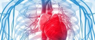
The pulmonary (inhalational) form of anthrax, or shepherd's disease, leads to death within 48 -72 hours. Death can be caused by complications:
- acute cardiovascular failure;
- pulmonary edema.
This feature of the development of the pulmonary form of the disease was used in the development of biological weapons designed to cause a person to inhale the spores.
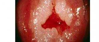
Intestinal form
This clinical form of pathology is extremely rare. It is distinguished by its exceptional severity. The incubation period ranges from 48 hours to 5 days after infection.
The patient complains of:
- general weakness;
- nausea and uncontrollable vomiting with bile;
- severe abdominal pain;
- lack of appetite;
- bloody diarrhea;
- increase in abdominal size;
- ulcers on the mucous membranes of the mouth and pain when swallowing;
- carbuncles in the throat;
- swollen lymph nodes;
- temperature rise up to 40 degrees Celsius;
- severe headache and dizziness.
The disease provokes a sharp increase in lymph nodes in the neck and jaw. Visually, the mucous membranes become bluish, the vessels in the eyes (on the sclera) become red.
The combination of lesions in the oropharynx and enlarged lymph nodes blocks the patient’s ability to swallow, causing massive bleeding from the mouth. An infected person breathes heavily, and compression of the airways can cause suffocation. Death due to increasing symptoms can occur very quickly.
Septic form
It is rare and can form as a complication of the critical course of various clinical forms of pathology. In this form of anthrax, pathogenesis is formed by the penetration of Bacillus anthracis into the bloodstream, causing multiple lesions of all major organs of the human body.
Multiple damage to the body by an extremely toxic bacterium causes infectious shock.
The main manifestations of the disease will be:
- fever and rapidly rising temperature up to 41 degrees;
- labored breathing;
- frequent shallow breathing;
- severe pain in the abdomen and chest;
- indomitable cough with production of foamy sputum;
- bloody diarrhea;
The rapid development of pathology quickly ends in the death of the patient.
Timely recognition of the disease and its clinical form is extremely important for assessing the patient’s condition and determining further treatment.
Mechanism of disease development
When it enters through damaged skin, the respiratory or digestive tract, the pathogen is captured by blood cells and transferred to regional lymph nodes. At the nodes, spores germinate into vegetative cells capable of producing exotoxins. If the immune system is unable to contain the infection at the level of the lymph nodes, it breaks through into the bloodstream. In most cases, this leads to the development of infectious-toxic shock. Congestion and thrombosis occur in the vessels, the liquid part of the blood passes into the tissues, and areas of necrosis (destruction) form in the organs due to thrombosis of the vessels feeding them. Multiple organ failure develops, the body is unable to cope with the infection, and death occurs.
Diagnostic methods
Diagnosis of anthrax combines:
- collecting anamnesis with comparison of epidemiological data;
- assessment of symptoms that characterize clinical forms of anthrax;
- conducting laboratory studies of biological fluids.
Differential diagnosis with diseases that cause carbuncles-like formations is important:
- bubonic plague;
- carbuncles caused by inflammation of several nearby hair follicles;
- syphilis in the first stage;
- erysipelas.
In other forms of ulcers it is differentiated;
- pneumonia;
- tularemia;
- pneumonic plague;
- dysentery.
A distinctive feature of the carbuncle caused by the ulcer bacillus is its painlessness - the patient does not feel any painful symptoms, even if the sore is punctured or otherwise injured.
When collecting an epidemiological history, the doctor finds out the possibility of the patient’s contact with contaminated meat or animal corpses.
All manifestations of ulcers are characterized by changes in the blood count:
- increased erythrocyte sedimentation rate;
- decreased leukocyte levels;
- increase in the number of lymphocytes.
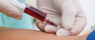
To carry out laboratory diagnostics, the following are taken for examination and culture:
- fluid discharged from blisters, carbuncles, or ulcers;
- urine and feces;
- mucus produced when coughing;
- blood.
For comparison, samples are taken for research:
- soil;
- water;
- animal meat;
- wool and pieces of skins;
- other.
When diagnosing anthrax and confirming the pathogen, bacteriological and bacterioscopic analysis is used - growing and studying the bacilli. Laboratory mice and rats are used to test highly contaminated material. When they are exposed to a charcoal stick, they die within 24 hours, and an ulcer forms at the site of penetration of the infectious agent.
To study samples from which it is difficult to isolate the pathogen (wool, hair), the Ascoli reaction is used.
Additionally assigned:
- chest examination (CT) if a pulmonary form of the disease is suspected;
- puncture in case of suspected CNS lesions.
To get the correct result you must:
- all materials for the study are collected before the start of antibiotic treatment;
- obtaining the necessary material is carried out under completely sterile conditions;
- a sufficient amount of material should be collected for research;
- For safety, the samples obtained must be stored in a special environment and containers.
Based on the diagnosis, treatment for anthrax is prescribed. Its goal is to destroy the toxin and bacillus, helping the body recover from illness.
Diagnosis of the disease
All patients undergo a general blood test, a general urinalysis, a biochemical blood test, and an electrocardiogram.
Depending on the clinical symptoms, laboratory tests are supplemented by a survey X-ray of the abdominal organs, chest, and, if necessary, ultrasound, lumbar puncture, pleural puncture, CT, and MRI. The basis for specific diagnosis of the pathogen is bacterioscopic, bacteriological methods, PCR, ELISA.
For research, they take the discharge of ulcers, the patient’s blood, sputum, vomit, and inoculate them on nutrient media. During microscopy and Gram staining, characteristic large immobile gram-positive rods are found, located one after another in a chain with a common capsule. On nutrient media, bacterial colonies exhibit characteristic growth.
The PCR method determines the presence of DNA, and ELISA determines specific antibodies against the pathogen (Ig M, Ig G). It is also possible to perform the Ascoli thermal precipitation reaction to detect anthrax antigens.
There is such a method as a biological sample. Its essence lies in the fact that laboratory animals (mice, rats) are infected with secretions from the patient. Animals die. Smears are made from the discharge obtained from animals (exudate from the abdominal cavity), and then a pure culture of the pathogen is detected under microscopy.
A general blood test reveals an increase in the total number of leukocytes due to band neutrophils, an increase in the number of lymphocytes, and an acceleration of ESR. The ECG shows the appearance of sinus tachycardia, rhythm and conduction disturbances. If the lungs are affected, a plain X-ray of the chest organs reveals the presence of infiltrates and effusion in the pleural cavity.
For diagnostic purposes, it is possible to perform an allergy skin test with anthraxin (anthrax allergen). In most patients it is positive either from the first days of illness or by the end of the seventh day. For quick diagnosis, the sensitivity of the isolated culture to a specific bacteriophage is determined.
Treatment
The life of an infected person depends on the speed of identifying the disease and the timeliness of assistance provided - without treatment, death is ensured in 100% of cases with pulmonary and septic forms of the disease, 50% with intestinal, 20% with skin.
Treatment of anthrax is complex and includes the use of antibiotics and immunoglobulin. The antibiotic is selected according to the form of the disease:
- for the pulmonary form of the pathology, Ciprofloxacin is used at a dosage of 500 mg. (tablets or capsules); Doxycycline 100 mg can be used for 2 months; if you are sensitive to drugs of these groups, use Amoxicillin at a dosage of 500 mg for 60 days; Regardless of the contact, the vaccine is used.
- for the skin form of an ulcer, the same scheme is used; additional treatment is required for the ulcer itself, which will progress with the growth of a scab;
- additionally, for all forms, the same antibiotics can be used intravenously together with Meropinem; Rifampin; Vancomycin; Clarithromycin.
For anthrax treatment, the following can be used:
- blood or plasma transfusion, drug infusion;
- artificial lung ventilation devices;
- means for artificial blood purification.
Treatment with a single antibiotic to prevent resistance to the pathogen is not recommended, although this ability has not been clinically proven.
It is irrational to deny the possibility of developing particularly resistant strains - most likely, military biologists have developed such samples, but they have not yet demonstrated themselves clinically.
Burial of the dead
The corpses of people who die from anthrax are not autopsied. Only in cases of extreme necessity, an autopsy is performed by a doctor with the strictest subsequent disinfection of the room and all items, including gowns, gloves, shoes, etc. Burial of those who died from anthrax is permitted in a regular cemetery, but special measures are always taken. In cases where the corpse has not been opened, it is tightly wrapped in plastic film and only then placed in a coffin. When burying a corpse that has undergone an autopsy, additional measures are taken - for example, a layer of dry bleach is poured under a plastic film at the bottom of the coffin.
Prevention of anthrax
For persons who have a high risk of contracting the disease during professional activities, vaccination is used. This:
- military;
- veterinarians;
- employees of textile enterprises processing imported wool;
- SES laboratory assistants.
Modern vaccines require repeated doses to create lasting immunity. Side effects after administration of the drug cannot be ruled out. Nowadays a live vaccine is used, which is prepared from a specific strain of ulcer, the prevention of which by vaccination is the only effective means against infection.
Preventive actions
Preventing the spread of this pathology involves influencing the source and mechanism of transmission. Points where outbreaks of the disease were previously recorded (farms, tracts, etc.) are subject to special recording. Specialists monitor the incidence of diseases among animals. Each case is investigated to establish the causes and carry out a set of preventive measures.
The sick person is hospitalized. A nurse or nurse is assigned to care for him. In the cutaneous form, the patient is discharged after a scar forms at the site of the ulcer. People who died from this infectious disease are not autopsied.
Relatives and the sick person are not separated. The patient's immediate environment is monitored until his complete recovery. If people have been in contact with material contaminated with spores, then emergency prevention is indicated.
Brief facts about the history of the disease
Some researchers claim that anthrax is described in the Bible as the fifth and sixth Plagues of Egypt.
The anthrax bacillus was first isolated by the French Daven and Reyet; at the end of the 18th century it was first isolated as a pure culture. The idea of using a live vaccine to prevent pathology belongs to Pasteur, which was created by veterinarian Ivan Lange. Now the number of infections in Russia does not exceed 50 per year.
Active work on the Siberian bacillus to create biological weapons began after the First World War simultaneously in different countries. Isolated areas were used to test intermediate samples.
The most famous anthrax island is Gruinard. The tests were carried out by the Porton Down laboratory on sheep on the personal orders of Winston Churchill. The secret of the successful experiment was kept for many years, until in 1981 the militant terrorist group Dark Harvest Commando sent letters with contaminated soil from the island to the UK Chemical Defense Center and representatives of the Conservative convention in Blackpool, which led to the death of several people.
The measures taken by Great Britain were unprecedented - the entire topsoil was removed from the island, and tons of formaldehyde was poured onto the remaining soil. Since 1990, the island has been considered safe.
The USSR has been working on the creation of bacteriological weapons since its creation in the 20s of the last century. The Russian island of death was Vozrozhdeniya Island in the Aral Sea, where experiments were carried out on monkeys, colic and prisoners. With the collapse of the USSR, experiments were curtailed, and the finished weapon Antrax-836 was partially destroyed.
Pathogenesis
The entrance gates for the exciter are:
- even slightly damaged skin (the most common method);
- mucous membranes of the respiratory system;
- gastrointestinal tract.
The bacterium penetrates the tissue and at the point of penetration releases a special substance - bacterial exotoxin. At this site, an inflammatory focus occurs, noticeable swelling and necrosis (necrosis) of the surrounding tissues. Subsequently, a carbuncle appears, in the center of which dead tissue is observed. Meanwhile, the pathogen moves through the lymphatic system to the nearest lymph nodes, causing their inflammation. Anthrax in the cutaneous form is rarely accompanied by sepsis. Blood poisoning is possible if the route is airborne.
Penetrating into the human body, the anthrax bacterium releases a dangerous substance, exotoxin, which causes tissue necrosis.
Prevention
Prevention of the disease includes the following measures:
- vaccination of people at risk;
- animal vaccination;
- pet control;
- control of transportation and storage of meat products;
- early identification of patients and their timely hospitalization (in order to prevent the spread of the disease, people who were in close contact with the patient are also isolated);
- carrier isolation;
- the corpses of infected animals are burned.
Epidemiology
In places where it is endemic, Pendensky ulcer shows a clear dependence on the time of year. It is the first cases that appear in the fall, then their number increases from September to November, after which the diseases begin to occur less frequently and stop in January or February. Everywhere, the increase in cases of the disease coincides with the rainy season. The disease is observed equally in old people and children, in men and women, natives and foreigners, the latter even showing a slightly greater tendency to the disease.
Cases of introduction of Pendin ulcer into areas where it is not usually found are very rare and have never caused endemic spread.
The carriers of the disease are mosquitoes Phlebotomus sergenti, Phlebotomus papatasii, etc. Sources of infection are sick people, dogs, gerbils.
Vaccination
Anthrax is a serious, deadly disease that can be prevented by vaccination.
Modern types of vaccines:
- Inactivated. Contains a weakened pathogen incapable of reproduction. Today, such a drug is used extremely rarely.
- Alive. It consists of weakened spores, which provoke a mild course of the disease without a pronounced clinical picture. After recovery, patients have stable, reliable, lifelong immunity.
- A combined vaccine containing components of inactivated and live vaccines.
Most countries use only live and combination vaccines. Characteristics of live vaccine:
- Available in dry form
- cutaneous route of administration,
- double administration,
- the effect occurs two weeks after the manipulation.
Combined vaccine:
- comes in dry, liquid form,
- administered only subcutaneously,
- single administration,
- protection occurs a week after the procedure.
The number of people who are required to be vaccinated:
- laboratory technicians working with ulcer spores or ulcer bacteria,
- veterinarians,
- persons working in the processing of animal wool and skin,
- slaughterhouse workers,
- meat processing plant personnel.
Side effects of vaccination:
- fever,
- pain in the head
- malaise,
- lymphopathy.
Contraindications to the procedure:
- exacerbation of infections, inflammations,
- oncology,
- hematological diseases,
- HIV AIDS,
- skin lesions
- childhood,
- pregnancy,
- lactation.
Clinical picture
Initially, at the site of development of the pimple, a pinpoint hemorrhage is noticed, surrounded by a narrow red rim, and then an inflammatory nodule is formed, similar to acne or folliculitis. This initial change may disappear without a trace, or a small yellowish or brownish-red nodule appears, which gradually increases to the size of a large nodule. In the center, at the top of the node, sometimes a blister or abscess appears, or light peeling begins, and then a dense, firmly seated dark brown crust. As the node on the periphery increases, the crust also increases. If the latter is removed, then under it they find a completely round ulcer with steep edges, from which pus is released under pressure. In the immediate vicinity of the first node, similar nodules develop, which partly merge with each other so that a round or oval plaque is obtained, the central part of which consists of an oblong, crusty ulcer, and the peripheral part of 6-10 small ones, touching and partly merging with each other infiltrates of a pale or brownish-red color. This plaque is very characteristic of a Pendin ulcer.
In the circle of the primary infiltrate, very small miliary pustules are visible with the naked eye or a magnifying glass, which open and turn into crater-shaped, roundish ulcers that become crusty. Due to the increase in the periphery, these small ulcers merge with each other and with the central ulcer, so that a deep, large, round or oval infiltrate is obtained with an irregular, round or oval crust in the center. If you remove the crust, you will notice an ulcer with steep, sharp, densely infiltrated, light or brownish-red edges and a pinkish-red bottom of glassy, reddish granulations, which separates liquid, serum-like or blood-stained pus, characterized by a torpid character and is non-painful. At this stage, the Penda ulcer, without increasing particularly in size, remains for months; only rarely does it undergo more rapid decay and take a more malignant course.
After existing for several months, the ulcer begins to clear, scars from the center and heals, leaving behind a dense, constricting scar surrounded by a pigmented border. In rare cases, it is abortive and the infiltrate resolves without reaching ulceration.
Pendine ulcer is rarely single, more often it appears in several copies and even in large numbers, simultaneously or one after another; it is located almost exclusively on exposed parts of the body, on the face, the back of the hands and feet, and on the extensor surface of the forearm and lower leg. It never occurs on mucous membranes.
Sometimes returns and secondary infection are observed, but usually after a single illness, a certain immunity to the disease remains.
Therapy methods
The fight against the disease in question is complex and multi-stage:
- bed rest,
- balanced diet,
- etiological therapy (course of penicillin and fluoroquinolone antibiotics),
- administration of antiulcer immunoglobulin (ready-made antibodies),
- local therapy of the skin form (use of antiseptics, sterile dressings without surgical treatment),
- infusion treatment, intensive detoxification (in severe cases with the use of hormone therapy).
Severe forms of the disease, the development of complications (infectious-toxic shock, brain swelling, respiratory failure), a direct indication for transferring the patient to the intensive care unit. Antibacterial therapy eliminates the cause of the disease. The causative agent of anthrax is sensitive to penicillins. In case of intolerance to this drug, the following is prescribed:
- tetracyclines,
- macrolides,
- fluoroquinolones.
Effective antibiotics:
- Penicillin, a medicine with a broad spectrum of action. Destroys bacteria because it has a bactericidal effect. The drug is usually administered parenterally in doses of up to 4 million units. For complicated septic forms, the daily dose is increased 4-5 times. Course of therapy, up to 10 days.
- Tetracycline, a broad-spectrum drug with a bacteriostatic effect (stops the growth and development of bacteria). It is usually consumed up to 6 times a day for a week.
- Erythromycin, a representative of macrolides. Has a strong bacteriostatic effect on pathogens. It is prescribed four times a day for a week.
- Ciprofloxacin, a fluoroquinolone drug. It has a wide spectrum of action, creating a bactericidal effect. It is prescribed parenterally by drip three times a day for 2 weeks.
- Levofloxacin is classified as a fluoroquinolone. A broad-spectrum medicine that reliably destroys all pathogenic microbes. It is administered dropwise twice a day for 2 weeks.
- Doxycycline is a tetracycline. Stops the growth and development of bacteria. In the first days of the disease, the drug is prescribed four times a day, then the frequency of administration is gradually reduced to 2 times a day.
Hormone therapy
For general strengthening, reduction of fever and intensity of inflammation in severe cases of the disease, the use of glucocorticosteroids is indicated. Tablet forms of Prednisolone and Dexamethasone are often prescribed at a dosage of 90-120 mg per day. With septic Siberia, their dosage increases several times.
Infusion therapy is the parenteral administration of liquid solutions. Such therapy is aimed at replenishing and maintaining the volume and condition of body fluids. It relieves intoxication symptoms.
For anthrax, the following groups of solutions are prescribed:
- colloids,
- crystalloids,
- hematological drugs.
Colloidal preparations include:
- Poliglyukin. Consists of dextran and saline solution. It has a significant anti-shock effect, replenishes fluid reserves in case of dehydration, loss of plasma or blood. The dosage of the medication is often selected individually, depending on the complexity of the disease and the patient’s condition.
- Reopoliglyukin. Consists of dextrans and glucose solution. Quickly replenishes the deficit in circulating blood volume, improves blood flow in small vessels, and minimizes the risk of thrombosis. Administered parenterally, by drip.
Crystalloid drugs
This group includes physiological sodium chloride solution. This is an isotonic solution that allows you to maintain a stable osmotic pressure of plasma and extracellular fluid. Using this product, you can replenish the required level of sodium and chlorine in case of loss of plasma and blood.
Ringer's solution, a complex agent including sodium, chlorine, potassium and calcium. It is often taken for severe losses of plasma and intercellular fluid. The maximum daily dose of medication is sometimes up to 3 liters.
Glucose solution is a hypotonic solution containing dextrose. It is successfully used for detoxification and replenishment of fluid volume in the body. The effect of the drug on heart contraction, metabolism, and oxidative processes in the body has been proven.
Hematological drugs are presented in the form of Albumin. Fresh frozen plasma consists of hemostatic factors, proteins, carbohydrates, salts and fats. Often used for plasma loss, blood loss, and significant intoxication. This is the main source of immunoglobulins, so a group compatibility test must be performed before administration.
Albumin is a preparation of a complete blood component. Maintains the stability of its colloid osmotic pressure. When the drug is administered, an increase in BCC is achieved (fluid is attracted and stored in the vessels). In addition, albumin supports the normal functioning of the body by providing complete nutrition.
Use of immunoglobulin
This product is ready-made antibodies against the pathogen obtained from the plasma of vaccinated horses. This allows you to create and maintain immunity to the causative agent of the disease. It has been proven that the medicine has a detrimental effect on the anthrax bacillus and has a pronounced antitoxic effect.
With its help, it is possible to cure the disease by preventing the development of pathology. When treating ulcers, the dosage of the medication is selected depending on the severity of the patient’s condition:
- for mild anthrax, up to 20 ml of medicine is used,
- for moderate severity, up to 40 ml,
- 60-80 ml of the drug per day, for severe illness.
Immunoglobulin is injected parenterally into the upper part of the buttock. This protein drug can cause allergies. Therefore, before therapy, a sensitivity test to horse protein is required. A small amount of the drug (diluted) is administered intradermally. After 30 minutes the reaction is assessed. A small skin reaction (papule up to 0.9 cm) indicates the possibility of the procedure.
A positive drug test (papule more than 1 cm in diameter) is a direct contraindication to the procedure. But in especially severe cases it is still administered, but after strict administration of glucocorticoids. Immunoglobulin is not a panacea for the disease. Good results can be obtained by using the product together with antibiotics.
For urgent prevention of anthrax, immunoglobulin is prescribed according to the following scheme:
- adults, 20 ml,
- teenagers, 12 ml,
- children under 14 years old, 8 ml.
Prophylactically, the drug is administered once. Indications for emergency immunization:
- contact with a sick animal,
- contact with material or product containing anthrax spores,
- a man was cutting up the carcass of a sick animal,
- eating contaminated meat.
Topical use of ointments
In medicine, there is no consensus on the need for local therapy. Some experts believe that treating carbuncles and rashes can cause generalization of the process, others insist on the need to treat skin ulcers.
Some popular remedies for carbuncles of ulcerative nature:
- Antiseptics are used to wash pustules and ulcers. Chlorhexidine, Miramistin, Decasan, Furacilin are often used. Manipulations inhibit bacteria and prevent concomitant infections. It is important to carry out treatment three times a day.
- Antibacterial ointments are used to apply antibiotic dressings. The most commonly used are Penicillin ointment, Levomekol, Gentamicin ointment, and Erythromycin ointment.
- Glucocorticosteroids fight itching, swelling, and pain. Combination drugs of antibiotics and hormones are often prescribed: Trimistin, Pimafucort.
- Regenerating ointments, preparations that accelerate reparative processes and skin restoration. Panthenol, Bepanten, Sudocrem are often used.
Histological changes
On sections from young, not yet ulcerated nodes, when stained with Giemsa and hematoxylin according to Van Gieson, several layers are distinguished. Under the epidermis there is granulation tissue, which contains numerous plasma cells, round cells, some leukocytes, eosinophilic and mast cells and consists in general of 3 layers, which are clearly distinguishable under low magnification, but under high magnification they pass into each other without sharp boundaries. If we call the epidermis the first layer, then underneath it lies a second layer, consisting of dilated papillae, in which there are larger or smaller clusters of macrophages containing parasites. The third layer contains granulation tissue, which passes downwards into the fourth layer, consisting of cell-rich granulation tissue with nodules containing giant cells.
The epidermis is partly normal, partly thinned so much that it consists of only a few layers of cells. The Malpighian layer is preserved, in places flattened and thinned by dilated papillae and cell masses pressing down from below. There is no hair, sweat glands are present only in very scanty quantities. Between the cells of the epidermis there are small numbers of leukocytes. The cells of the Malpighian layer are often swollen and contain vacuoles. Proliferation of the epithelium is often observed, which in the form of thin rods and branching cords penetrates into the third and even fourth layer. The papillae are partly only infiltrated with small cells, while partly they are dilated, voluminous, permeated with plasma cells and macrophages. The latter are large, mononuclear cells, which are densely packed with the parasites described above. They have a round, oval, multifaceted or irregular shape and are equipped with processes; their protoplasm is granular and contains many small vacuoles, or one or several large vacuoles. The nucleus is round, oval or slightly curved, with sharp outlines, clearly defined nuclear bodies and small grains. These macrophages originate from cells that are located among them in varying numbers and, in their position and appearance, represent enlarged connective tissue cells or endothelial cells of the lymphatic vessels. These cells have the same large nucleus as macrophages; their protoplasm increases in mass, acquires a granular or vacuolar character and swells even more after the introduction of parasites. These cells, macrophages and transitional forms, lie next to each other, often several at a time, in the crevices of the connective tissue. Macrophages are quite similar to the large cells found in the liver and spleen during kala-azar. In the protoplasm of macrophages there are protozoa, sometimes in small, sometimes in very large quantities.
In the 3rd layer there is an abundance of newly formed vessels, infiltration with round and plasma cells and single or small groups of macrophages with a meager number of parasites. In addition, it contains infiltrates in the form of nodules, consisting of round and epithelioid cells and containing in the center a giant cell with numerous oval nuclei located at the edges and fine-grained protoplasm, which gives off processes between the surrounding cells.
Finally, in the 4th layer there are round and plasma cells, and between them fibroblasts and thin fibers, indicating the beginning of healing, which, like scarring, proceeds from the base and from the edges towards the surface. Elastic fibers die in the infiltrate and are preserved only in normal skin tissue.
Diagnosis and treatment
The diagnosis can be made, in addition to the clinical picture, based on the presence of parasites. In this regard, it should be noted that in the ulcer and in its compartment, leishmania is found only in scanty quantities. The best results are obtained when injecting into pimples that have not yet ulcerated: the blood flowing from such injections contains a very large number of parasites.
Treatment of Penda ulcer is carried out with the help of monomycin, as well as solyusurmin, glucantim, metacycline, doxycycline, and antimalarial drugs.