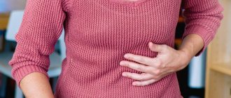
Rapidly and rapidly developing inflammation of the gallbladder is called acute cholecystitis. The disease, which occurs for the first time, ends with complete recovery with adequate treatment. When attacks recur, a chronic course of the disease is diagnosed. With age, the possibility of developing cholecystitis increases; women are more susceptible to pathologies than men. At risk are pregnant women, taking hormonal medications and obese people.
Causes of formation of gallstones
Medical scientists cannot say exactly why a chemical imbalance occurred in the body of a particular person, causing the formation of gallstones and a constant exacerbation of cholecystitis. However, there are still general characteristics by which one can determine whether a person has a predisposition to gallstone disease, and these are:
- Excess weight (especially in women). It has been noticed that the thicker a woman’s waist, the greater her chances of developing cholelithiasis and acute cholecystitis. Obese children are at risk and may periodically suffer from exacerbation of cholecystitis, while children with normal weight almost never complain of pain in the right hypochondrium.
- Pregnancy . If a woman has been pregnant at least once, she is also at risk.
- Diets . Gallstones are a complication of rapid weight loss and the habit of constantly losing weight and gaining weight.
- Pregnancy protection . At risk are women taking oral contraceptives and undergoing estrogen therapy in high doses.
- Heredity . Genetic predisposition to cholelithiasis and, as a consequence, to exacerbations of cholecystitis.
- Poor nutrition . Lovers of fatty foods often suffer from cholelithiasis and acute cholecystitis.
- Floor . Women get sick more often than men.
- Age. People over 60 years of age are more likely than younger people to suffer from gallbladder diseases.
- Increased sugar. Diabetics are predisposed to the formation of gallstones.
- Taking certain medications . Sometimes acute cholecystitis is caused by medications that lower blood cholesterol levels.
Diagnostics
Diagnosis of acute cholecystitis begins with taking an anamnesis. First of all, the doctor is interested in the following complaints:
- Nature and localization of pain in the right hypochondrium. In this case, the duration of the attack can be more than 30 minutes;
- Presence of nausea and vomiting;
- Increased body temperature;
- Hepatic colic (observed in 50% of patients).
After this, a visual inspection of the skin is carried out and the abdominal cavity is palpated. The following signs indicate the development of pathology:
- Murphy's symptom is an involuntary holding of breath when pressing on the area of the right hypochondrium. In this case, the patient experiences severe pain.
- Abdominal muscle tension.
- An increase in the size of the gallbladder is palpable in 40% of patients.
- Yellowing of the skin is not a specific symptom and is observed only in 10% of patients.
The next stage of diagnosis is laboratory tests. These include:
- Complete blood count - reveals an increase in the number of leukocytes. The higher this indicator, the more extensive the inflammatory process.
- Biochemical blood test - shows an increase in the norm of C-reactive protein, alkaline phosphatase and specific liver enzymes.
- A general urine test is effective only if the patient’s condition sharply deteriorates. The presence of protein and casts may indicate the development of necrosis and severe intoxication. Also, in the presence of jaundice, traces of bilirubin are found in the urine.
To make a final diagnosis, instrumental studies are carried out. Most often, experts prescribe:
- Ultrasound of the gallbladder is the simplest and most accessible method. Using ultrasound, you can detect stones in the bladder, foci of inflammation and thickening of the walls of the organ. It is worth noting that during the procedure, most patients experience Murphy's symptom.
- MRI - prescribed when ultrasound is ineffective. The procedure is also performed on pregnant women if pathology is suspected.
- Scintigraphy - during the procedure, special preparations based on low-toxic radioactive substances are introduced into the patient’s body. This allows you to effectively “highlight” even the most minor changes in the structure of the organ. The procedure is completely safe, but is performed extremely rarely due to technical difficulties.
- X-ray examination is almost never prescribed for acute cholecystitis. If there are stones in the bladder, X-rays are only 10-15% effective, provided that the formations contain calcium. However, with the help of radiation, cavities with gas can be detected in the walls of the bladder, which indicates emphysematous cholecystitis.
Treatment of acute cholecystitis is prescribed based on diagnostic results. If necessary, the patient may undergo emergency surgery.
Other causes of acute cholecystitis
In addition to cholelithiasis, the cause of the development of acute cholecystitis can be:
- Abdominal injury.
- Surgery on the abdominal organs.
- Tumor. Many people do not know that acute cholecystitis is also caused by a tumor, a neoplasm that can be solid or filled with liquid. The word "tumor" does not mean cancer. Tumors can be benign, precancerous, or cancerous.
Benign tumors do not cause harm to health “by default,” but if they put pressure on nerves or blood vessels, other, very unpleasant changes occur in the body. Thus, even a benign tumor can stop the flow of bile from the gallbladder and thereby cause an attack of acute cholecystitis.
Forecast
The prognosis for uncomplicated forms of acute cholecystitis, provided timely medical care is provided, is generally favorable. Acute non-calculous cholecystitis usually ends in complete recovery and only in a small percentage of cases becomes chronic; the likelihood of chronicity of acute calculous cholecystitis is much higher.
The prognosis sharply worsens with the development of complications (peritonitis, peri-vesical abscess, empyema). The probability of death in this case is, according to various sources, 25–50%.
https://youtu.be/u9vBEVbaNo0
Risk factors
A risk factor is a condition or situation that increases the risk of developing a disease.
Crohn's disease
One of the main risk factors for the development of acute cholecystitis is Crohn's disease, a permanent condition in which the gastrointestinal tract is constantly inflamed. Crohn's disease can affect any part of the intestine, including the anus. People suffering from this disease experience constant fatigue, frequent diarrhea, discomfort and abdominal pain.
About a fifth of patients have close relatives with this disease. From this we can conclude that Crohn's disease can be inherited. In addition to genetic predisposition, Crohn's disease is caused by an incorrect response of the human immune system to certain foods and especially medications, and this disease is also provoked by a poor environment.
Symptoms of Crohn's disease depend on which part of the intestine is affected. When the intestinal walls are damaged, the following is usually noted:
pain at the level of the right lower abdomen;
- ulcers in the intestines, which sometimes bleed and it is by the blood in the stool that the patient understands that something is wrong with his intestines;
- mouth ulcers;
- diarrhea – can be mild or severe. Sometimes mucus, blood, and pus are noticeable in the stool;
- false urge to stool;
- Fatigue is a common feeling for patients with Crohn's disease;
- loss of appetite;
- weight loss due to poor appetite;
- anemia (associated with anal fissures). Bleeding from the rectum and anal fissures are not so dangerous, since not much blood is usually lost, but the fissures themselves are very painful and bring patients many unpleasant hours.
Obesity
Another risk factor for acute cholecystitis is obesity. Despite the fact that everyone reads and writes about obesity, most people do not even understand what it is and do not know that there is obesity, which is not visually visible. To determine the presence or absence of excess weight, several factors should be correlated, such as age, muscle-to-fat ratio, height, gender and bone density.
Based on these indicators, it sometimes turns out that the ideal weight of two people of the same height can vary greatly.
However, one thing is undeniable: excess weight is dangerous to health.
Physical work during pregnancy
This risk factor is not as rare as one might think. Pregnant women are often forced to work physically. However, such stress damages the gallbladder, and acute cholecystitis may develop several weeks after birth.
Similar chapters from other books
Letter 6 Pinworms. Scabies. Opisthorchiasis. Botkin's second symptom
Letter 6 Pinworms. Scabies. Opisthorchiasis. Botkin’s second symptom Dear Olga Ivanovna! Good day to you and best wishes. I have been studying parasitic diseases for many years. When I read your books, a lot of what I thought was confirmed. I recently read about
Full symptom
4. Full symptom - “A chair stands firmly only if it has at least three legs.” A well-evaluated symptom must also have at least three pillars. However, a chair with four legs is even better. A complete symptom is made up of four elements. He
Is ophthalmopathy a complication, symptom or disease?
Is ophthalmopathy a complication, symptom or disease? In diseases of the thyroid gland, changes in the eyes occur. These disorders are accompanied by single signs or multiple signs. They occur behind the eyeball, but can also involve the eye itself,
Not a disease, but a symptom!
Not a disease, but a symptom! Misconceptions that contain some truth are the most dangerous. Adam Smith Let's start with the main and obvious. Cough is not a disease, but just a symptom[10] of a specific disease. There is no cure for cough! Treat the disease that caused it
Botkin's disease (hepatitis A)
R. D. Sokolovskaya BOTKIN'S DISEASE
R. D. Sokolovskaya BOTKIN'S DISEASE Epidemic hepatitis, or, as this disease is more often called, Botkin's disease, is caused by a special virus. Fifteen years ago, this disease was considered non-contagious and was called catarrhal jaundice, and popularly simply jaundice.
COURSE OF BOTKIN'S DISEASE
COURSE OF BOTKIN'S DISEASE After the causative agent of epidemic hepatitis enters the body of an adult or child, the disease does not immediately occur. Some time passes before the first signs of the disease appear. This latent period of the disease, called
Pain as a symptom of a disease
Pain as a symptom of a disease A person acquires most of his ailments with age or as a result of diseases inherited from his parents. And he consults a doctor only when he feels pain that he cannot bear. For a specialist, pain comes first
Botkin's disease
Botkin's disease An infectious disease that affects liver tissue. If the diagnosis is made in a timely manner and treatment is carried out in a hospital, then after treatment the liver should have little or no concern. But it’s better to help our worker, and this is quite easy to do in
Tinnitus is an alarming symptom
Tinnitus is an alarming symptom. Cardiovascular diseases affect more than just the heart. They also work by ear. Sometimes, however, the hearing in older people is within normal limits, but pain in the ears periodically occurs. Doctor's examination and rinsing procedures are not
Hepatitis A, Botkin's disease
Hepatitis A, Botkin's disease The hepatitis A virus has an acid-resistant envelope. This helps viruses that come from contaminated food and water to pass the acidic protective barrier of the stomach. The hepatitis A virus is stable in the aquatic environment, so hepatitis A epidemics often have
Symptoms of acute cholecystitis
- Pain localized under the right lower edge of the ribs.
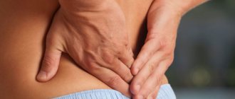
- Aching pain spreading down the lower back.
- Your right shoulder may hurt.
- The person feels nauseous and sometimes vomits.
- Increased sweating of the back.
- Anxiety.
The patient's condition worsens if biliary colic is added to inflammation of the gallbladder. This happens only in one case, when a gallstone enters the bile duct, and through it into the ducts of the duodenum.
This situation can be provoked by eating fatty foods - the pain begins two hours after eating. It can be continuous, for 24 hours, or paroxysmal.
It happens that acute cholecystitis is accompanied by infection of gallstones. At the same time, the patient has a fever and constantly trembles. The vast majority of cases of acute cholecystitis, complicated by infection of gallstones, end with surgery, during which the stones are removed.
Symptoms
The severity of symptoms depends both on the form of the pathology and on external factors. Patients experience the strongest sensations after consuming fatty foods and alcohol, physical activity, stress and overexertion. The most common symptoms of acute cholecystitis are:
- Severe pain in the right hypochondrium. Most often the attacks are colicky in nature. The pain can radiate to the collarbone, back, neck, shoulder blade or affect the left hypochondrium area. It is important to note that the severity of pain may decrease with the development of the gangrenous form of cholecystitis due to the death of nerve endings.
- Nausea and vomiting. There are traces of bile in the vomit.
- Feeling of bitterness in the mouth.
- Increased body temperature. Indicators can range from 37.5 degrees to 40 degrees.
- Uncontrolled holding of breath when pressing on the right hypochondrium (Murphy's symptom).
- Painful sensations when tapping the lower ribs on the right side (Ortner's symptom).
- Yellowing of the skin is observed in 10% of cases with swelling of the bile ducts or their blockage with stones;
- Increased size of the gallbladder.
Diagnosis of acute cholecystitis
Very often, a preliminary diagnosis of “acute cholecystitis” is made by emergency doctors. This is due to the fact that the disease begins suddenly and proceeds very violently. Usually, the patient’s relatives call an ambulance, and doctors, after collecting an anamnesis and palpation examination, understand what kind of misfortune they are dealing with.
Then, regardless of whether the patient is sent to the hospital or remains treated at home (very rarely), a series of diagnostic procedures are prescribed.
Blood analysis
Using a general analysis, the number of leukocytes in the blood is determined. An increased number of leukocytes indicates the presence of inflammation in the body, and if there is also a high level of bilirubin and alkaline phosphatase, this is obvious evidence in favor of acute cholecystitis.
Computed tomography (CT) or ultrasound
This test allows the doctor to see how the gallbladder looks at the time of illness, and whether there are ulcers, tumors, etc. on its walls.
HIDA scan
This test allows you to examine the liver, gallbladder, bile ducts and small intestine. The doctor can monitor the production and flow of bile from the liver into the small intestine and determine whether there is a blockage and, if so, where there is a blockage.
Features of symptoms for various types of cholecystitis
Stoneless
In approximately 20% of the population, the inflammatory process develops without the presence of stones. This can often be found in children and elderly men. The difference between the pathology and calculous cholecystitis is in the very mechanism of pathogenesis of the disease and the absence of stones in the gall bladder. For several years, patients do not even suspect the presence of inflammation. And the feeling of heaviness in the area of the projection of the liver and gall bladder after eating large quantities of fatty fried foods does not entail a trip to the doctor. Therefore, it is most often diagnosed accidentally, during an ultrasound examination of other organs, or during planned hospitalization for other diseases.
Calculous
The reason, as we found out, is stones that occur when bile stagnates and salts increase in it.
Catarrhal
It is the most favorable form of the disease. It is characterized by the presence of a large, enlarged bladder, the wall of which is hyperemic and thickened.
Phlegmonous
The gallbladder becomes covered with fibrin films and increases in size. The contents of the organ are purulent exudate. The mucous membrane is inflamed, swollen, reddened.
Treatment of acute cholecystitis
Immediately after diagnosis, the patient will be advised not to eat not only solid, but also liquid food for some time. The body will be “fed” with intravenous drips.
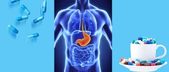
Medications include analgesics and antibiotics. There are never any problems with analgesics; the patient and his relatives understand that if a person has something in pain, they need to relieve the pain by giving him an injection. It's a different matter when it comes to antibiotics.
Once upon a time, at the dawn of the “era of antibiotics,” these dosage forms were prescribed and enthusiastically taken at the slightest manifestation of inflammation. In recent years, another trend has emerged - people do not want to take antibiotics, rightly believing that this leads to a decrease in immunity and other problems in the body. This opinion is partly correct, but there are times when these medications, when used correctly, save lives.
The word “antibiotic” can be deciphered as “against bacteria”. That is, a medicine that destroys or slows down the growth of bacteria. It is clear that this is a very powerful medical remedy, and it should be prescribed when the body's natural defenses are not enough to cope with the infection. Acute cholecystitis is just such a disease.
How is antibacterial treatment prescribed for acute cholecystitis?
For patients with acute cholecystitis, antibiotics are prescribed either orally (by mouth) or by injection. In most cases, the effect is visible within a few hours after the start of treatment. However, even if improvement occurs on the second day, the course of antibiotics should be completed. If this is not done, if treatment with antibacterial medications is necessary in the future, there is a high probability that they will not work.
It is especially important to remember that these medications should be taken exactly as prescribed by your doctor. If the doctor prescribes taking the drug one hour before meals or two hours after meals, then this is what you should do. Some antibiotics should absolutely not be taken with some food products (for example, tetracycline and dairy products are incompatible) - these rules should not be ignored, otherwise cholecystitis will worsen even more.
Side effects of treating acute cholecystitis with antibiotics:
- Diarrhea. Improper use of antibiotics causes an increase in sugar levels in the intestines. Bacteria take hold in the intestines and “thrive” on sugar. As a result, a person begins to experience constant diarrhea.
- Fungal infection of the mouth, digestive tract and vagina. “Good” bacteria that people need live in the mouth, in the digestive tract and in the vagina; with antibacterial treatment, they also die, and a fungus “comes” in their place.
Surgical treatment of cholecystitis
Once aggravated, cholecystitis will worsen again and again. Therefore, sooner or later, doctors recommend surgical removal of the gallbladder. If acute cholecystitis is aggravated by perforation of the gallbladder, then the operation is performed urgently.
Usually, after resection of the gallbladder, there is no deterioration in the quality of life, perhaps sometimes episodes of diarrhea will occur (since without the gallbladder, bile from the liver flows directly into the rectum), but no more. The main thing is that a person does not indulge in excess in drinking and eating.
Heaviness in the stomach: diagnosis and treatment of the symptom in combination with other signs of dyspepsia
Our readers successfully use Monastic Tea to treat gastritis and ulcers.
Seeing how popular this product is, we decided to bring it to your attention. Read more here... Every person periodically experiences discomfort and discomfort in the abdominal area, especially after eating. But most often, such symptoms remain without proper attention. Heaviness in the stomach is a sign of transient or permanent functional failure of the gastrointestinal tract. Heartburn, belching, nausea and increased gas production - all of them are included in the concept of dyspepsia syndrome and occur in various diseases.
Functional dyspepsia
Functional dyspepsia (FD) is understood as the periodic occurrence of complaints of:
- heartburn (“burning” pain behind the sternum), frequent belching;
- nausea, much less often – single vomiting;
- aching pain in the projection of the stomach;
- bloating due to increased gas formation;
- bowel dysfunction in the form of constipation.
With normal functioning of the gastrointestinal tract, the food mass from the stomach cavity gradually moves further into the duodenum. Having gone through all the stages of digestion in the small and large intestines, the chyme (bolus of food) is safely excreted. With dyspepsia, this process slows down significantly, causing pain, abdominal discomfort and bloating.
An unpleasant aspect of medical practice is that patients seek help from a doctor already with serious disorders. They try to treat relatively minor ailments on their own, guided by advertisements they see.
Having turned to a pharmacist for help, patients often only eliminate the obvious symptoms without trying to find the true reasons that caused the gastrointestinal tract to malfunction.
What can be recommended for people suffering from dyspepsia? Symptoms such as pain, bloating, severe gas formation are gradually eliminated with an integrated approach:
- For some time, antacids are used to protect the gastric mucosa (Phosphalugel, Almagel);
- pain syndrome can be relieved with antispasmodics of the No-shpa type;
- a course of prokinetics (Domperidone, Duspatalin) is also indicated;
- it is important to normalize nutrition (the frequency of food consumption should be at least 5 times a day; exclude spicy and fatty foods, smoked foods, and confectionery products from the diet);
- limit intake of alcoholic beverages, quit smoking.
More details about the causes, types, clinical picture and treatment of functional dyspepsia can be found in the article.
Poor nutrition
Digestive problems can occur in completely healthy people. In some cases, such phenomena go away on their own. However, with frequent occurrence of heaviness in the stomach, a thorough examination and, if necessary, treatment are extremely necessary. The reasons for heaviness and discomfort in the stomach area in this case are as follows:
- Eating insufficient amounts of vitamin-containing foods can cause significant problems in the gastric mucosa;
- uncontrolled eating, large portions cause stretching of the stomach cavity, which makes the digestion process incomplete;
- Frequent consumption of rough, fried and fatty foods disrupts gastrointestinal motility and increases the activity of digestive enzymes.
In this case, dyspeptic symptoms appear: nausea, belching and increased gas formation after eating.
Discomfort in the abdominal area can occur if a person eats rarely, but at the same time his portions are large. It is quite difficult for the body to cope with such a volume of food, so slow and incomplete digestion of the food mass occurs. This pathology is characterized by pain, belching, bloating and nausea.
Thus, the patient needs to modify his diet:
- eat food often, in small portions;
- limit harmful and irritating foods to the stomach wall;
- do not overeat, eat in the evening no later than 4 hours before bedtime.
In some cases, discomfort in the gastrointestinal tract can be caused by low-quality products that can cause acute food poisoning. The patient begins to be bothered by belching, pain in the stomach and umbilical region, nausea, turning into vomiting. Experts recommend doing a gastric lavage with boiled and salted water, and taking medications that help remove the infection from the body (sorbents).
Belching after eating
Belching is not normal and appears as a result of:
- Eating large amounts of food quickly (large food pieces and swallowed air irritate the wall of the esophagus and stomach, causing regurgitation of food and air bubbles).
- Talking while eating.
- Frequently drinking carbonated drinks, which also cause bloating.
- Sudden bending forward after eating.
In order to get rid of it, it is necessary to exclude all the provoking factors listed above and take prokinetics (Domrid) for 2-3 weeks.
Gastritis as a cause of stomach discomfort
With various types of gastritis, inflammation of the gastric mucosa occurs due to increased or insufficient production of gastric juice and infection with H. Pilory. The disease also occurs as a result of poor nutrition and stressful situations.
Gastritis may cause the following symptoms:
- heartburn (with increased stomach acidity);
- nausea (especially after eating);
- belching sour;
- sharp or dull pain in the projection of the stomach;
- excessive gas formation.
Treatment of the disease and, as a result, bloating due to gastritis, depends on the type of gastritis and may include:
- antibiotics (penicillins, macrolides) to eradicate pathogenic bacteria;
- antacids that reduce acidity and protect the mucous membrane (Ranitidine, Nolpaza, Phosphalugel);
- pancreatic enzymes (Pancreatin);
- normotonics (Duspatalin)
- diet, which is mandatory.
Chronic cholecystitis and pancreatitis
During the period of exacerbation, these diseases are accompanied by nausea, pain on the right or left side, gas and bloating. The causes of discomfort lie in stagnation of bile and insufficient production of enzymes by the pancreas.
Treatment of the disease begins with a strict diet, which includes a large amount of plant foods. Enzyme preparations such as Mezim and Pancreatin are also prescribed. Drug therapy involves long-term use of such drugs, after which the patient experiences noticeable relief. The pain that tormented him, increased gas production and frequent bloating disappear.
Diet for acute cholecystitis
In case of acute inflammation of the gallbladder, accompanied by intense pain in the right hypochondrium and other negative symptoms, in the first 2 days the patient is prescribed a fasting diet, which allows to reduce severe inflammatory symptoms. During this period, warm drinks are allowed, in small portions, 2-3 glasses a day (rosehip decoction, weak sweet tea, sweet fruit and berry juices diluted with water).
In the next two days, pureed food (mucous rice, semolina or oatmeal soups and porridges, mousses, jelly, low-fat milk and compotes) is introduced into the diet. All products can be consumed in small portions, 6 times a day. To enhance the outflow of bile, the patient is prescribed plenty of fluids (2-2.5 liters of liquid).
After 3-4 days, the patient is transferred to diet No. 5-c (ground food without salt). It includes cottage cheese, steamed fish and lean meat, wheat crackers, mashed potatoes and carrots, steamed omelet, low-fat kefir.
After 5-10 days, diet No. 5-a is prescribed, and after pain relief, diet No. 5 is prescribed.
More about pathology
Obstructive cholecystitis is not an independent disease, but in most cases develops against the background of an acute course of the calculous form of the inflammatory process or cholelithiasis.
Acute calculous (obstructive) cholecystitis is a variant of cholelithiasis (cholelithiasis), in which inflammatory damage to the gallbladder is accompanied by the formation of stones of various shapes and volumes: from microscopic to the size of a chicken egg. On average, up to 10% of the adult population experiences this disease, while the proportion of people who have crossed the 40-year mark is larger. Women, due to their hormonal background, are susceptible to the disease much more often than men. But in children, such a disease is extremely rare. What provokes acute calculous cholecystitis, how does the disease manifest itself and what to do about it?
Possible complications of acute cholecystitis
If left untreated, acute cholecystitis can lead to:
- fistula (pathological channels between the gallbladder and duodenum);
- biliary peritonitis (leakage of bile into the abdominal cavity);
- gallbladder perforation (rupture of the gallbladder wall);
- gangrene of the gallbladder (impaired blood circulation in the walls of the gallbladder, and as a consequence of this, the death of sections of the walls);
- gallbladder abscess (abscess on the wall of the gallbladder).
Which doctor should I contact?
To avoid chronicity of the process and complications of cholecystitis, consult a gastroenterologist, therapist, family doctor and ultrasound specialist. Consultation with an endoscopist and surgeon may be required. Contact the doctors of the Botkin.pro medical video consultation service. See how our doctors answer patient questions. service doctors for free, without leaving this page, or here. Consult your favorite doctor.
Prevention of cholecystitis
Acute cholecystitis will never develop if:
- Eat regularly, three times a day, at the same time.
- Lead an active lifestyle, and thereby prevent the development of gallstones. It doesn’t have to be any kind of sport; you can do simple exercises five times a week for 30 minutes.
- Never lose weight quickly. Rapid weight loss is harmful to the gallbladder and provokes the formation of gallstones. You should lose weight slowly!
- Maintain normal body weight. The closer the weight is to ideal, the lower the risk of developing acute cholecystitis.
Questions from patients – answers from Botkin.pro doctors online:
Stone for ruminant fur in children 1 rik
Good afternoon, a consultation with a qualified doctor is required before removing a stone measuring 5-6 mm in a woman. mikhuri child 1 r_k. When the child was 1 month old due to diarrhea (bilirubin in the month of 107.8) was sent to the unit and there was a stagnation of the worm (sludge), which had melted empty at the fireplace, bathing the url, ursofalk on Trying 6 months did not give any results. The drug is hardened because due to dysbacteriosis there was no infusion of this drug. At 6 months we caught hepel + heper. Child 1 year old, currently not accepting anything. On the remaining ultrasound, the stone is found at the neck of the cervix, the liver is 1 cm larger than normal. What are we supposed to do, how to be kind, where to go, what kind of analysis to create??? Prohannya help. If necessary, I will send all ultrasound scans and analyzes that have been carried out throughout this period.
Zaplotnaya Anna AlekseevnaHepatologist
Sign up for a video consultation
Write to the doctor
It is very difficult to give advice to such a small child. In adults, surgical treatment is used in such cases. You definitely need a consultation with a pediatric surgeon. To begin with, it can be done remotely
Reduced hemoglobin when gallstones dissolve
I have gallstones, I dissolve them under the supervision of a doctor.
At one point the temperature rose and lasted 10 days. The field is her weakness. Tests show that hemoglobin has dropped, but iron is normal. What could it be? Shamray Dmitry Viktorovich OncologistSign up for a video consultation
Write to the doctor
I do not recommend that you “dissolve stones” in your gall bladder. You will waste time and money. The stones will not dissolve. Even if they stop bothering you, after a while there will be an attack of biliary colic.
Diffuse liver changes and cholecystitis
Hello, an ultrasound of the abdominal cavity diagnosed diffuse changes in the liver and cholecystitis.
Please tell me what this is and what the symptoms might be? Shamray Dmitry Viktorovich OncologistSign up for a video consultation
Write to the doctor
“Diffuse changes in the liver” - this is what uzologists write to everyone. Regarding cholecystitis, I would like to clarify: are there stones in the gall bladder? This greatly influences treatment tactics.
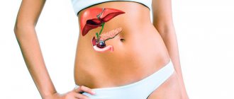
Causes
Cholecystitis occurs more often against the background of cholelithiasis, which develops due to the loss of contractility of the gallbladder. This organ serves as a reservoir where bile produced by the liver accumulates. Since bile contains a lot of cholesterol, when its density increases or when it stagnates, cholesterol crystals precipitate, forming stones. However, other factors can also cause acute cholecystitis:
- an infection that provokes inflammation due to bile retention and impaired drainage function;
- atrophy or sclerosis of the bladder walls;
- penetration of E. coli, staphylococci, streptococci and other bacteria;
- after pancreatic juice gets inside the walls of the gallbladder, which corrodes them;
- disruption of the outflow of bile as a result of elongation and bending of the gallbladder, the presence of stones;
- old age provokes vascular changes in the walls of the organ, which lead to cholecystitis;
- an acute attack often develops due to a food factor: spicy, fatty foods, overeating, which leads to spasm of the sphincter of Oddi.
Diagnosis of complications
All complications of cholecystitis require a comprehensive diagnosis, which combines the results of instrumental and physical studies.
The modern classification divides cholecystitis into catarrhal, phlegmonous and gangrenous, each of which requires specially selected treatment. The main diagnostic methods used to determine complications of acute cholecystitis are:
- studying the patient's medical history;
- palpation of the abdominal wall;
- Ultrasound of the abdominal organs;
- use of computed tomography;
- general and detailed blood test;
- biochemical examination of urine and blood.
How does the disease manifest itself?
The most obvious signs of cholecystitis in adults are dull pain and biliary colic, which is cramping in nature. The localization of pain is approximately the same - the right hypochondrium. Sometimes the pain can radiate to the right arm or back.
In clinical practice, the following symptoms of acute cholecystitis are noted:
- paroxysmal pain;
- chills;
- an increase in temperature, signaling the presence of infection;
- jaundice caused by stagnant bile;
- vomiting does not bring relief;
- pain radiates to the shoulder blade, neck, epigastric region.
The clinic and the treatment measures used depend on the stage of the disease. The classification of acute cholecystitis includes three stages according to the severity of the disease. Cholecystitis can be: catarrhal, phlegmonous, gangrenous.
- Catarrhal . The most harmless form of inflammation. Localization of pain in the epigastric region and right hypochondrium. Nausea, vomiting that does not bring relief. Biliary colic, which is cramping in nature. The clinic is erased, which makes early differential diagnosis difficult. diagnosis, additional research is required. Catarrhal cholecystitis is the easiest to cure.
- Phlegmonous . The clinic becomes more pronounced - the pain intensifies, the nature of the sensations is acute. Obvious signs of illness simplify the differential. diagnostics Pus mixed with bile accumulates in the organ cavity, and the temperature rises. Phlegmonous cholecystitis can easily become chronic, since changes in the tissues of the gallbladder at this stage are irreversible.
- Gangrenous. The most dangerous form of cholecystitis, since at this stage tissue death and gangrene begin. There is a risk of organ rupture with subsequent infection of the entire abdominal cavity. The clinic is deteriorating very quickly. The purulent contents of the gallbladder, spilled into the body cavity, can lead to death; this is a critical condition of the body; the patient requires emergency care and surgical intervention.
Diagnostic methods
Before treating cholecystitis, the doctor will prescribe a differential. diagnosis of the disease. Differential diagnosis is necessary so as not to confuse one disease with another that has similar external signs. This is especially important if there is a suspicion that surgery is necessary. Surgery may not be necessary at all if a similar clinical picture is observed in another disease that is easier to treat. Then conservative treatment is prescribed.
To determine cholecystitis, the following differential methods are used. diagnostics:
- blood biochemistry;
- ultrasound examination (the biliary system is checked for the presence of stones and pus);
- cholecystocholangiography is a specific diagnosis of acute cholecystitis.
You should contact a gastroenterologist for referrals. He will also take a history of cholecystitis. The doctor is interested in the full clinical picture of the disease: symptoms, date of the first attack of pain, features of your diet and lifestyle. The nature of the symptoms is important for determining the stage: sometimes the lion's share of examinations is missed due to a threat to life, and surgical intervention and removal of the gallbladder are immediately prescribed.
https://youtu.be/4xw4JtF9hnI
Treatment
Complicated cholecystitis must be treated so that the disease does not progress to the chronic stage without exacerbation. Treatment is carried out in a hospital setting, and the main emphasis is on the use of antibacterial therapy. Antibiotics suppress bacterial flora and are also an ideal tool for preventing bile infection. To relieve a person of pain during an attack of cholecystitis, doctors prescribe antispasmodics. If severe intoxication of the body has occurred, detoxification therapy is carried out.
Treatment of acute cholecystitis with non-surgical methods includes mandatory dietary requirements. On the first day after the attack, the patient is on complete fasting, and in the subsequent days he must adhere to a strict diet. Ursodeoxycholic or chenodeoxycholic acid can be used to dissolve stones. To maintain the normal functioning of other organs, hepatoprotectors and choleretic drugs are prescribed. This therapy for cholecystitis can be carried out for a long time - up to 2 years, but the possibility of relapse remains.
Complications of chronic cholecystitis
The chronic course of the inflammatory process is characterized by alternating periods of remission and exacerbation of the disease. There are not as many complications of chronic cholecystitis as in the acute form, but they all require surgical treatment. These include:
- reactive hepatitis;
- chronic duodenitis;
- pericholecystitis;
- reactive pancreatitis;
- chronic stagnation of bile;
- cholelithiasis;
- deformation of the affected organ;
- formation of adhesions and fistulas.
Reactive pancreatitis is an acute inflammatory process of an aseptic nature, which is localized in the pancreas. Characterized by rapid development of symptoms:
- heartburn;
- nausea and vomiting;
- severe girdle pain in the abdomen;
- increased gas formation;
- fever;
- signs of intoxication of the body.
Reactive hepatitis is a secondary diffuse liver injury. Expressed by moderate symptoms, such as:
- increased weakness;
- fast fatiguability;
- decreased or complete lack of appetite;
- heaviness and discomfort in the projection of the gallbladder, i.e. in the area under the right ribs;
- increase in liver size;
- jaundice.
Pericholecystitis is an inflammation that is localized in the peritoneum covering the gallbladder. Symptoms include severe pain in the area under the right ribs, as well as the appearance of a bitter taste in the mouth.
Gallstone disease is a pathological process that is accompanied by the formation of stones of various sizes in the gallbladder or bile ducts. The disease is accompanied by:
- biliary colic;
- intense pain in the projection of the affected organ;
- yellowness of the skin and mucous membranes.
Pancreatitis
Against the background of acute cholecystitis, pancreatitis is formed - inflammation of the pancreas. With a mild pathological process, the organ can still be restored; at advanced stages, stones appear in the organ, tissues die, become infected or encapsulate.
Acute pancreatitis is characterized by intense pain; when trying to lie on your back, the pain increases several times. The pain is worse after eating, consuming fatty foods, or drinking alcohol.
The patient suffers from nausea, uncontrollable vomiting, his body temperature rises, the skin and sclera become icteric. Signs of a serious digestive disorder may appear:
- bloating;
- heartburn;
- cyanosis on the body;
- hemorrhages in the navel area.
For diagnostic purposes, the patient is prescribed a blood and urine test. Instrumental analyzes help to identify structural changes: MRI, ultrasound, MSCT.
What to take for cholecystitis complicated by non-calculous or calculous pancreatitis? The pain syndrome is relieved by novocaine blockades and antispasmodics. If necessary, if stones appear, fluid accumulates, tissue dies, emergency surgery is performed.
Choleretic remedies and folk methods are powerless. In the postoperative period, a gentle diet is prescribed.
Prevention
Prevention consists of following a healthy diet, limiting the consumption of alcohol, large amounts of spicy, fatty foods. Physical activity is also encouraged - physical inactivity is one of the factors contributing to the stagnation of bile and the formation of stones.
It is better to eat according to the schedule, at least every 4 hours. Be sure to drink enough fluid (from one and a half liters), and do not overeat at night. Obesity, intestinal parasites (roundworms, lamblia), and severe stress are also unfavorable for the health of the gallbladder.
Home therapy and prevention
- You can relieve the symptoms of cholecystitis with the following medicine. You need to take a glass of horseradish roots and mix with 1.2 liters of water. The product is infused for a day, then filtered and heated. You need to take 2 tbsp. l. 20 minutes before meals. The course of treatment is 5 days.
- You need to take 500 ml of beet juice, mix with the same amount of aloe juice (preferably a young plant), carrot juice and black radish. The same amount of honey and vodka is added to the ingredients. The medicine is shaken, poured into a bottle, and the lid is tightly closed. Keep in a dark place for 2 weeks, take half an hour before meals.
- The herbal medicine contains celandine, peppermint, dandelion roots, tansy flowers, cinquefoil rhizomes (a large spoonful of the mixture per glass of water). The medicine is infused for 30 minutes, filtered, and taken half an hour before meals. The course of admission is 20 days. Herbal medicine neutralizes pain due to cholecystitis.
Prevention of the disease is proper nutrition, giving up bad habits, and leading an active lifestyle. If a person is hypodynamic, bile will stagnate in the body, and this will lead to diseases. You should eat often, but in small portions! After removal of the gallbladder, you need to adhere to diet 5a: it includes steamed dishes. For any problems with the liver, drinking plenty of fluids is recommended: you can drink 1 tbsp of water. Once every 2 hours.
Causes of complications
The consequences of acute and chronic cholecystitis are numerous. May be prerequisites for concomitant complicated diseases. Complications can be caused by:
- Gallbladder infection;
- Infection of the tissues of the biliary organ with waste products of harmful microorganisms;
- Inflammation in the peritoneum, fistula formation;
- Inflammation of the pancreas.
The stage of acute cholecystitis must be diagnosed in time, so as not to allow it to develop into a chronic stage. The examination is carried out by a doctor. Complications can occur quickly. In acute cholecystitis, the patient’s lack of action to prevent the disease leads to undesirable consequences. In chronic cholecystitis, non-compliance with the diet provokes the occurrence of the listed symptoms. Possible parasitic cholecystitis.
Treatment of obstructive cholecystitis
The main goals of treatment are to eliminate the cause of the pathology, alleviate symptoms, prevent the disease from progressing to a more complex form and prevent the development of complications. Depending on the research results, the treatment method is selected. In the initial stage, therapy is carried out with the help of medications, and when complicated forms are diagnosed, surgical intervention is prescribed.
Drug treatment
Conservative therapy involves taking the following medications:
- analgesics (Baralgin);
- antibiotics in the form of intramuscular injections (Erithomycin, Ceftriaxone);
- antimicrobial (Nitroxoline, Biseptol);
- anthelmintics (Vermox, Nemozol);
- antispasmodics (Papaverine, Buscopan);
- detoxification (Reamberin);
- antiemetics (Cerucal, Metoclopromide);
- immunomodulators (Imunofan);
- drugs whose action is aimed at dissolving gallstones (Chenofalk, Litofalk).
In rare cases, using only drug treatment, it is possible to solve the problem, so non-invasive operations (choledochostomy, nasobiliary drainage, cholecystomy, etc.) and radical surgery are prescribed to crush stones. Medicines are used in the postoperative period to prevent the development of inflammation and relapse.
Surgical intervention
It involves removing the gallbladder. There are 3 types of cholecystectomy:
- Laparoscopy. It is carried out using a laparoscope and surgical instruments, which are inserted through small punctures (up to 1 cm) in the abdominal wall. The laparoscope is equipped with a small camera and allows you to monitor the progress of the operation by displaying an image on the monitor. The advantage of laparoscopy is the absence of scars. The rehabilitation period in this case is reduced to 4 days, since the risk of complications is minimal.
- Laparotomy. The abdominal cavity is opened in the area of the gallbladder and the affected organ along with the stones is removed. A scar remains on the abdomen, the length of which is 20-25 cm. Laparotomy is used mainly when there is no possibility of a more gentle operation or complications are suspected.
- Percutaneous. Prescribed if there are contraindications to the above operations. In this case, a drainage system is introduced into the patient's affected area through a small incision on the anterior abdominal wall and stones, bile and pus are pumped out. In the postoperative period, it is necessary to follow the diet and all doctor’s recommendations for taking medications and maintaining a lifestyle, which will reduce the risk of complications and relapse.
The operation of choice for acute calculous cholecystitis is cholecystectomy: laparoscopic or open in combination with corrective interventions on the extrahepatic bile ducts (if necessary). The surgical approach for open cholecystectomy is an upper midline laparotomy or an oblique incision in the right hypochondrium.
The choice of method for removing the gallbladder depends on the experience of the surgeon, the technical equipment of the operating room, and the nature of inflammatory changes in the gallbladder and surrounding tissues. According to generalized literature data, laparoscopic cholecystectomy in patients with acute cholecystitis can be performed in 73.8-97.2%.
Contraindications to surgery
The following are considered contraindications:
- inflammatory infiltrate or abscess in the gallbladder area;
- expansion of the common bile duct (more than 8 mm);
- the thickness of the gallbladder wall is more than 1 cm;
- “shrinked” gallbladder;
- increased levels of bilirubin and amylase in the patient’s blood;
- pulmonary heart diseases in the stage of sub- and decompensation;
- uncorrectable disorders of the hemocoagulation system;
- widespread peritonitis;
- third trimester of pregnancy;
- biliodigestive and biliobiliary fistulas;
- portal hypertension syndrome;
- unclear anatomical situation in the area of the gallbladder neck and hepatoduodenal ligament.
However, due to the constant progress of endoscopic surgery, contraindications to laparoscopic cholecystectomy for acute calculous cholecystitis are constantly narrowing. But keep in mind that the consistent rule of modern gallbladder surgery is immediate conversion to open cholecystectomy in case of difficulties in manipulation in the subhepatic space, rough adhesions and extensive dense infiltration in the area of the gallbladder that is not amenable to blunt preparation, and unclear anatomical situation.
The results of cholecystectomy for acute calculous cholecystitis largely depend on the condition of the bile ducts. Therefore, all patients with this disease must undergo a comprehensive examination of the biliary tract before surgery: ultrasound, ERCG, intravenous cholecystocholangiography.
If it is not possible to carry out a comprehensive examination of patients in the preoperative period (emergency surgery), the condition of the biliary tract should be assessed during the operation. The most informative method for intraoperative assessment of the patency of the bile ducts during laparoscopic cholecystectomy is intraoperative cholangiography.
Indications for the operation
Indications for its implementation in patients with acute calculous cholecystitis are:
- history of jaundice with a common bile duct diameter of up to 7-8 mm according to ultrasound;
- dilation of the extrahepatic bile ducts more than 7-8 mm, discovered during surgery;
- the diameter of the cystic duct is more than 2 mm;
- small (up to 1-2 mm) stones in the gallbladder;
- changes in the anatomy of the extrahepatic bile ducts and difficulties in preparation in the area of Calot’s triangle;
- lack of visualization of the bile ducts due to swelling of the hepatoduodenal ligament;
- impossibility of using ERCG in the preoperative period if there are appropriate indications.
If a pathology of the biliary tract or major duodenal papilla is established, an appropriate corrective operation is performed. Thus, in patients with stenosis of the papilla of Vater and choledocholithiasis, endoscopic pagkillosphincterotomy is performed in the postoperative period. In the case of cicatricial stricture of the common bile duct, a transition to open surgery is performed with the formation of one of the options for biliodigestive anastomosis.
Diet
Treatment of any disease is not complete without following a diet, the purpose of which in case of obstructive cholecystitis is to reduce acidity and secreted bile. To do this, you need to exclude the following foods from your diet:
- flour, sweets;
- smoked, fried foods;
- sour berries and fruits;
- liver, lard, fatty meat;
- mushrooms, legumes, cabbage, garlic and onions;
- citrus;
- pickles, canned food, marinades;
- fresh bread;
- products with cocoa, coffee;
- alcohol and carbonated drinks.
Cooked dishes should not include spices, vinegar or hot seasonings. They should be served boiled, stewed or baked.
As first courses, preference should be given to pureed vegetable soups or broths prepared with lean meat (turkey, rabbit, chicken, veal).
For the second course, it is recommended to eat boiled porridge (buckwheat, wheat, oatmeal) with cutlets or meatballs.
From dairy products you can eat low-fat sour cream, kefir and cottage cheese. Fresh fruits and vegetables will be very useful.
The following food rules must be observed:
- the daily dose of vegetable fats should not exceed 80 g;
- food is consumed in small portions 5-6 r. in a day;
- You need to drink 2-2.5 liters of liquid per day;
- preference is given to protein foods;
- The calorie content of the daily diet is 2000 calories;
- limiting carbohydrate foods;
- dishes are served warm;
- minimal salt intake;
- ban on alcohol.
A return to your usual diet should be discussed with your doctor; the introduction of new foods is allowed no earlier than a month after the operation.
Folk remedies
Alternative medicine is used as an auxiliary treatment, since it is not able to solve the problem on its own. The choice of a folk remedy and its dosage must be discussed with a doctor in advance, since improper treatment can aggravate the course of the disease. Let's imagine the following recipes used for obstructive cholecystitis:
- a decoction of buckthorn bark, dandelion, barberry root and chicory. The ingredients are taken in the proportion 1:2:2:2. The crushed plants are poured with 250 ml of boiling water and left to simmer over low heat for 30 minutes. The product is consumed warm before bed, 1 glass;
- dandelion infusion. 40 g of dry plant roots are poured into 0.5 liters of water. Bring the mixture to a boil and remove from heat after 10 minutes. After cooling, the broth is filtered. Drink ½ glass before meals;
- decoction of mint and celandine. The ingredients are mixed in equal parts. Pour 2 tbsp. the resulting collection add 250 ml of boiling water and leave in a water bath. After 30 min. the product is filtered. You need to drink the decoction 2 times. per day (morning and evening) 250 ml;
- green tea should be consumed at least 1 cup per day hot;
- tea from wormwood and horsetail. Herbs taken in equal proportions are poured with boiling water. Take morning and evening;
- tincture of birch leaves. 3 tbsp. leaves, pour 0.5 liters of boiling water and leave in a thermos for 8 hours. Use 3 r. per day, dividing the resulting tincture into 3 parts.
Sources
https://okgastro.ru/zhelchnyy-puzyr/516-obturatsionnyj-kholetsistit https://zpechen.ru/bolezni/obturacionnyy-holecistit https://zhkt.expert/holetsistit/obutratsionnyy.html https://zhktinfo.ru /holecistit/ostryj-kalkuleznyj-holecistit/ https://med-books.info/66_farmakologiya_832/ostryiy-holetsistit-72961.html