Deep vein thrombosis (DVT) is a pathological condition characterized by the formation of a blood clot in the lumen of a vessel, impairing blood flow. Most often, such changes occur in the veins of the lower extremities; other vessels are rarely affected by this disease. The pathology is dangerous due to the possibility of a blood clot breaking off, which, spreading with the bloodstream, can cause blockage of smaller vessels, including the arteries of vital organs. This condition can cause disruption of blood supply and even death. Thus, deep vein thrombosis of the lower extremities can provoke pulmonary embolism.
Causes
Varicose veins are a serious risk factor for the development of acute phlebothrombosis.
So far, the exact reasons for the development of thrombosis have not been established. It is known that the following triad of factors can contribute to clogging of venous vessels:
- high blood clotting;
- slowing blood flow;
- damage to the venous wall.
Various diseases and conditions can contribute to the occurrence of the above factors:
- varicose veins;
- smoking;
- long bed rest;
- pregnancy and childbirth;
- C-section;
- taking certain medications that increase blood viscosity (for example, oral contraceptives);
- atherosclerosis;
- injuries of vascular walls (including those associated with frequent venous punctures);
- compound fractures;
- surgical interventions on joints and abdominal operations;
- obesity;
- infections;
- long trips or air flights;
- physical inactivity;
- alcohol abuse;
- malignant neoplasms;
- heart valve pathologies;
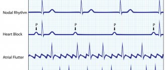
- arrhythmias;
- heart failure;
- old age.
What to do if you have a blood clot in your leg
As soon as the first signs of a blood clot in the leg are noticed, the patient should immediately be provided with bed rest, complete rest and call an ambulance. It is impossible to predict the future fate of the patient, because sometimes death occurs within a few minutes. To save the patient, the doctor makes a decision based on the current situation. The location of the thrombus is important. If a person is managed to be taken to the hospital, the following measures will be taken to save his life:
- surgery to remove the stuck clot;
- installation of a venous vena cava filter, which is capable of intercepting a detached thrombus;
- injection of a large amount of anticoagulant into the vessel (Heparin is often used).
Although deep vein thrombosis is a disaster, blood clot rupture in the lower extremities is rare. For this to happen, three reasons must come together:
- Inflammation of the veins . Even the initial degree of varicose veins signals pathology. The presence of spider veins on the legs is already a mild inflammatory process. He needs timely therapy so as not to wait for a blood clot to form.
- Slowing blood flow . Occurs with a sedentary lifestyle. Without the work of the muscular system, there will be no normal tone of the venous walls. You don't have to do strength training or running. You need to walk regularly and learn to breathe from your stomach to help your blood circulation.
- Increased blood clotting . As a result of improper nutrition, blood viscosity increases and clots form. To liquefy, it is necessary to include in the diet foods such as beets, garlic, oatmeal, eggs, sunflower seeds, and sour milk products. In addition to a special diet, you can additionally take medications (Aspirin).
Classification and stages
Depending on the location of the blockage of the venous vessel, experts distinguish the following types of thrombosis:
- subcutaneous – damage to the superficial veins occurs;
- deep – blockage develops in deep veins;
- ascending - in addition to blockage of the venous vessels, the patient develops additional pathologies in the lymphatic system (lymphostasis, limangoitis), it is very difficult and without treatment in 90% of cases causes death.
Depending on the type of blood clot, the following types of vein thrombosis are distinguished:
- parietal - the blood clot is located near the venous wall;
- occlusive - a thrombus completely blocks the lumen of the vein;
- floating - a blood clot is attached to the venous wall on one side only, and the other end is in motion and can come off;
- mixed - combines the characteristics of previous varieties.
During acute venous thrombosis, two stages are distinguished:
- compensation – no pronounced hemodynamic disturbances are observed, pain and discomfort occur periodically, sometimes the temperature rises for no apparent reason, the duration of this stage can vary from 24 hours to 1 month;
- decompensation – hemodynamic disturbances occur, pain becomes intense, swelling appears, skin color changes and limb mobility becomes difficult.
Prevention of thrombosis
There are simple rules that, if followed, will help avoid thrombosis even for people at risk:
- weight control;
- increasing physical activity, especially if the work involves staying in one position for a long time;
- performing a foot massage;
- walking for at least 30 minutes a day;
- before going to bed you should take a contrast shower;
- after the operation, begin to perform feasible physical exercises as soon as the doctor allows;
- quitting smoking and alcoholic beverages;
- when giving injections into a vein, it is important to alternate vessels so that the same one is not constantly injured;
- use of products made from compression knitwear in the presence of varicose veins;
- before or after surgery, especially in bedridden patients, the use of Warfarin, as well as Aspirin, to prevent blood clots in people prone to the disease.
Prevention of thrombosis involves following a diet; changing the diet is aimed at achieving optimal weight, strengthening the vascular wall, and thinning the blood. There are a number of recommendations developed by nutritionists and intended to prevent relapses of thrombosis:
- the volume of fluid drunk should be at least 2 liters per day;
- It is important to include fruits and vegetables in the diet;
- enrichment of food with antioxidants: vitamin C (rose hips, citrus fruits), E (beans, nuts);
- consumption of foods rich in flavonoids: rutin (spinach, buckwheat, raspberries);
- sufficient intake of copper (seafood);
- products that reduce blood viscosity (onions, garlic) should also be included in the diet;
- limiting animal fats in food, baked goods, sweets, coffee, alcohol.
Timely seeking medical help is of great importance. At the first signs of thrombotic lesions of the superficial veins, you should undergo examination. In the initial stage, the disease is easier to treat and the risk of developing all possible complications is reduced.
Symptoms
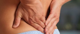
In the initial stages of development, vein thrombosis can manifest itself as minor and rare pain, sensations of fullness in the muscles and heaviness in the legs. Sometimes pain occurs in the lumbosacral region or lower abdomen on the side of the thrombosis. Usually these symptoms do not cause much concern, and the patient consults a doctor only at the beginning of the acute stage of the disease.
As thrombosis progresses, the following symptoms suddenly occur:
- sharp pain in the leg;
- stiffness and difficulty in movements;
- swelling (the leg increases in size, its soft tissues become denser);
- the skin on the leg turns blue (sometimes becomes blackish or pale milky);
- local increase in temperature (fever in the leg);
- noticeable visual expansion of the saphenous veins;
- low-grade fever, weakness, adynamia.
All of the above manifestations are caused by stagnation of venous blood below the site of thrombus formation. If the vessel is completely blocked, the patient's hemodynamics are disrupted, swelling increases and gangrene may develop. At all stages of the disease, when a blood clot breaks off, the development of pulmonary embolism, stroke, and thromboemolius of other organs is likely.
Acute venous thrombosis always develops suddenly, and its manifestations can become maximal from the first day of the disease. The nature and severity of symptoms depend on the following factors:
- location of the blocked vessel;
- diameter of the affected vein;
- type of blood clot;
- rate of blood clot formation;
- the likelihood of a reflex circulatory disorder in neighboring vessels;
- tissue sensitivity to hypoxia;
- severity of collateral circulation.
What does a blood clot look like?
To see a blood clot in a vein in the leg, you need to carefully examine and feel the lower limbs. If redness or hardening is found in the area of the arteries, pain on palpation, then we can talk about thrombophlebitis. Sometimes the temperature in a compacted area increases significantly. Often, symptoms of a blood clot in the leg are not present at all, but the neoplasm is visually visible through small swelling and bluish areas, as in the photo.
Ileofemoral venous thrombosis
This type of venous thrombosis is distinguished separately, since this disease is very severe, progresses quickly and is associated with a high risk of developing pulmonary embolism. Ileofemoral phlebothrombosis is caused by blockage of the iliofemoral segment and is characterized by intense pain, severe swelling of the entire leg and severe general condition of the patient. The affected limb turns blue and the saphenous veins dilate. When the outflow of blood completely stops, the patient quickly develops gangrene.
Rehabilitation
After the illness, the patient enters a period of postthrombophlebitic disease. In the absence of the necessary rehabilitation measures, there is a risk of developing symptoms of chronic venous insufficiency: edema, varicose veins, trophic disorders, as well as the likelihood of recurrent thrombosis. In this regard, it is important to constantly monitor the patient's condition.
A set of rehabilitation measures after deep vein thrombosis:
- course of coagulants;
- use of phlebotonics 1.5 months annually;
- maintaining moderate physical activity;
- eliminating bad habits;
- physiotherapy.
A set of rehabilitation measures allows you to maintain the patient’s quality of life and avoid relapse of the disease.
Thrombosis of the inferior vena cava
This type of thrombosis is also characterized by a severe course and a high risk of complications. When the inferior vena cava is blocked by thrombotic masses, both legs of the patient swell, and 80% of patients develop renal failure, accompanied by the appearance of blood in the urine. In cases of blockage of the liver segment, liver failure develops, complicated by Budd-Chiari syndrome. In the future, the patient may develop severe inferior vena cava syndrome.
Procedures to eliminate blood clots
There are certain procedures for successfully treating thrombosis:
- Clamping of the saphenous veins.
- A procedure that allows blood clots to be removed using a tube is thrombectomy.
- Stenting.
If it is necessary to remove a large vein, general anesthesia is used. After the operation, he is prescribed a special diet and rest.
Diagnostics
Duplex scanning of veins plays an important role in the diagnosis of acute venous thrombosis.
To detect venous thrombosis, the following studies are performed:
- Dopplerography and duplex scanning of veins - allows you to identify the location and extent of thrombosis, assesses the quality of blood flow and the condition of the venous walls;
- X-ray contrast venography – performed if the results of an ultrasound scan are questionable or if a blood clot is located above the groin;
- MR angiography – performed when the results of previous studies are questionable;
- impedance plethysmography - performed when thrombosis of veins above the knee is suspected, carried out using a cuff that is inflated with air and provides temporary occlusion of the veins to measure changes in their filling before and after deflation of the cuff;
- X-ray of the lungs – performed if pulmonary embolism is suspected;
- blood tests (coagulogram, D-dimer, culture for sterility) - are carried out to determine blood clotting indicators and identify infections.
Diagnostics. How to recognize thrombosis
Diagnosis of thrombosis consists of 3 main components:
- examining the patient and conducting functional tests;
- laboratory research;
- instrumental studies.
Inspection and functional tests
The doctor clarifies the presence and nature of the patient’s complaints, conducts an examination and identifies signs of thrombosis. Functional tests used:
- Lowenberg: a tonometer cuff is applied above the knee joint, pressure is inflated when it reaches 100 mmHg. The appearance of pain is typical; on the healthy leg there are no unpleasant sensations.
- Brodie-Troyanova-Trendelenburg: the patient should lie on his back, raising the affected leg, the doctor expels blood from the veins with massaging movements in the direction from the fingers upward, a tourniquet is applied to the middle part of the thigh, then the patient should stand up. Rapid filling of the vessels below the compression site indicates venous dysfunction.
- Hackenbruch test: the area where the great saphenous vein flows into the femoral vein is clamped, the patient must cough. The sensation of a push created when blood is reflected from a clot indicates pathology.
Laboratory research
A coagulogram is used for diagnosis. This type of study displays the state of the coagulation system.
Coagulogram indicators:
- clotting time;
- bleeding time;
- prothrombin index;
- prothrombin time;
- plasma fibrinogen.
Since thrombosis is often caused by infectious and oncological diseases, in some cases the patient is sent for examination to identify concomitant pathologies.
Instrumental diagnostics
To establish a diagnosis of vascular thrombosis, a wide range of instrumental studies are used, which make it possible to determine the presence of an organic lesion and its location. Among them:
- Angiography is an x-ray diagnostic method that allows you to assess the condition of blood vessels. A puncture is made in the vein and an X-ray contrast agent is injected through it. Then an x-ray or computed tomography is performed. Thanks to the injected substance, the features of blood flow through the affected vessel are visible on the x-ray.
- Doppler ultrasound allows you to both determine the presence of the disease and evaluate how effective the treatment is. The method is based on the reflection of ultrasonic waves from the blood and the difference in vibration frequency when passing through different tissues. This method has no contraindications and does not cause discomfort to the patient. Using Doppler ultrasound, you can assess the condition of the vessels, valve apparatus and determine the location of the pathological focus.
- Duplex scanning of veins is an informative research method that reflects the condition of blood vessels. Using scanning, you can not only determine the condition of the wall and valves, but also visualize the vessels along their entire length. General information about the condition of the veins can be obtained in 10 minutes, and a detailed study takes up to 50 minutes. Before duplex scanning begins, the doctor examines the patient and identifies areas that require special attention. It is necessary to supplement the study with functional tests.
- Rheovasography is a study that allows you to assess the state of blood flow, as well as the filling of the veins at rest and during exercise. Rheovasography is not one of the main methods, since obtaining reliable results is influenced by many factors: external (microclimate in the room for the procedure) and internal (presence of concomitant diseases).
Treatment
The main objectives in the treatment of acute venous thrombosis are aimed at restoring blood flow in the affected vessel, preventing the progression of edema, the development of gangrene of the limb, preventing pulmonary embolism and other complications. If damage to the deep veins is detected, the patient is urgently hospitalized in a specialized angiosurgical hospital or general surgery department. Patients with superficial vein thrombosis can be observed on an outpatient basis.
Depending on the clinical case, treatment can be conservative or surgical. If the risk of thromboembolism is high, bed rest is prescribed. All patients with venous thrombosis are recommended to wear compression stockings (the tightness of the stockings should be determined by the attending physician) and follow a diet.
All patients with venous thrombosis are advised to take anticoagulants. These medications are the most effective at preventing progression of the disease. Patients are sequentially prescribed direct (nadroparin, dalteparin, enoxaparin and other low molecular weight and unfractionated heparins) and indirect (phenyline, acenocoumarol, warfarin, ethyl biscoumacetate) anticoagulants. When choosing a drug, contraindications to its use must be taken into account.
To improve blood circulation and thin the blood, patients with venous thrombosis are prescribed:
- clopidogrel;
- rheopolyglucin;
- ticlopedin;
- pentoxifylline;
- phleboactive agents: Troxevasin, Escusan, Detralex, etc.
To eliminate pain and reduce platelet aggregation, it is recommended to take non-steroidal anti-inflammatory drugs:
- diclofenac;
- ketoprofen;
- ibuprofen, etc.
If infections are detected or there is a high risk of developing (for example, AIDS, diabetes, etc.), antibiotic therapy is indicated for the patient.
To eliminate venous thrombosis, hirudotherapy may be recommended as an addition to treatment. The saliva of medicinal leeches contains substances that help eliminate inflammation of the venous walls, destroy blood clots and prevent the formation of new blood clots. When prescribing hirudotherapy, the doctor must take into account possible contraindications to this method of treatment. The number of sessions is determined by the clinical case.
Sometimes conservative measures are not enough to eliminate thrombosis and prevent its complications, and then patients undergo surgical operations, which can be performed either planned or urgently. The following methods can be used for this:
- installation of a vena cava filter - a special metal device in the form of an umbrella is installed in the lumen of the inferior vena cava temporarily or permanently, the operation is performed endovascularly (through the lumen of a venous vessel) and is carried out to prevent thromboembolism (for example, with floating blood clots);
- thrombolysis - intervention is performed when it is necessary to remove large blood clots (prescribed infrequently due to the high risk of bleeding), carried out using a special catheter into which a drug that destroys the clot is injected;
- venous angioplasty - a balloon is inserted into the area of narrowing of the vessel, which, after inflating, expands its lumen; a stent is installed in the place of narrowing of the vein;
- venous bypass - during the intervention, incisions are made outside the narrowed part of the venous vessel, to which a venous graft (taken from the patient’s thigh or synthetic) is sutured, ensuring blood flow in the area affected by thrombosis;
- thrombectomy - the operation is performed classically or endovascularly; under angiography control, the doctor identifies the location of the blood clot, makes a small incision and removes the blood clot using a special catheter.
After the operation, the patient is prescribed drug therapy.
Diagnosis of thrombophlebitis
To date, 2 effective methods for diagnosing the condition of blood vessels have been identified. Specialists rely on the result of a duplex scan and a blood test for D-dimer.
When the description of the duplex study is not credible, then radiopaque venography will be performed. Also, this technique is used if the patient is suspected of having thrombosis above the inguinal fold.
In order for the location of the thrombus to be accurately determined in the image, contrast will be administered through a vein . This simple method is more informative than ultrasound. Special cases require MRI or CT angiography .
Due to the fact that the manifestation of the disease may be similar to other pathological processes, additional examination is suggested. For example, only the differential diagnostic method will help exclude the presence of a Baker's cyst, Buerger's disease, or acute embolism.
Often, soreness in the calf muscles occurs due to neurological processes in the sciatic nerve. Such pain haunts the patient constantly; problems with sensitivity and slight tissue atrophy can be noted.
With tumor processes, arthritis, lymphostasis, myalgia and myositis, a similar clinical picture may be observed. For this reason, only a comprehensive examination will help find the true cause of inflammatory processes and blockage of veins.
Diet
To improve the rheological properties of blood, a patient with venous thrombosis should drink up to 2.5 liters of water per day.
All patients with venous thrombosis are advised to follow a special diet and take a sufficient amount of fluid (up to 2.5 liters per day). Proper nutrition can improve the rheological properties of blood, reduce swelling and improve the condition of vascular walls.
The following foods that increase blood clotting should be excluded from the menu:
- foods high in vitamin K and C: green vegetables and fruits, spinach, nettle, sorrel, walnuts, rose hips, currants, citrus fruits, bell peppers, chokeberries, etc.;
- fatty meats;
- sausages;
- canned meat;
- fatty dairy products;
- mayonnaise;
- spicy, fried, smoked, sweet and salty dishes;
- confectionery products with margarine, butter and cream;
- pastry products;
- coffee;
- alcoholic drinks.
Products rich in polyunsaturated fatty acids (Omega 3 and Omega 6) and vitamin E have a beneficial effect on the condition of blood vessels and blood. In addition, the diet should include foods that prevent flatulence and constipation, in which blood stagnation in the lower part of the body increases.
For vein thrombosis, the following products should be included in the daily menu:
- fish fat;
- fatty fish: salmon, pike perch, mackerel, cod;
- lean meat (1-2 times a week);
- seafood: squid, mussels, crabs;
- low-fat fermented milk products;
- cashew nuts;
- cereals;
- legumes;
- vegetable oils: olive, cedar, flaxseed, corn, soybean, wheat germ, etc.
- asparagus;
- corn;
- onion;
- garlic;
- horseradish;
- pepper;
- pumpkin and sunflower seeds;
- melons and watermelons;
- foods rich in fiber: cabbage, carrots, etc.
For vein thrombosis, it is recommended to prepare dishes by boiling or steaming.
Reasons for the development of pathology
Thrombosis of the lower extremities can occur for various reasons:
- A blood clot may appear due to the presence of infectious diseases, pustular formations, and the spread of pathogens. This is due to the fact that bacteria destroy the integrity of blood vessels and negatively affect blood circulation.
- Mechanical injuries of the lower extremities, which lead to the destruction of blood vessels.
- Blood clotting too quickly.
- Diseases of the cardiovascular system.
- Genetic predisposition to the formation of this disease.
- Passive lifestyle. Sedentary work, minimal movement during rest periods.
- Previous strokes or heart attacks.
- Hormonal disbalance. Taking hormonal drugs.
- Elderly age.
- Slow blood circulation.
Once the cause of the blood clot has been identified, it must be eliminated before proceeding with the main treatment.
How to treat the disease
To cure venous thrombosis, you need to act comprehensively and follow all the doctor’s recommendations, without giving preference to self-medication. In most cases, the patient is prescribed bed rest with a sick leave certificate. The specialist also decides whether the patient should stay in a hospital, or whether home treatment will be sufficient.
Not everyone knows which doctor treats thrombosis and where to go to treat such a disease. You can make an appointment with a vascular surgeon or a more specialized specialist - phlebologist.
At the initial stages of the development of the disease, the pathology can be managed with the help of drug treatment. The doctor may prescribe indirect anticoagulants, which prevent further development of the blood clot by stimulating the release of certain enzymes.
To directly dissolve blood clots in the vessels, thrombolytics or enzyme preparations are prescribed. In addition to this, hemorheologically active agents may be prescribed that restore normal circulation and thin the blood. To eliminate inflammation and pain in the affected vein, nonspecific anti-inflammatory drugs are used.
In addition to drug therapy, it is often necessary to bandage the affected area to stimulate normal blood circulation. In addition, in consultation with your doctor, you can use traditional medicine recipes aimed at eliminating the symptoms of thrombosis.
A specialized diet may also be useful, which will allow you to lose excess weight, thereby reducing the load on your legs, as well as strengthen the walls of blood vessels and reduce blood viscosity.
In cases where conservative treatment remains powerless and the disease progresses, surgical intervention may be prescribed. There are several operations that can be used in this case. Some of them are aimed at installing additional devices that can protect the patient from possible risks and complications, while the main effect will subsequently be achieved by conservative therapy. Other operations involve removing a blood clot or the entire affected area of the vein, if the cause of the pathology is varicose veins.
Forecast
Depends on the type and position of the blood clot. Occlusive thrombosis is not dangerous, since the thrombus is tightly attached and completely blocks the blood flow. A parietal thrombus is also not dangerous, but it can continue to grow and become floating. The probability of a floating thrombus breaking off is very high.
Proximal vein thrombosis in approximately half of cases is accompanied by pulmonary embolism, often asymptomatic . After a few months, in most patients, venous blood flow is restored, but weakness of the valve apparatus develops.
In distal veins, thrombi often resolve on their own, but thrombosis can spread to the proximal veins.
Within several years after the disease, more than half of patients develop postthrombotic syndrome , and in the absence or ineffectiveness of treatment, asymptomatic PE. Chronic venous insufficiency worsens the quality of life and leads to disability.
Forms and types
Thrombosis is distinguished by etiology, place of formation and types of blood clots:
- Proximal thrombosis is localized in the popliteal or femoral vein and causes pain in the leg, swelling and tenderness when palpating over the affected veins. But sometimes its first manifestation is pulmonary embolism.
- Distal thrombosis affects the calf veins. There is moderate pain and tenderness in the lower leg, but sometimes there are no symptoms. There is usually no swelling.
- Ileofemoral thrombosis occurs in the iliac and femoral vessels. Pain is felt along the inner thigh, calves, and groin. The leg swells noticeably from the foot to the groin. Palpation in the projection of the main veins of the thigh and groin is painful.
Thrombi often form in the femoral vein, somewhat less frequently in the popliteal and gastrocnemius, and in the inferior vena cava - relatively rarely.
By origin, thrombosis can be stagnant (with varicose veins, external compression of blood vessels, internal obstructions to blood flow), inflammatory and associated with thrombophilia.
There are also occlusive, parietal and floating thrombosis . A floating thrombus is diagnosed in approximately 10% of cases. It is attached to the venous wall at only one end and seems to dangle in its lumen, so the likelihood of tearing off is very high.
Preventive measures versus treatment and disease
Preventive measures are suggested by the following actions:
- the fastest possible restoration of activity after operations, with gymnastics and exercise therapy;
- wearing compression stockings, using elastic bandages;
- taking anticoagulants;
- prevention of everything listed in the causes and factors of the development of the disease.
For air transport passengers, it is worth thinking about the fact that implementation of the proposed measures is becoming extremely necessary. In addition, it is recommended that: clothes should be loose, drink plenty of water, do exercises and knead the legs and muscle areas with massage movements.
The acute form of DVT, affecting the lower extremities, is a real threat to human health and life. If symptoms with severe pain, swelling, and cyanosis appear, it is simply dangerous to delay treatment. Only with timely hospital treatment under the supervision of health workers can PE be prevented. In addition to medications, the effectiveness of prevention is achieved through continuous participation in active life, especially with daily exercises.
About the causes and factors of the possible development of acute TVH
There are a number of primary causes, including hereditary blood clotting disorders. Secondary processes can arise solely due to the influence of external factors. R. Virchow managed to understand this issue of thrombus formation. The reasons were then given, expressed:
- irritation of the vessel;
- coagulation disorder;
- change in blood flow.
These processes are marked in medicine by Virchow's triad.
Several risk groups should be noted regarding DVT with PE:
- Low risk includes those who are under 40 years of age and have undergone minor surgical procedures.
- The moderate risk category is represented by people aged forty...sixty years who have undergone any surgery.
- The group at very high risk is older people aged 60 and above, after undergoing leg surgery, hip fracture or other injuries with complications.
So, there is a close relationship between acute thrombosis and previous fractures that violated the integrity of the tubular bone of the leg, as well as serious surgery on the joint or in the peritoneal area. And the problem can develop over time in the early and late postoperative periods. Moreover, with a longer duration of surgical intervention in the area of the hip or knee joints, the risk of diagnosing acute venous thrombosis is 30...50%. In the case of a minor intervention lasting no more than 30 minutes, the risk level does not exceed 10%.
Among the main factors in the formation of DVT there are reasons due to:
- prolonged immobility with a post-stroke state, bed rest, constant physical inactivity, as well as frequent air travel;
- obesity with altered fibrinolytic activity;
- general anesthesia, supplemented with muscle relaxants, turning off the muscle pump;
- in women at different stages of pregnancy followed by the postpartum period, which is due to altered hemostasis and compression of blood vessels as the fetus grows;
- taking certain medications, estrogen-based contraceptives;
- oncology with tumor processes, DVT, Trousseau syndrome;
- thrombophilia caused by impaired hemostasis;
- viral infection;
- age-related disruptions in the body with weakening of blood circulation, weakening of the vein wall, and increased blood viscosity;
- former DVT of young years.
It should be noted that there is a congenital form of a genetic defect, which gives rise to thrombosis. This is possible with Leiden mutation, deficiency of antithrombin III with proteins S and C.
Causes and risk factors
Typical causes of an intravascular clot are explained by 3 main factors (Virchow’s triad):
- traumatic damage to the wall of an artery or vein of any origin (external influence, inflammatory process, action of atherosclerotic plaque);
- slowing down blood flow;
- imbalance between the processes of thrombogenesis (blood clotting) and fibrinolysis (clot resorption).
Thrombosis risk factors are of great importance for the implementation of intravascular problems:
- atherosclerosis;
- varicose veins;
- arterial hypertension;
- diabetes;
- obesity;
- prolonged fasting and dehydration;
- bearing a fetus;
- long-term uncontrolled use of hormonal drugs and medications;
- oncological pathology;
- acute or chronic infection with penetration of microbes into the bloodstream;
- any surgical intervention;
- long history of smoking.
A combination of several conditions and diseases is especially dangerous (varicose veins and smoking, high blood pressure due to obesity and diabetes, surgery and atherosclerosis, as the main risk of atherothrombosis).
Symptoms and diagnosis
Any person who is predisposed to developing this disease needs to know how thrombosis manifests itself. This will allow you to recognize symptoms in the early stages and consult a doctor in time to begin timely treatment. To reduce risks, you need to be regularly examined by a doctor.
If the symptoms of vein thrombosis are still pronounced, the patient may observe the following changes in his condition:
- pain throughout the entire pathologically dilated vein, which intensifies with any, even minor, loads on the affected area;
- heaviness and fatigue of a body part with a blood clot;
- swelling and swelling;
- redness of the skin, which is usually localized over the area in which the blood clot has formed;
- increased skin sensitivity;
- causeless convulsions.
In most cases, a blood clot forms in a dilated vein, but there may be exceptions when the pathology develops in an apparently healthy person. Other noticeable signs of the development of the disease include a thickening felt throughout the vein, the appearance of a visible venous network, as well as the unchanged state of the vein when pressing on it. It should also be taken into account that in different patients, pathology may manifest itself differently depending on the characteristics of the body and the presence of other diseases.
Venous thrombosis: symptoms, causes, treatment
Thrombosis is a disease in which large blood clots form, blocking blood vessels and interfering with normal blood circulation in the body. This pathology is one of the complications of varicose veins, which explains the fact that a blood clot most often occurs in the lower extremities. Venous thrombosis can and should be treated, so it is important to immediately recognize the symptoms of the pathology and contact the right doctor who will select the appropriate treatment methods.
Folk remedies
When everything is clear with drug treatment and medical procedures, you need to learn how to treat blood clots on the legs at home. To do this, you can use folk remedies:
- Onions mixed with honey.
- Infusions based on white acacia, hop cones and verbena.
- Raw ginger.
- Freshly squeezed cherry juice, fruit drink.
- Decoctions from carrot tops.
- Compresses based on bodyaga.
- Dried nettle decoctions. Take twice daily.
Thrombosis: types, risk factors, treatment and prevention of vascular blockage
One of the common causes of impaired blood flow through the vessels is thrombosis. The formation of an intravascular blood clot is caused by a combination of unfavorable factors, and the pathogenesis of thrombosis depends on the severity of pathological changes in the blood. The type of vessel in which the occlusion occurred is of great importance for the prognosis of the disease. At the examination stage, it is important to promptly identify signs of a blood clot in the body, and the effectiveness of treatment largely depends on the stage of thrombosis.
Classification of thrombosis
According to the attachment of the plaque to the wall, thrombosis can be:
- parietal - the thrombus is attached to the wall, does not interfere with blood flow, is less dangerous;
- occlusive - the vein is completely blocked;
- mixed - the blood clot moves up and down the vein;
- floating - a thrombus that runs along the wall, its tip floats in the lumen of the vein, and is easily able to break off and enter small vessels, clogging them;
- multifocal thrombosis - blood clots appear in any place.
There are also certain types of thrombosis in the lower extremities themselves: damage to the superficial and deep veins, thrombosis of the arteries of the legs, ileofemoral thrombosis of the legs.
How does a blood clot form in blood vessels?
The formation of blood clots occurs due to three mechanisms that depend on environmental factors. It is rare that only one factor is responsible for vascular occlusion - usually several.
Changes in the inner wall of the vessel
Repairing or sealing damage inside blood vessels is the task of the coagulation system. However, in some cases, an “extra” clot forms. Possible causes of damage:
- Injuries to the walls of blood vessels during surgery.
- Metabolic disorders.
- Hormonal disorders.
- Previous surgery (joint replacement).
- Age-related changes.
- Coronary artery bypass grafting (surgery to replace damaged vessels).
- Obesity, disability, infections.
- Smoking: Inhaled carbon monoxide damages the walls of blood vessels, which are further narrowed by nicotine.
Blood flow is too slow
If blood flows too slowly, blood clots form. Often the body is able to dissolve it. If this does not happen, a large clot will form that will block the blood vessels. Various circumstances lead to slow blood flow. Among the common ones are:
- Varicose veins: increases the likelihood of thrombosis several times.
- Prolonged physical inactivity, such as while sleeping, after surgery, or while wearing a cast. However, even shorter phases with limited movement, such as during a long flight, can increase the risk of thrombosis. The reason is that blood flow in the legs is stimulated by the activity of the calf muscles. If leg muscles remain inactive for a long time, blood flow slows, causing clots to form quickly.
- External pressure, for example: due to a tumor or during pregnancy. Here, the enlarged uterus may press more and more against the veins in the abdomen, especially in the last trimester of pregnancy.
- Heart problems. Cardiac arrest or atrial fibrillation can cause blood clots to build up and slow it down.
Changes in blood clotting
Typically, there is a balance in the blood between coagulation and anticoagulant factors. Exogenous and endogenous factors influence this balance, so the blood coagulates faster. Important factors include:
- Congenital bleeding disorder in infants.
- DIC syndrome.
- Birth of a child.
- Cancer. Tumors produce substances that stimulate blood clotting in the body. Tumor therapy is often associated with an increased risk of thrombosis.
- Changes in hormonal balance, as this affects blood clotting and the elasticity of vessel walls.
- Age. Associated with hormonal changes. There is a decrease in physical activity,
- Infections or inflammations because related processes in the body increase the tendency of blood to clot. Illnesses such as influenza or pneumonia are often associated with prolonged bed rest and physical inactivity.
- Fever contributes to a lack of fluid.
- Overweight.
- An acute lack of fluid leads to “liquefaction” of the blood.
What contributes to thrombosis?
- long-term immobility (hospital stay, serious illness, long travel);
- severe injuries;
- operations (especially orthopedic);
- use of certain medications (hormonal, antitumor);
- previous episodes of venous thromboembolism;
- obesity;
- heart failure;
- heavy smoking;
- pregnancy and postpartum period.
The risk of thrombosis also increases with certain diseases, such as cancer, heart disease, inflammatory bowel disease, rheumatologic and hematologic diseases. In addition, some people are genetically burdened with a tendency to thrombosis or congenital thrombophilia. In these people, venous thrombosis may occur repeatedly, and it may also affect relatives.
Thrombosis usually affects the deep veins of the lower extremities, but also occurs in the veins of the pelvis and upper extremities.
Basic methods for diagnosing pathology
At the first manifestations of acute thrombosis, you should immediately contact a medical institution to consult a doctor and conduct an examination of the body. Examination of the patient allows you to confirm or refute the doctor’s assumption about the presence of pathology in the body.
Diagnosis of deep vein thrombosis can be made using non-invasive and invasive methods.
Most often, radionuclide scanning, phlebography, thromboelastography, duplex ultrasound scanning, abdominal x-ray, blood test for the presence of leukocytes, laparoscopy and thermogram are used during the examination.
Even without special equipment, the location of blood clots and the degree of development of the disease can be determined using special tests. These types of studies are samples
Latest information: Is it possible to swim with varicose veins and do water aerobics in the pool?
- Homans;
- marching test;
- Pratt;
- Lowenberg's sign.
Thrombosis can be determined even at home. True, this kind of information cannot be considered one hundred percent probability. Most often used for preliminary diagnosis.
Under the influence of what reasons does it occur?
The disease is caused by many factors. There are two groups of reasons, which may be:
- Primary, depending on a hereditary predisposition to disruptions in the process of blood clotting (that is, a person from birth has weak vein walls, weak valve function, varicose veins).
- Secondary, appearing under the influence of external factors.
The reasons of the second group include:
In some situations, clots form when there is no vascular damage
- injury and fracture of the leg, surgery on the lower extremities. In these cases, bleeding occurs and a high level of thromboplastin is formed, which changes the structure of the blood, leading to the formation of clots;
- paresis or paralysis of the legs. In such conditions, the muscles atrophy, and the blood flows slower, and platelets stick together;
- excess body weight leads to the production of leptin, which provokes blood clots;
- oncological diseases cause increased blood clotting;
- disturbances at the hormonal level are fraught with disturbances in the blood flow and its thickening;
- various septic processes occurring as a result of burns, blood poisoning, tuberculosis, the formation of ulcers, cause the appearance of toxins, due to which thrombotic substances increase.
In addition, the risk of thrombosis increases in the presence of the following factors:
As a result, oxygen access to this area is limited, and metabolic products accumulate here
- sedentary lifestyle;
- excessive physical activity;
- pregnancy;
- diabetes mellitus;
- disturbances in cardiac activity;
- old age;
- smoking;
- the use of medications that promote blood thickening;
- long-term treatment with bed rest.
Prognosis and consequences of the disease
A disease such as thrombosis leaves an imprint on the rest of a person’s life. The patient must constantly follow a diet, do special exercises and be constantly monitored by specialists. You should not rely on traditional medicine; following the doctor’s recommendations will bring much more benefit.
In many cases, the patient must take blood thinning medications.
Prognosis in the case of this disease also depends on where the blood clot, which is a source of threat to the patient’s health, was found. For example, with occlusive and parietal thrombus, periodic examinations in the clinic are sufficient. These are the least dangerous types of blood clots, but monitoring with a doctor will help ensure that the clot has not started migrating. Thrombosis in the proximal and distal veins is also considered dangerous. The process of a full rehabilitation cycle takes a long time in this case, but even if the recommendations are fully followed, it is ineffective.
An advanced stage of vascular blockage will usually require ongoing therapeutic measures.
Dealing with thrombosis is difficult, but efforts can be made to prevent the disease. Focus on physical activity, walk outside, eat vitamin K and C, wear comfortable, loose clothing, and drink plenty of fluids. This is most relevant for people who constantly feel tired after a working day. Swelling of the upper limb in the morning or general malaise and weakness are also good reasons to think about visiting a doctor.
Development of the disease in intestinal form
Mesenteric or intestinal thrombosis is classified as an arterial form of the disease. Develops due to blockage of the mesenteric artery.
Older people are most often affected. Pathology can equally likely affect the body of both women and men.
Latest information: What is contraindicated for varicose veins on the legs?
The main reasons that cause thrombosis of this subtype are considered to be heart disease that develops against the background of atherosclerosis; in addition, the occurrence of the disorder is facilitated by myocardial infarction and endocarditis.
Atherosclerosis is one of the most dangerous diseases leading to the activation of blood clot formation processes. The lack of adequate and timely treatment leads to the development of thromboembolism, which can lead not only to disturbances in the functioning of the circulatory system, but also to death.
Intestinal thrombosis is recognized by some characteristic signs of pathology:
- severe abdominal pain;
- rapid heartbeat;
- vomiting and nausea;
- diarrhea;
- constant increase in body temperature.
Only an experienced doctor will be able to make the correct diagnosis, since over time these symptoms of thrombosis may be similar to intestinal obstruction and it can easily be confused with other diseases.
Prediction of consequences in acute deep vein thrombosis
Predictions are made based on the location of the blood clot. The presence of occlusive thrombosis is less dangerous, because the body of the thrombus is closely connected to the walls of the vessel, blocking it completely. You should also not expect consequences from a parietal thrombus. However, there remains a possibility that the latter will take the form of a floating one, which has a high level of risk of separation. Thrombophlebitis with all the ensuing consequences is also possible.
In the case of thrombosis in the proximal vein, pulmonary embolism develops without symptoms. Only after months will the blood flow be restored. But the valve apparatus suffers, seriously weakening. With thrombosis of the distal vein, resorption of the stagnation is possible. But in the future, a problem in the proximal vein may develop. Over the years, the disease can reveal itself in such a way that it acquires a state of chronic failure. This leads to a deterioration in the quality of life with subsequent disability.
Diagnostic and treatment methods
Various techniques are used in the diagnosis of thrombosis, the simplest of which are functional tests. They differ depending on the area being studied. Thus, to determine thrombosis of the superficial veins of the leg, the Hackenbruch test is used. It is performed by pressing on the point where the great saphenous vein flows into the femoral vein, and the patient must cough. With thrombosis, a reverse wave of blood appears, which is pushed away from the clot.
REFERENCE! Functional tests are a method that can be used for preliminary self-diagnosis of thrombosis. However, a more complete picture can only be obtained based on instrumental methods.
The most informative methods for studying blood vessels are based on the use of contrast agents. On ultrasound (Dopplerography of blood vessels), you can see a round formation that impedes the movement of blood. On the one hand, there is excessive filling of the vessel with blood.
Treatment is prescribed individually, depending on the location of the blood clot and its size. The treatment regimen may include the following methods:
- direct coagulants - Heparin (thin the blood);
- indirect coagulants - prevent the formation of prothrombin;
- hemorheologically active drugs (Reosorbilact) - improve blood movement through the vessels;
- non-steroidal anti-inflammatory drugs;
- surgical removal of the thrombus.
The best way to prevent thrombosis is proper nutrition and moderate physical activity. With age, the walls of blood vessels become less elastic, so it is worth periodically undergoing routine examinations. The prognosis depends on many factors, including the timeliness of medical care.
Signs of thrombosis
Symptoms of vein thrombosis depend on the location of the clot. For example, if the upper and lower extremities are affected, physical activity decreases, and if the pulmonary artery is blocked, doctors do not rule out signs of hypotension, loss of consciousness, or coma.
Limbs
The first signs of thrombosis of the veins of the lower extremities appear with excessive physical exertion and disappear on their own after a short rest. Later, the disease manifests itself in the resting stage. Since the initial signs of thrombosis are reversible and can be successfully treated, the pathological process should not be started. Otherwise, the acute form of the disease is modified into a chronic, already incurable, prone to relapse. Symptoms:
Deep vein thrombosis of the extremities
Arterial thrombosis of the arms and legs
The formation of a blood clot in the lumen of a vessel disrupts local blood circulation. Women are more often susceptible to this disease than men. This is due to high levels of female hormones, pregnancy, and other factors. At first, the pathological process is asymptomatic, and its diagnosis is difficult. In 80% of cases, vein thrombosis can cause pulmonary embolism (a fatal complication). Symptoms:
Femoral and iliac
Brachial and subclavian
Central retinal vein
Pulmonary artery
If there is a disruption in the flow of blood from the veins of the legs and pelvis, blockage of the pulmonary artery is observed. The consequences of such a pathological process depend on the size and number of clots received, the reaction of the lungs, and the activity of the body’s thrombolytic system. When a small blood clot enters the pulmonary artery, there are no symptoms at all. Medium and large blood clots provoke the following conditions:
- cyanosis and pallor of the skin;
- enlarged veins in the neck;
- sharp pain in the chest, reminiscent of a heart attack;
- cough with blood in the sputum;
- difficult, rapid breathing;
- decreased blood pressure;
- general weakness;
- wheezing;
- confusion, loss of consciousness.
Brain vessels
Blockage of cerebral vessels impairs cerebral circulation, which is fraught with hypoxia, the formation of foci of necrosis of the meninges with the development of ischemic stroke. Atherosclerotic plaques are most often localized in the carotid, vertebral or intracerebral arteries. Symptoms of cerebral thrombosis:
- facial asymmetry;
- more frequent attacks of headaches;
- decreased visual acuity with episodes of blindness;
- speech disorder;
- nausea;
- dizziness;
- impaired coordination of movements;
- myalgia, muscle cramps;
- memory loss;
- loss of skin sensitivity;
- stroke-like symptoms.
The symptoms of cavernous sinus thrombosis are especially dangerous. The disease, in the absence of timely treatment, leads to complete blindness, stroke, and dysfunction of the pineal gland. The prognosis is unfavorable, the development of a coma is possible.
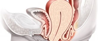
Hemorrhoids
The pathology most often progresses in women after pregnancy and childbirth. The formation of blood clots in the veins of the hemorrhoid is considered a dangerous complication of hemorrhoids, and the likelihood of rectal bleeding is high. Symptoms of the pathological process:
- acute pain and itching of the anus;
- burning and swelling in the anus;
- injuries of hemorrhoids with subsequent infection;
- high body temperature, fever;
- protrusion of hemorrhoids;
- rectal bleeding.
Danger and complications
The main danger is pulmonary embolism. Complete blockage causes instant death, partial blockage causes heart failure .
As a cause of sudden death, pulmonary embolism ranks third after coronary artery disease and stroke. After thrombosis of the proximal veins, chronic venous insufficiency often develops. Venous pressure increases when moving, valves do not work well. Venous stagnation is manifested by trophic disorders : hyperpigmentation, dermatitis, induration, and in severe cases, trophic ulcers. Venous insufficiency can be suspected by swelling of the legs, depending on the position of the body.
More than half of patients develop postthrombotic disease. Almost a third of such patients become disabled .
Find out more about the disease from the video: