Every visit to the clinic causes us some anxiety, or, more simply, doubt. We do not know how to behave during the examination, what food can be taken before the procedure, and what actions are generally contraindicated. So, MRI is no exception. Let's take a closer look at some of the steps and nuances before an MRI. We will definitely assure you that the examination is completely harmless if you follow all the doctor’s recommendations and instructions. Carefully study the questionnaires that the clinic provides to familiarize yourself with your characteristic characteristics of the body. Specialists will definitely tell you how to prepare for an MRI of the pelvis.
Is it possible to eat food before the examination?
The internal organs of the small pelvis include the digestive tract. Therefore, eating, or rather the question of time, how many hours before you can eat food, is urgent for many patients. Sometimes, due to shyness, we do not ask additional questions to the doctor, which are important in the examination. This is how food intake depends on the type of examination. What can you eat before an MRI of the pelvis and is it possible to eat before an MRI at all? During your consultation with your doctor, you will be told in detail what you should not eat before an MRI and what foods cause excess gas. These products include: carbonated water, cabbage, kefir, fermented baked milk, sour cream, etc. According to medical statistics, a good way to prepare the body is a slag-free diet before an MRI of the pelvis.
Are there alternative diagnostic methods?
Currently, diagnosing the human body involves choosing one of the methods: ultrasound, computed tomography, x-ray or MRI. It is worth noting that magnetic resonance imaging has many advantages:
- The doctor gets the opportunity to study the required organ in as much detail as possible;
- The pictures have good clarity;
- The ability to notice even the slightest deviations in the structure of tissues and organs;
- Maximum information content regarding the diagnosis of cancer, etc.
Perhaps only hysteroscopy can compare with MRI, but its use requires penetration into the tissue through surgery. In comparison with hysteroscopy and other methods, Magnetic resonance imaging is a non-traumatic and absolutely safe procedure, which is carried out in a fairly short time and is popular all over the world.
What features of the female body should be taken into account
Every woman, starting at about 12 years of age, begins puberty. Every month a girl, and subsequently a woman, undergoes a menstrual cycle. And accordingly, she wonders whether it is possible to do an MRI during menstruation? Doctors recommend conducting the examination from the 6th to the 12th day. The patient can independently calculate the day of her cycle when it is better for her to go to the clinic for examination. MRI of the pelvis during menstruation is contraindicated, since blood flow into the pelvic area during menstruation increases, as a result of which diagnostic results may be unreliable.
Description
This examination method allows for a thorough analysis of the pelvis.
In this way, the doctor can see how the disease progresses from the inside, what happens to the organs. Based on the results of the study, the correct diagnosis is made and a treatment regimen for the patient is prescribed.
What does an MRI of the pelvis show in women? Thanks to this method, it is possible to scan the internal genital organs. Another MRI of the pelvic organs in women shows the urinary system
This examination is also carried out for guys. It makes it possible to identify pathologies in the male reproductive system, for example, in the seminal vesicles, as well as diseases of the prostate gland, urinary tract and rectum.
MRI of the pelvis with contrast - preparation for examination
A contrast agent allows you to increase the clarity of the image in the picture. MRI with contrast is most often prescribed to people suspected of having pelvic cancer. This method allows you to detect the presence of a malignant neoplasm at the initial stage.
The patient is administered a drug (based on the metal gadolinium) intravenously, having previously calculated the permissible dose and rate of administration. The contrast agent enters the body synchronously with the examination procedure. We dare to assure you that this is a completely safe substance.
However, there is a contraindication to the use of contrast. Pregnant and lactating women fall into this category. In general, in rare cases they are prescribed an MRI. But if there is still a need for an examination, you should definitely inform the doctor about the presence of such factors, so as not to harm the fetus inside the womb or the child during lactation. You can feed the baby only after several days after the procedure.
Preparation
Now we’ll tell you how to prepare for an MRI of the pelvis in women? We have already found out what this study shows. There are certain rules for preparing for an MRI. These steps vary depending on the type of MRI. But there are also general recommendations that must be followed before an MRI of the pelvic organs is performed in women. What this examination shows was described at the beginning. So, the basic rules:
- Diet. It is important to avoid eating foods that cause gas a couple of days before the procedure. Such products include vegetables, fruits, peas, and drinks containing gas.
- Immediately before the procedure, you should drink “No-shpa” and “Papaverine” an hour before the procedure. These are drugs that relieve spasms.
- If you plan to undergo a procedure with contrast, you should not eat food before the examination. In the case where an MRI is planned to be done without the above-mentioned remedy, a light meal is allowed.
- If the bladder is examined, it must be full. That is, you cannot go to the toilet.
- When examining other organs, the bladder should be empty.
- Before examining the rectum, you should have a bowel movement. If the patient is constipated, an enema should be given. This is necessary for the intestines to become empty.
- For women who are having their ovaries or other organs of the female reproductive system examined, it is important to come on certain days of the menstrual cycle. Namely, from 6 to 9 days.
at a consultation with a doctor
A magnetic resonance imaging scanner is a tube surrounded by a magnet. During operation, a magnetic field is created that disables all metal electrical appliances (if the patient has any). Therefore, when filling out a form at the clinic, do not neglect this issue. Be sure to tell your doctor if they are present in your body. Before the MRI procedure, you should remove all metal-containing items (watches, belts, etc.).
One important detail that includes preparation for a pelvic examination is bowel movement. If you suffer from constipation, you need to do an enema, since intestinal contamination can affect reliable results.
We have already said that a pelvic examination includes the organs of the digestive tract. In order to prevent peristalsis (bowel movement), it is recommended to take two tablets of NO-shpa or Buscopan a few hours before the MRI. This will reduce the shrinkage of the hollow tube walls.
Doctors recommend performing an MRI of the pelvis with a full bladder. It is advisable not to urinate two hours before the procedure. But there is no need to drink additional water.
What data can be seen on an MRI machine?
the uterus, is examined first.
. Cysts, fibroids of various etiologies, polyps, and inflammation of the internal mucous membranes can be found in it. MRI can detect congenital malformations of the uterus.
The condition of the fallopian tubes can be assessed in detail by an MRI machine. Adhesions or inflammation may be found in them. Small tubes cannot be detected using ultrasound.
Bladder examinations can reveal kidney stones and inflammatory processes. The urethra is checked for pathologies.
MRI for infertility allows one examination to identify developmental abnormalities and establish all functional disorders, which cannot be done in a short time with ultrasound.
An MRI of the pelvis may show abnormal ovarian development. Endometriosis is well identified with this study. Magnetic resonance imaging can easily detect a malignant or benign formation.
How long will it take for a pelvic MRI and preparation for the examination?
It is impossible to answer this unequivocally. Time frames can range from several days to a week. It is MRI of the pelvis that requires longer preparation than other examinations. This depends on several factors: for example, in women, on the menstrual cycle. The rest of the preparation is aimed at normalizing the functioning of the intestines, since its condition affects the reliability of the results.
Remember that a pelvic MRI is not an operation and you will not experience any pain. Remain calm and follow the advice of specialists. Listen carefully to the consultant doctor and ask additional questions if something is not clear to you. Take the procedure seriously, and then your examination results will be most useful for making a diagnosis.
Indications and contraindications for MRI of the pelvic organs in women
Like every type of diagnosis, MRI of the pelvic organs has its own indications and contraindications that you should know
Magnetic resonance imaging of the pelvic organs is justified if you have the following complaints:
- painful menstruation, change in the nature of discharge (appearance of clots, pus, unpleasant odor);
- no desired pregnancy during sexual activity without contraception for 12 months;
- irregular menstrual cycle;
- menarche does not occur in a girl over 15 years of age;
- pain, pain when urinating, hematuria (blood in the urine);
- painful sensations during sexual intercourse, the appearance of bloody discharge subsequently;
- unexplained pain in the lower abdomen;
- irregular bowel movements, tendency to constipation or diarrhea,
- the appearance of blood in the stool.
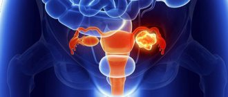
MRI of the pelvic organs in women shows:
- endometriosis;
- malignant and benign neoplasms of the uterus and its cervix, ovaries, bladder, large intestine;
- inflammatory processes of organs in the area of interest;
- fistulas;
- vascular pathologies;
- inguinal hernias;
- post-traumatic consequences, including iatrogenic injuries during surgery or an invasive diagnostic procedure;
- developmental anomalies of the pelvic organs;
- hematometer (accumulation of blood in the uterine cavity);
- interstitial cystitis;
- bladder stones;
- changes in the distal ureters;
- enlarged lymph nodes;
- pathological processes in adjacent tissues and pelvic bones, etc.
All of the above and the ambiguous results of other studies are considered as indications for magnetic resonance scanning.
MRI of the pelvic organs in women is justified in cases of chronic pelvic pain syndrome of unknown etiology. The procedure allows for differential diagnosis between a number of pathological processes and establishing the cause. The specialist takes into account the clinical picture as a whole: the most intense pain often affects women with impaired development of the genital organs, which prevents the outflow of menstrual blood. In chronic inflammatory diseases of the pelvic organs and endometriosis, the symptoms are less pronounced. Dyspareunia (painful sexual intercourse) is suspicious for endometriosis, retrodeviation of the uterus (incorrect anatomical position), chronic salpingoophoritis, adhesive disease, which is clearly visible on tomograms. The sensitivity of MRI in the diagnosis of pelvic varicose veins accompanied by congestion is higher than that of laparoscopy, ultrasound and computed tomography.
MRI of the pelvic organs in women does not use X-rays to create images and does not affect the reproductive organs. In the ovaries, which are included in the area under study, follicles mature, followed by the release of the egg. Oocytes, unlike sperm, cannot renew themselves, and therefore X-ray irradiation is undesirable for them.
The diagnostic procedure cannot be performed in the following situations:
- There are metal objects in the body. An insulin pump, pacemaker, cochlear implant, vascular clips, orthopedic structures, etc. lead to the appearance of defects on tomograms. This makes the study impractical. The magnetic field can disrupt the operation of devices and provoke serious complications, for example, arrhythmia if a pacemaker breaks down. The movement of metal fragments causes trauma to surrounding tissues. Dental structures (bridges, metal-ceramic fillings, implants) are not an obstacle to MRI of the pelvic organs. It is better to remove removable dentures during the diagnosis. If a woman has an intrauterine device with metal elements installed as a contraceptive or therapeutic agent, it is preferable to do a CT scan.
- Pregnancy in the first trimester. In world practice, there is no evidence of the harm of a magnetic field on a developing embryo, but for preventive purposes, a woman carrying a child, MRI in the early stages of gestation is performed exclusively for health reasons. In the future, when the formation of fetal organs and systems is completed (2-3 trimesters of pregnancy), diagnosis is possible.
- Obesity. If the body weight is above 120 kg, magnetic resonance scanning cannot be performed for technical reasons: the subject will not fit into the tomograph ring.
- Inability to be in a supine position while maintaining maximum immobility. Spinal curvature, tremor accompanying a number of pathologies (for example, Parkinson's disease), and mental disorders can become a serious obstacle to performing MRI of the pelvic organs.
- Claustrophobia. To avoid a traumatic situation for patients who are afraid of closed spaces, MRI is performed on open-type machines. In some cases, before the diagnosis begins, sedatives are administered to put the patient into medical sleep.
Contraindications to contrast include:
- a history of a generalized allergic reaction to the administration of a paramagnetic agent (extremely rare);
- kidney diseases accompanied by end-stage chronic renal failure with a decrease in glomerular filtration of less than 30 ml per minute.
What are the complications?
We found out how MRI of the pelvic organs is performed in women and what indications there are for its use. Now let's talk about complications. This examination, as a rule, does not have any side effects. However, when undergoing a procedure using a contrast agent, a small proportion of people experienced a burning sensation at the site where the catheter was inserted. Also, some patients began to choke or cough. Typically, these manifestations occur in people who have allergic reactions in their bodies. They either did not know about them or hid them from the doctor.
Conducting a survey
We figured out why MRI of the pelvic organs is done in women (indications). How is the procedure done? Now we will describe the stages of implementation.
When an MRI is planned for a girl, it will be determined whether she is pregnant or perhaps breastfeeding.
Next, the patient is advised of his actions in the event of fear, fear or deterioration in health during the examination.
After the conversation, the patient needs to change into clothes made of cotton fabric and remove all items with metal inserts. Next, the person is asked to lie down on the scanner table. His limbs are fixed with special devices. This measure is necessary in order to avoid sudden involuntary movements during the examination. Once the patient is ready for an MRI, he is moved directly into the scanner. This is a tunnel in which a person will be.
Examination of men
What does an MRI of the male pelvic organs show? The examination can confirm or refute the presence of the following pathologies in the body:
- Oncological diseases of the testicles.
- Malignant cells in the bladder.
- Prostate cancer.
In addition, magnetic resonance scanning can show pathologies such as tumors and bone fractures, arthrosis.
Having figured out how and what an MRI of the pelvis in women shows, let’s talk about how the examination is carried out?
First of all, the doctor talks to the patient. The doctor asks him if there are any implants or stimulants in his body. The specialist also asks about the presence of allergic reactions in the patient.