Cancer is a malignant tumor process that grows from epithelial tissue. These tissues are located not only on the skin surface, as many mistakenly believe, but also line the surfaces of the reproductive system, urinary and air tracts, digestive tract, etc.
All these structures communicate in a certain way with the external environment, harmful and carcinogenic substances, which increases the likelihood of malignant oncology. There are several types of cancer, but squamous cell carcinoma affects the squamous epithelium.
What is squamous cell skin cancer
This type of cancer, such as squamous cell skin cancer, can appear in any area of the skin. A distinctive feature of all types of this pathology is the speed of development.
The basis for the formation of squamous cell skin cancer is the spinous layer, which includes keratonocyte cells. As a rule, malignancy affects unprotected areas that are more exposed to ultraviolet rays. Most often, the lesion is localized on the lower lip.
The malignant neoplasm is characterized by an aggressive course, in which active infiltration of the lower layers of the skin occurs and the spread of metastases.
To a greater extent, the pathology affects people with fair skin. Typically, the pathology manifests itself in older people aged 60-65 years. The occurrence of the disease in children is genetic in nature.
Non-keratinizing squamous cell carcinoma
Squamous cell non-keratinizing cervical cancer is cytologically characterized by less pronounced polymorphism of tumor cells, which are located separately or in the form of complexes.
The cells are round or oval in shape, large, with a pronounced increase in the nuclear-cytoplasmic index, mononuclear, but multinucleate cells are also found. The location of nuclei in cells is predominantly central.
A distinctive feature of this form of cancer is nuclear polymorphism. The chromatin network is fine-grained, but there are nuclei in which the chromatin has a coarser structure. The nuclei are stained mostly normochromically and contain 1-3 often hypertrophied nucleoli.
The cytoplasm of the cells is represented by a narrow homogeneous rim, mostly basophilic in color. Mitoses are rare. Quite often large “naked” nuclei are present.
Diagnosis of this form of tumor is more difficult compared to keratinizing cancer.
During a cytological examination, one should find the structures of cell accumulations in which all the characteristic features characteristic of a malignant neoplasm are observed.
Classification
Pathology has its own varieties. When classifying, various factors are taken into account.
Among them are:
- size ;
- the rate of its growth;
- differentiation ;
- level of keratinization.
The above criteria are important in treatment. Thanks to these indicators, it is possible to predict the success of therapy, avoid relapse, and improve the quality of life of patients.
Squamous cell carcinoma can be low- and highly differentiated, keratinizing or not. Also in the medical literature you can find a description of the four stages of the disease.
Based on the direction of growth, cancer is divided into endovite and exophytic forms. In the first case, cancer cells spread deep into the skin, and in the second case, its outer layer is affected.
It is important to classify the tumor according to the degree of deepening or invasiveness. It is customary to distinguish between pre-invasive and invasive forms.
Cancer in situ
This preinvasive type is distinguished by the absence of deepening of the process into the tissue. The cells are atypical. According to specialists, the tumor is classified as stage zero cancer.
Carcinoma in situ is preceded by various types of keratoses. If precancerous conditions are not treated, they will degenerate into a malignant neoplasm.
Erythroplasia Keira
This preinvasive form is a plaque localized on the genitals. The boundaries of oncological education are clear. The pathology is characterized by slow development.
Statistics show that this form of cancer is usually diagnosed in men who have not circumcised the foreskin of the penis. Over time, the pathological process becomes invasive with metastases.
Bowen's disease
This pre-invasive condition is characterized by the appearance of plaques on the body that are characterized by slow growth. Their surface is covered with crusts and flakes off. Provoking factors for the development of pathology include long-term treatment with drugs containing arsenic.
Invasive form
This pathology develops in open areas of the skin and lips. In 70% of cases it is facial cancer. The mucous membranes of the genitals and anus may also be affected.
Often, the trigger for the development of the disease is the presence of a pre-invasive form, a burn scar, a trophic ulcer, or altered skin pigmentation. Malignant neoplasms are more often diagnosed in fair-skinned people.
The invasive form is characterized by metastases. In this case, cancer is usually divided into highly and poorly differentiated. In the first form, keratinization is noted, and in the second, carcinoma develops.
keratinization
Squamous cell keratinizing skin cancer is characterized by a solitary nodular growth or plaque on the face or body. The morphological structure of the papule is characterized by density and horny layers. The edges are raised. The tumor may be round or polygonal and pink, yellow or red.
Carcinoma
Carcinoma is a tumor that is usually located in the face, ears, and bald areas of men. In women, it develops in the lower leg area.
On this topic
- Oncodermatology
Prognosis for skin basal cell carcinoma
- Natalya Gennadievna Butsyk
- December 10, 2020
During a medical examination, symptoms of insolation are usually revealed:
- dry skin;
- presence of freckles;
- expansion of capillaries;
- pigmentation.
In the presence of metastases, palpation reveals an increase in nearby lymph nodes. There are no painful sensations. If a cancerous tumor has formed in the area of trophic ulcers or scars, then diagnosing the disease becomes more difficult.
Basalioma
This form of cancer is a small nodule, which is distinguished by a smooth surface and a “pearl belt”. In some cases, a brown tumor develops in the form of a flat plaque, the edges of which are raised. The tumor does not metastasize; it develops slowly. If left untreated, it damages the skin and nearby tissues.
Squamous cell carcinoma
Squamous cell carcinoma is a malignant neoplasm. This type is characterized by a high degree of spread of metastases.
As a rule, the tumor is localized in the face, ears, lower lip, and upper arms. On palpation, hardness is noted. The formation may become covered with ulcers and crusts. It grows faster than basal cell carcinoma.
Poorly differentiated non-keratinizing form
This type involves the presence of papules or nodes. The skin becomes covered with a rash, the red granules of which are easily injured and grow.
The morphology of the papules is fleshy. Visually, they look like ulcers with soft edges and necrosis at the bottom. Often the granules become crusty. This cancerous growth is usually localized in the genital area. Very rarely it occurs in the face and torso area.
The pathology is provoked by Keir's erythroplasia or Bowen's disease. During a medical examination, the softness of the oncological formation is noted. It is distinguished by its irregular shape.
Metastases spread to nearby lymph nodes. When examining the patient, a low rate of differentiation of skin cells and the absence of keratinization are revealed.
Causes and risk factors
There are some factors that contribute to the occurrence of squamous cell carcinoma:
- heredity (genetic predisposition);
- smoking, drinking alcohol;
- UV radiation;
- ionizing radiation;
- taking immunosuppressants;
- poor nutrition;
- work in hazardous production;
- bad ecology;
- infections;
- age.
- Genetic predisposition can be expressed through the following factors:
- failure in the antitumor defense system. If for some reason a mutation occurs in an antigen that stops cell division, a cancerous process may begin;
- disruption of antitumor immunity. The immune system reduces its protective functions due to gene mutations constantly occurring in the body, and this entails the creation of favorable conditions for the development of tumor cells. Gene mutations are inherited;
- disruption of carcinogen metabolism. If a mutation occurs in genes that promote disinfection and rapid removal of carcinogens, then the risk of tumor development increases.
- Tobacco smoking, both active and passive, increases the risk of developing this type of cancer in the oral cavity, respiratory system and gastrointestinal tract. When burned, not only nicotine enters the body, but also other combustion products of tobacco - phenols, cadmium, formaldehyde, benzene - the danger of which has long been proven. All these harmful substances, absorbed by the mucous membranes of the oral cavity and respiratory tract, have a local carcinogenic effect, and spread through the bloodstream throughout the body, contributing to the formation of cancerous tumors in various organs;
In addition, there are other types of tobacco - chewing, snuffing, their use can increase the risk of oncology of the lips, tongue, and nasopharynx organs.
- A component of all alcoholic beverages, ethyl alcohol can cause the development of malignant tumors.
The risk of cancer increases in those who combine alcohol with smoking (or other means of using tobacco).
- UV radiation with prolonged exposure has a harmful effect on human skin, as a result of which various genetic mutations can occur, and this, in turn, leads to the growth of cancer cells and reduces antitumor protection. With prolonged exposure to UV rays, antitumor immunity may not be able to cope with the resulting mutated gene, and this leads to the development of squamous cell carcinoma.
- Ionizing radiation (gamma rays, x-rays, helium and hydrogen nuclei). The action of such radiation has a damaging effect on the genome of the cell, which leads to mutation. The human immune system is also affected, which means the risk of cancer increases several times.
- Taking immunosuppressants, i.e. medications (azathioprine, mercaptopurine, etc.), which are used for various diseases and have a depressing effect on the body’s defenses, as well as on antitumor immunity.
- Poor nutrition. The presence in the diet of a large amount of fatty foods and very spicy, salty foods increases the risk of oncology of the gastrointestinal tract and kidneys. A balanced diet, on the contrary, hinders development;
- Working in hazardous industries involves frequent contact with harmful substances (through breathing, direct contact through the skin). The longer such contact, the greater the risk of developing cancer.
- Bad ecology. The risk of developing cancer increases significantly for those people who live near industrial enterprises.
- It has already been scientifically proven that certain types of infections (viruses) can provoke the appearance of squamous cell carcinoma. These viruses are considered:
- human papillomavirus (multilayer koilocytosis), which is capable of causing the development of benign tumors on the skin and mucous membranes - papillomas, condylomas, and by causing various intraepithelial types of neoplasia, becomes the cause of cervical cancer;
- HIV (human immunodeficiency virus) affects the human immune system, which can lead to the development of AIDS and a decrease in the body's antitumor defense.
- Age. As a person ages, the function of the immune system decreases and becomes impaired and the processes of recognition of mutated cells deteriorate, which means the risk of squamous cell carcinoma increases.
In addition to the above risk factors that contribute to the occurrence of squamous cell carcinoma, there are so-called precancerous conditions. Although they are not malignant neoplasms themselves, they increase the chances of developing cancer. These precancerous conditions are divided into obligate and facultative.
Obligate conditions include:
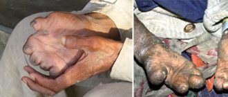
- xeroderma pigmentosum, a disease transmitted by an auto-recessive type. It occurs in children aged 2-3 years and is manifested by redness of the skin, ulcers, and wart-type growths. This disease occurs due to intolerance to UV rays, which, when entering the skin, damage DNA, leading the cell to mutation and the occurrence of oncology;
- Paget's disease. More often, females are susceptible to this disease. The main locations of the disease are the armpits and genitals. It looks like redness with clear boundaries and a surface that can be either moist or dry and flaky. This skin lesion can develop over several years and develop into squamous cell carcinoma;
- Bowen's disease. Outwardly it looks like several small red spots that may be on the surface of the body. Over time, a scaly red-brown plaque appears in the area of the neoplasm. As the disease progresses to squamous cell carcinoma (squamous cell carcinoma), the surface of the plaque begins to ulcerate.
Facultative precancerous conditions include diseases in which the appearance of squamous cell carcinoma is not necessary, but the risk of its occurrence is quite high.
Such diseases include:
- cutaneous horn. It is hyperkeratosis (thickening of the stratum corneum of the epidermis). The development of oncology in this case can occur in 7-15% of cases;
- actinic keratosis. The main reason for the appearance is ultraviolet rays that affect bare skin. The age of patients is after 60. The risk of this cancer is up to 25% of cases;
- keratoacanthoma. Age category – after 60 years. Located on the skin of the face or the back of the hands in the form of a round formation with a depression in the center with horny masses;
- contact dermatitis. Occurs when the skin is exposed to chemicals and is characterized by local inflammation, swelling and redness. With a long process, the formation of squamous cell carcinoma is possible.
The formation of secondary foci (metastases) of squamous cell carcinoma can occur in several ways. Squamous cell carcinoma can metastasize through the lymphogenous, hematogenous and implantation routes.
Lymphogenous metastasis occurs in about 98% of squamous cell cancer, which occurs with the help of lymph nodes, hematogenous - cancer spreads through the bloodstream and can move to almost any organ (about 2% of cases), implantation - upon contact with neighboring organs, when the tumor grows into organ tissue.
Causes of pathology
Experts believe that the main reason for the development of squamous cell cancer is an unfavorable genetic background. But the disease can be either hereditary or acquired. There are a number of provoking factors that cause the appearance of a tumor.
These include:
- elderly age;
- light tone ;
- no use of sunscreen ;
- work in hazardous industrial production;
- diseases ;
- decrease in defenses .
There are also a number of diseases that precede the appearance of cancer. Without proper treatment, they can degenerate into a malignant neoplasm. Among them, dermatoses, pustular lesions of the skin, and ulcers should be noted.
Poorly differentiated (immature) cancer
Poorly differentiated, or immature, cancer (small cell, basal cell, spindle cell, anaplastic cancer). Histologically, immature forms of cancer consist of sheets, cells and strands of tumor cells without any tendency to differentiate.
The cells are sharply polymorphic, of different shapes and sizes, with large, sometimes small and hypochromic or hyperchromic nuclei.
Often tumor cells have a light, seemingly empty cytoplasm (clear cell cancers); in some cases, elements of poorly differentiated cancers acquire an elongated shape and form cords running in different directions. These cancers may resemble spindle cell sarcoma.
Cytological examination of poorly differentiated cancer reveals pronounced anisocytosis. In the total mass of cellular elements, along with cells with a diameter of less than 10 microns, there are giant and often multinucleated cells.
Round or oval cells predominate. The nuclear cytoplasmic index is not always increased. The cytoplasm of the cells is stained basophilically, it is abundant and vacuolated. The chromatin is coarsely reticulate or fine-grained, hyperchromatically colored. The nucleoli in the nuclei are clearly visible. Mitoses in the preparations are relatively common; “naked” nuclei are present.
Symptoms
The initial stages of the malignant process, as a rule, do not cause any concern to the patient. Gradually, the neoplasm takes on an asymmetrical shape. It is characterized by tuberosity and an increase in diameter.
The following symptoms are noted:
- soreness in the area affected by the tumor;
- increased volume of lymph nodes;
- presence of edema;
- itching and burning of the skin;
- lack of sensitivity of the affected area;
- painful sensations in the area of growth formation;
- loosening or softening of the tumor;
- constant feeling of fatigue;
- hyperemia of the skin near the affected area;
- lack of appetite;
- bleeding .
A clear sign of carcinoma should be noted. In the presence of this type of tumor, an unpleasant odor emanates from the cancerous tumor.
Symptoms of the disease
Oncology is treated depending on the location and stage of the disease. It is important to identify the problem in time, since the disease is prone to rapid metastasis. First, doctors surgically remove malignant tumors, and then carry out therapy to destroy the remains of cancer cells.
Symptoms of the disease depend on the location and which organ is affected. All types of this oncology have common clinical signs that characterize the characteristics of its growth.
Depending on the method of growth, cancer can be divided into the following forms:
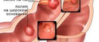
- the exophytic form (papillary) is characterized by the appearance of a nodule, clearly limited from the surrounding tissues. It quickly increases in size and becomes similar to cauliflower inflorescences of a red-brown hue. The surface of the tumor is lumpy, with a visible depression in the middle. This tumor is attached to the surface of the mucosa or to the skin with a thin stalk or a wide base. Over time, the entire surface of the tumor ulcerates, turning into an endophytic variety;
- the endophytic form (infiltrative-ulcerative) has a small primary nodule, which quickly ulcerates and in its place one large ulcer appears. This tumor is irregular in shape, has dense raised edges, a rough bottom, which is covered with a whitish coating with an unpleasant odor. The ulcer almost does not change in size, as the tumor spreads deep into the tissues;
- the mixed form combines all of the above characteristics.
Diagnostics
If you have the above symptoms, you should immediately contact a specialist. The oncologist will conduct a proper examination of the changed area of skin and collect anamnesis. The patient will have to undergo a series of examinations.
Initial examination
It is carried out using a special optical device, a dermatoscope, which allows one to obtain an enlarged image of the affected area of the skin. The specialist analyzes the condition of all layers of the epidermis and studies the structure of cells.
This method makes it possible to identify a tumor, assess the degree of cell differentiation and indicate the depth of damage to the layers of the skin.
Siascopy
Siascopy is a progressive method for examining skin tumors. It is carried out using siascanners. A three-dimensional image of the tumor is observed by a specialist on a monitor screen.
Siascopy makes it possible to examine the affected area of skin without screening tissue for further study in the laboratory.
Additional diagnostic methods
Histological examination of neoplasm particles makes it possible to characterize the structure of spinalioma. Instrument-based diagnostics are used by oncologists to detect secondary cancerous lesions and examine lymph nodes. Typically, MRI, CT and radiography are used.
Survival prognosis
A patient’s standard of living after cancer depends on the type of cancer and the speed of its development. At the first stage, the forecast is 90%, at the second it drops to 60%, and at the third it drops to 30-35%, and at the fourth it does not rise above 10%. If cancer is detected at an early stage and the patient is provided with timely and comprehensive treatment, then the prognosis is 98% that the cancer is completely cured. Therefore, with any manifestation of neoplasms on the skin, you should consult a doctor for timely removal of dermatitis.
Squamous cell carcinoma develops from keratinocytes (one of the types of epidermal cells - stratified squamous keratinizing epithelium). Affects the skin and mucous membranes.
Treatment
The use of one or another therapeutic method in the presence of squamous cell skin cancer depends on the stage of the pathological process. When applying a certain method of treating a tumor, the oncologist takes into account the type of cancer lesion.
Experts give a positive prognosis only if the patient has an initial form of the disease. If the area of the pathological process is large and metastases are diagnosed, then the prognosis is poor. In this case, the general condition of the patient’s body and his age play an important role.
Surgical intervention
The method that has the highest level of effectiveness is surgical. During surgery, the tumor and nearby tissues located at a distance of 1-2 cm from the cancerous lesion are excised.
Effective implementation of this operation requires a preliminary microscopic examination of the cells. During the operation, excision is performed using a neodymium or carbon dioxide laser. This technique allows you to reduce blood loss to a minimum.
Electrocoagulation
If the size of the tumor does not exceed 1-2 cm, and there is no deep growth of the cancerous tumor, then it is removed by electrogulation, curettage or laser. Healthy tissue is captured at a distance of 5-10 mm from the cancerous tumor.
Cryodestruction
If the tumor is superficial and the degree of penetration into the depth of the tissue is small, then cryodestruction is used. It involves capturing intact tissue located at least 2-2.5 cm from the tumor. This procedure is carried out after a biopsy, which helps determine the nature of the malignant neoplasm.
Chemotherapy
This method is among the most effective. It is used both for relapses and for late stages of the process and extensive skin lesions. If a patient is diagnosed with basal cell carcinoma, then the use of a cytostatic agent during chemotherapy is justified.
Chemotherapy makes it possible to preserve the surface of the skin around the affected area. The impact is aimed only at oncological neoplasms.
X-ray therapy
For more serious skin lesions, integrated treatment approaches have proven themselves to be effective. Close-focus X-ray therapy has high results.
Large tumors are irradiated with an electron beam. After such an intervention, a course of radiation therapy is indicated, which is prescribed to patients with relapse of the pathological process, the presence of metastases, or a contraindication for surgical intervention. Photodynamic therapy involves the use of otosensitizers.
How is the treatment carried out?
Treatment of squamous cell carcinoma is selected on a case-by-case basis and involves the use of radical therapy while preserving reproductive and menstrual functions and the reproductive system as a whole. The following treatment methods can be used:
- surgical;
- chemotherapy;
- radiation therapy;
- combination therapy.
The choice of treatment method depends on the location of the carcinoma, its size, the degree of neglect of the process and the general condition of the patient.
Of the surgical techniques in the early stages, conization in combination with curettage is most often used; in more complex situations, hysterectomy is performed; in addition, excision of lymph nodes is possible, etc. If cervical cancer has spread to the vagina, then a radical hysterectomy is performed with removal of the uterus, its cervix, part of the vagina and all appendages. Surgical techniques can be combined with radiation and chemotherapy before or after surgery. It is important to remember that it is impossible to cure a cancerous tumor with medications and traditional methods.
Complications
A cancerous tumor is characterized by the presence of metastases. It penetrates the tissues, promoting their destruction. Swelling on the face spreads to the ears, eyes and sinuses. Patients lose hearing and vision. Often a person is struck by sinusitis or meningitis.
Metastases affecting the face affect the lymph nodes located in the neck, armpits and groin. A clear sign of their involvement in the pathological process is their density and large size. When palpated, they do not hurt and are mobile.
Over time, the lymph node fuses with the tissue, which contributes to the loss of its mobility. The patient complains of pain. The process ends with the destruction of the lymph node. An ulcer forms on the area of skin above it.
Squamous cell carcinoma: classification, diagnosis and treatment
Cancer is a malignant tumor process that grows from epithelial tissue. These tissues are located not only on the skin surface, as many mistakenly believe, but also line the surfaces of the reproductive system, urinary and air tracts, digestive tract, etc.
All these structures communicate in a certain way with the external environment, harmful and carcinogenic substances, which increases the likelihood of malignant oncology. There are several types of cancer, but squamous cell carcinoma affects the squamous epithelium.
Squamous cell carcinoma is a malignant tumor process that develops from the epithelium of the skin or mucous tissues.
This type of cancer is characterized by an aggressive course with rapid development.
The oncological process begins in the skin or mucous layer, but very quickly spreads to local lymph nodes, neighboring tissues and organic structures, destroying their structure and undermining their activity. As a result, multiple organ failure occurs, leading to death.
Prevention
The main preventive measure, which is aimed against pathology, is timely treatment of dermatoses that precede cancer. Preventive measures involve eliminating risk factors leading to cancer.
The main measures include:
- timely treatment of diseases that provoke the development of cancer;
- ensuring skin protection from the harmful effects of ultraviolet radiation;
- the use of special creams to prevent skin drying;
- protection of scars from secondary damage;
- careful handling of chemicals that contain carcinogens;
- sunlight by people affected by skin cancer ;
- regular visits to an oncologist and treatment of precancerous conditions through cryodestruction;
- use of retinoids in ointments;
- monthly examination .
Peculiarities
Theoretically, a keratinizing type of cancer is possible in all organs and tissues, even in the absence of a keratinizing cell type. This is due to primary metaplasia, when initially normal cells are transformed into a keratinizing type, and then a malignant process develops.
In practice, there are several forms of squamous cell carcinoma, localized:
- on the skin;
- on the edge of the lips;
- in the oral cavity;
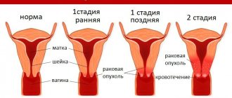
- cervical canal of the uterus;
- esophagus;
- larynx;
- bronchial tree;
- trachea.
The first three forms grow from keratinizing cells. An oncological neoplasm can grow exophytically, that is, with the formation of a dense nodule, or endophytically, when ulcerative defects appear.
Skin cancer
Most frequently reported. In 90% of cases it is of the keratinizing type. It develops mainly on open areas of the skin (face, hand, neck).
Local itching, pain, burning, swelling, changes in sensitivity, and redness are noted.
Lip border cancer
The lower lip is often affected and has a rapid, aggressive course. Locally manifested by swelling, induration, redness, pain, and ulcerative defects.
Oral cancer
The lesion is localized on the cheeks, gums, and palate. Symptoms include pain, increased salivation, bad breath, disturbances in chewing and speech.
About the disease
Squamous cell carcinoma with keratinization is an aggressive in nature, malignant neoplasm, formed from cells of the epithelial skin layers and mucous tissues.
The anomaly is characterized by the slow development of the situation - the time intervals from one stage of the disease to the next can be quite long.
Originating in the superficial epithelial layers, cancer gradually affects neighboring lymph node connections and then metastasizes to other organs and vital systems of the human body.
Very often, this clinical picture leads to the early onset of multiple organ failure and death.
In three of four situations when diagnosing the disease, it affects the area of the face and skull. Moreover, this type of tumor is more often detected in people with light skin types, those who do not tolerate exposure to ultraviolet radiation.
The complexity of the disease lies in its ability to grow quite deeply into the internal layers of tissue, right down to the skeletal muscles. If the disease occurs against the background of cancroid, formations may develop that, almost from the moment of inception, sprout deep internal shoots.
There are two types of cancer with keratinization:
In the first case, the compaction is a raised area above the surface of the skin, and is also characterized by dense structural filling and a massive base. In the second, the nodular formation quickly undergoes expression.
As the anomaly progresses, irreversible processes occur in the body that destroy vascular sections, soft and hard bone tissue.
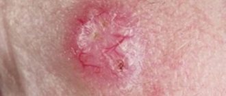
Initially, the compaction resembles a plaque, the consistency of which is denser than that of the skin. The shade of pigment at the site of the lesion becomes pink or reddish, and the lesion becomes covered with scaly, keratinized layers - hence the name of the disease.
A little later, the plaque is replaced by an ulcer, characterized by an uneven shape and blurred, ragged outlines.
In addition to the skin, the disease affects:
- Respiratory system - in 40% of detected cancer cases, it penetrates into the root zone of the lung over time and is extremely difficult to diagnose; The genitourinary tract – the cervix – is the organ most affected by a tumor of this form. Initially, the pathology resembles the papilloma virus or condyloma. With timely diagnosis, the prognosis for cure is quite optimistic; The cervical region is poorly identified and just as difficult to treat. The clinical picture and primary symptoms are extremely vague.
The main provoking factors that can cause the development of a squamous cell tumor with keratinization are:
- Incorrect exposure to the sun - direct rays are extremely aggressive and can deeply affect tissues, destroying their structure at the cellular level. At the same time, the qualitative content of the cells undergoes a change, causing their degeneration; Thermal or chemical burns - such phenomena completely break the structural molecular lattice, resulting in the appearance of abnormal, chaotically multiplying cells of a malignant nature, which, in fact, is cancer; Direct contact with hazardous chemical components - during the production process, a person may be forced to receive a certain toxic dose, which, accumulating in the body, gives rise to pathological formations;
Skin diagnoses - Paget virus, Bowen virus, xeroderma pigmentosum - the chronic course of these ailments can cause surface microtraumas and qualitatively change the content of tissues.
The usual processes of cell division in the lesion are disrupted and the process becomes uncontrolled. After a little time, this zone becomes malignant and a precancerous anomaly forms; Smoking – carcinogens, penetrating into the blood, create favorable conditions for the development of cancer processes in the most vulnerable places of the human body.
In this article we will tell you how to detect Bartholin gland cancer at the beginning of its development.
In order to better select a treatment plan for the disease, the disease is classified into the following stages:
- 1 – initial . The lesions are minimal, their size does not exceed 1-2 cm. Despite the fact that the base of the formation is not motionless, it is located within the affected area and does not injure neighboring tissues. There are no symptoms, metastasis is not detected;
2 – this stage is characterized by the rapid growth of the anomaly . The disease progresses, the area of distribution of mutating cells increases. However, this course cannot yet be considered active, since the tumor can be controlled.
In some cases, distant single metastasis and damage to lymph node connections located in the immediate vicinity may be observed; 3 – cancer spreads to most of the lymphatic system , affects surrounding formations, tissues and actively grows into neighboring organs and systems. Treatment at this stage is difficult, but there is still a chance for an optimistic prognosis. Symptoms are severe and often painful;
4 – the final stage of the disease, stage . Not only soft tissues, but also bone tissues and cartilage joints were subjected to irreversible processes. Even with a relatively small tumor, it is capable of producing multiple metastases.
Almost all joints become immobilized. The patient endures stage 4 extremely difficult. Treatment is no longer effective. The only thing doctors can do is to alleviate the symptoms and somewhat prolong the patient’s life.
Despite the fact that there are practically no symptoms at the initial stages of the progression of the anomaly, as it grows, the signs make themselves felt. The following manifestations of squamous cell keratinizing carcinoma are distinguished:
- Increased body temperature – characterized by a low growth rate, which accompanies the patient for a long period of time and is difficult to stop; Fatigue, weakness - even after a full rest a person feels tired. However, this condition is not associated with physical activity. Its reason is the increased load on the body due to a developing disease, the fight against which requires additional strength; Loss of appetite - the patient begins to eat less food, trying to save energy costs that are spent on the processes of its digestion; Pain – depending on the location of the anomaly, the degree of pain may vary, but almost always, starting from the second stage of the disease, discomfort accompanies the pathology to one degree or another; Swelling of the surrounding tissues - this is due to the negative impact of mutating cells on neighboring, still healthy fragments; Sensory disturbances at the site of the lesion - nerve endings become dull as inflammation spreads, their endings partially atrophy. Hence the decrease in reflex.
This article lists benign tumors of the esophagus.