Lymphoma is a tumor arising from lymphatic tissue. It refers to diseases of the hematopoietic system, but its primary focus is formed outside the hematopoietic organ - the bone marrow. The main accumulation of lymphoid cells is the lymph nodes. This is where this tumor most often occurs. This is the so-called nodal form.
But lymphoid cells (lymphocytes and their precursors) are found in almost all organs and tissues. Under certain conditions, mutations form in them and they begin to divide uncontrollably. This is how extranodal lymphoma develops (i.e., the neoplasm does not develop from the lymph nodes).
Primary gastric lymphoma is the most common extranodal variant of these tumors. There is also a secondary option, when the walls of the stomach are infiltrated by screenings of malignant cells from other locations.
Triggering factors
The most reliable cause of gastric lymphoma is Helicobacter pylori infection. This bacterium is found in 90% of patients with this type of tumor.
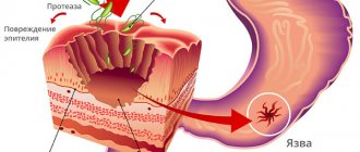
The gastric mucosa does not have any lymphoid focal formations; lymphatic cells in it are normally diffusely scattered throughout the lamina propria. Under the influence of prolonged inflammation caused by this bacterium, the proliferation (growth) of these cells leads to the formation of follicles and the development of atypia in them.
There are also other risk factors that can cause the development of lymphomas: hereditary predisposition, HIV infection, taking immunosuppressive drugs, exposure to various chemicals.
Causes of follicular gastritis
Follicular gastritis is not an independent disease; it is a consequence of the chronic form of this disease caused by Helicobacter pylori infection. Therefore, we list the causes of lymphoid gastritis, which can only contribute to its progression:
- addiction to fatty foods, fast food;
- eating on the go, dry, without thoroughly chewing food;
- long fasting;
- improper diet;
- ignoring healthy foods;
- constant stress, depression;
- nervous overload;
- frequent drinking and smoking;
- disorders in the autonomic system, vegetative-vascular dystonia.
Classification
Histological types of gastrointestinal lymphomas:
- MALT lymphoma (marginal zone B-cell lymphoma). This is the most common type of lymphoma in the stomach. It develops mainly in the antrum, and for a long time manifests itself with scant symptoms, hidden under the mask of gastritis. In 90% of cases it is associated with Helicobacter pylori. It is characterized by mild malignancy, almost no distant metastases, and can regress after eradication antimicrobial treatment.
- Diffuse large B-cell lymphoma (DLBCL). The second most common type has a worse prognosis.
- Follicular lymphoma.
- Primary Burkitt lymphoma is a very rare pathology.
According to the degree of malignancy, MALT lymphoma and follicular lymphoma belong to the indolent variant of the course, that is, less malignant. DLBCL and Burkitt's lymphoma are aggressive forms.
In addition to true lymphomas, pseudolymphomas are also distinguished. This is a benign infiltration of the mucous membrane, but malignant degeneration is possible.
What kind of gastritis is called lymphoid (follicular)
There is one form of the disease that stands apart, this is follicular gastritis. It is also called lymphoid. Usually this disease develops against the background of a pre-existing stomach disease. And most often the culprit is the Helicobacter bacterium, which settles on the mucous membrane, causing its inflammation.
Lymphocytes accumulate in these places to neutralize the pathogenic effects of bacteria. But it turns out that along with the healing effect, the opposite is also present - the follicles do not allow normal cells to produce full-fledged gastric juice. Lymphoid gastritis does not cause such suffering to patients as, for example, erosive or ulcerative gastritis.
Diet plays a special role in the treatment of this type of gastritis. As with any inflammation of the gastric mucosa, meals should be divided, literally every 3-4 hours. The food is gentle, that is, you need to exclude spicy and hot foods, eat steamed and baked foods. Since the disease can unnoticed develop into a malignant form, it is necessary to be examined by a doctor regularly.
But the following signs help to suspect something is wrong:
- frequent, but not severe pain in the epigastric region;
- heartburn;
- feeling of fullness and heaviness in the abdomen;
- nausea.
Lymphoma screening
- Mandatory full physical examination with palpation (palpation) of all accessible groups of lymph nodes (occipital, cervical, sub- and supraclavicular, axillary, cubital, inguinal), liver, spleen, examination of the tonsils,
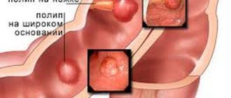
- Endoscopic examination (FGDS) with biopsy and subsequent cytological and histological analysis. On examination, lymphoma usually appears as a large ulcer or multiple mucosal ulcers. There are polypoid, nodular, infiltrative and mixed forms. The antrum is most often affected. Diffuse infiltration can masquerade as atrophic gastritis. Noteworthy is the significant thickening of the folds of the mucous membrane. In 20% of patients, the mucous membrane may not be changed.
- A biopsy is taken from several places in the suspicious area, if a diffuse lesion is suspected - at least 8-10 samples.
- Endoscopic ultrasound allows you to identify the depth of invasion (sprouting) of the wall and see the enlargement of the paragastric lymph nodes.
- CT or MRI of the abdominal cavity. Allows you to visualize the tumor, assess its spread to neighboring organs and identify pathological changes in the intra-abdominal lymph nodes.
- Ultrasound of peripheral lymph nodes - to exclude secondary damage to the stomach.
- CT scan of the chest.
- Test for Helicobacter pylori (HP). The most reliable is the presence of a microorganism in the biopsy specimen. Also used are tests for the presence of a specific antigen in stool, urease breath test, and antigen in the blood.
- Histological examination reveals accumulation of lymphoid tissue in the mucous membrane and submucosal layer, infiltration of the gastric glands. It should be noted that the histological diagnosis of MALT lymphoma is quite complex, because must be carefully differentiated from HP-associated gastritis.
- Immunohistochemical and genetic study of biopsy material. For example, the determination of translocation of chromosomes 11 and 18 is an unfavorable prognosis factor for antibiotic therapy.
- General clinical examination - general and biochemical blood tests, for markers of infectious diseases (HIV, hepatitis, syphilis), ECG, examination by medical specialists.
- According to indications - positron emission tomography (PET).
Diagnostics
Visualization of the affected area on the monitor screen will help determine the extent of the damage.
To make a correct diagnosis, a number of diagnostic procedures are prescribed. Research methods:
- Blood analysis. Shows the level of ESR and the presence of tumor markers.
- Endoscopy. Makes it possible to visually assess the condition of the organ mucosa.
- Biopsy. Determines morphological changes at the cellular level.
- Laparotomy. Using the procedure, biomaterial is collected for histological examination.
- X-ray. Based on the results, the relief of the mucosa, obstruction of patency and the stage of narrowing of the lumen are assessed.
- IHC. Determines the antigenic properties of malignant tumors.
Treatment of gastric lymphomas
To select a method, it is necessary to take into account the pathomorphological picture, stage of the disease, and its immunochemical characteristics. It must be said that there are still no uniform standards for the treatment of gastric lymphomas. For example, there is no generally accepted opinion about the need for surgery and the amount of chemotherapy. However, accumulated experience and scientific research form the basis for tactical recommendations.
Treatment of MALT lymphoma
In stage I MALT lymphoma, when the tumor is limited to the mucous and submucosal layer, complete remission can be achieved after HP eradication. A 2-week course of a combination of omeprazole, amoxicillin and clarithromycin is usually prescribed. After 3 months, endoscopic and histological control is carried out. In case of regression of lymphoma, dynamic observation is carried out.
If HP infection persists, second-line therapy is prescribed until complete eradication. Many studies have shown that regression of this type of tumor after antibacterial treatment is achieved in 80% of cases in patients with stage I of the disease.
In the absence of tumor regression, radiation therapy to the stomach and paragastric lymph nodes is indicated, SOD 30 Gy. If there are contraindications to radiation therapy, it is possible to supplement the cycle with rituximab. If there is no response to radiation, chemotherapy is prescribed.
Treatment of such lymphomas at a later stage includes systemic drug therapy according to the COP program (cyclophosphamide, Oncovin, prednisone), immunotherapy with rituximab, or local radiation. Surgery is also used, but the advantage of surgery over conservative tactics has not been proven.
Treatment of other histological types of lymphomas
For small follicular lymphomas without risk factors, watchful waiting and dynamic observation are recommended. Treatment begins only when the tumor grows.
For aggressive forms - diffuse large B-cell lymphoma and Burkitt's lymphoma, treatment is similar to stomach cancer and includes surgery, chemotherapy and radiation.
Causes and symptoms
Lymphoid gastritis is not an independent disease. It occurs against the background of existing pathologies in the epithelium of the mucous layer. In the vast majority of cases, the inflammatory process in the stomach begins due to infection of the body by the bacterium Helicobacter pylori.
For the development of inflammation and the growth of microorganisms, appropriate conditions are required. The following factors can trigger the activation of bacteria in the body:
- poor nutrition, prolonged fasting, abuse of unhealthy and fatty foods;
- nervous experiences, strong psycho-emotional stress;
- prolonged consumption of alcoholic beverages, frequent smoking;
- disorders of the body's autonomic system.
The symptoms of follicular gastritis are similar to those of other forms of gastritis initiated by the Helicobacter bacterium. The main features can be considered:
- pain syndrome that manifests itself in the morning, as well as some time after eating;
- sour belching, heartburn resulting from excess hydrochloric acid in the stomach;
- decreased appetite;
- dysfunction of the intestines, which is manifested by diarrhea and constipation.
With the development of the inflammatory process in the stomach, feelings of heaviness, distension and bloating appear. Nausea also appears, which is often accompanied by vomiting. Diarrhea can be followed by constipation, which happens quite often.
In advanced forms of the disease, the patient's condition worsens. Weakness and general fatigue of the body appear. The skin is pale and dry, and a white coating appears on the tongue. Decreased appetite leads to a decrease in the patient's weight.
Prevention of gastric lymphomas
The main method of preventing the development of gastric MALT lymphomas is the eradication of Helicobacter pylori, as the most proven risk factor.
It is advisable to monitor HP-associated pathologies (gastritis, peptic ulcer) with regular gastroscopy with mandatory biopsy of the mucous membrane, since it is difficult to distinguish gastric lymphoma from the external picture alone.
If lymphoma is diagnosed and eradication therapy is carried out, it is necessary to perform FGDS control with a biopsy after 2-3 months, and then the same observation every six months.
Disease clinic
Signs of the initial stages of lymphoma development are similar to gastritis or peptic ulcer. The first symptom of a malfunction of the organ is frequent heartburn. Then comes nausea, bloating, pain and disruption of the bowel movement. Typically, such symptoms do not cause concern to the patient, so diagnosis is delayed for a long time.
As the tumor grows, the discomfort increases, and the person notices changes:
- Epigastric pain.
- Nausea is accompanied by vomiting, often with blood.
- The functions of the pancreas and duodenum are impaired. A person is tormented by gases and stool disorders.
- Body weight decreases sharply.
- During night sleep there is profuse sweating.
- Lost appetite. The patient is satisfied with a small portion of food.
- Feces contain undigested food.
Any changes in the functions of the gastrointestinal tract, accompanied by pain, are diagnosed and treated by a gastroenterologist.
Forecast
The prognosis is determined by the type and degree of tissue proliferation. If the cause of this phenomenon is the response of the mucous membrane to the process of inflammation, then after treatment of the concomitant pathology the outcome is favorable. Gastric hyperplasia is not capable of causing cirrhosis of the liver: other, more dangerous pathologies lead to this disease. Oncological alertness is caused by lymphofollicular hyperplasia of the stomach. In some cases, it transforms into a malignant form.
Gastric hyperplasia is a pathology that is quite difficult to determine. Its symptoms are so secretive and nonspecific that the progression of the disease can be missed. Contrary to what some patients believe, the discovery of hyperplasia should not cause panic - it is in no way considered cancerous. The key to preventing dangerous complications is a timely visit to a gastroenterologist, treatment of concomitant diseases and, if necessary, periodic visits to an oncologist.
November 21, 2020, 2:53 0 24,133
Hyperplasia is a pathology in which cells in a separate part of an organ rapidly divide, causing the area of the organ to grow. Hyperplasia can occur in various organs and is a dangerous anomaly. Gastric hyperplasia is especially common.
IT IS IMPORTANT TO KNOW! Even “advanced” ulcers or gastritis can be cured at home, without surgery or hospitals. Just read what Galina Savina says and read the recommendation.
Very important! Savina G.: “I can recommend only one remedy for the quick treatment of ulcers and gastritis” read more.
Stages of the disease
Gastric lymphoma, like other types of this disease, has four stages. Each of them differs in severity and clinical picture. The first stage is the easiest; if the disease is detected at it, then the prognosis for treatment is almost one hundred percent positive. The situation is worst at the fourth stage of the pathology.
Below are the main signs for each stage of the disease:
- Stage 1: the pathological process is localized in the stomach. At stage 1a it is limited to the mucous membrane, at stage 1b it moves to deeper layers (muscular, serous).
- Stage 2: the lymph nodes lying nearby, as well as neighboring organs, are affected.
- Stage 3: distant lymph nodes are affected, and the tumor actively penetrates into neighboring organs. Lymph nodes are affected on both sides of the diaphragm.
- Stage 4: the lymph nodes located above the diaphragm are affected, as well as the lymph nodes located in the pelvis along the aorta.
At the third and fourth stages of the disease, the pathological process affects the liver, bone marrow, spleen, and other internal organs.