From this article you will learn about the disease hip dysplasia in children (CHD), what it is, and why early detection of this pathology in a child is very important. Symptoms, treatment methods, exercise therapy, set of exercises.
Author of the article: Nivelichuk Taras, head of the department of anesthesiology and intensive care, work experience 8 years. Higher education in the specialty “General Medicine”.
Article publication date: 06/26/2019
Article updated date: November 29, 2019
What is dysplasia? Hip dysplasia (HJD) is a congenital disorder of the normal structure of the hip joints (abbreviated as HJ), which increases the likelihood of developing dislocation of the femoral head from the acetabulum.
This problem occurs during fetal development, so symptoms and signs of underdevelopment can be detected immediately after the baby is born or in the first months of his life.
Much less often, the disease is detected in children over 1 year of age, when walking disorders are noticed. This article will focus specifically on children from 1 year of age who have already left infancy.
Hip dysplasia itself does not pose a risk to the child’s health, but its presence can cause serious disruptions in the functioning of the affected joints and sharply worsen the quality of life of a small patient.
Moreover, when this problem is detected late in children, traumatic and inconvenient treatment methods are used. In such cases, it is practiced to apply a rigid plaster cast in a non-physiological (unusual) position, the child is forced to remain motionless for 1–3 months. Surgeries to realign the femoral head under general anesthesia are also performed.
For hip dysplasia, a simple rule applies: the later the problem is identified, the more difficult the treatment and the worse its results. If this disease is detected after the age of 1 year, the baby and parents face a long and difficult road to recovery. Then long-term traction using a splint (up to 6 months) and (or) surgery may be required. Treatment along with rehabilitation takes at least a year, and it is not always possible to achieve a complete recovery.
If the problem is identified at an early age (up to a year), then it can be dealt with more gentle methods (using soft splints) and without surgery. The duration of treatment for congenital hip dislocation in children under one year of age is 3–6 months, the prognosis is favorable in 95% of cases.
The incidence of the pathology is approximately 1 case per 1000 people. Scientists also provide the following figures:
- Instability of the hip joint immediately after birth is observed in 1 out of 60 newborns.
- Within 1 week, the hip joint stabilizes in 60% of infants.
- Within 2 months, the hip joint stabilizes in 88% of infants.
- Only in 12% of children who have instability of the hip joint immediately after birth, this problem does not disappear on its own.
Pediatric traumatologists deal with the problem of hip dysplasia in children.
Degrees
Hip dysplasia is common in infants, occurring in 5-10% of children. Possible consequences in the future depend on a timely diagnosis. The joints of newborns are not yet fully formed, so the disease responds well to treatment.
This disease is a severe malformation in which all the constituent elements of the articular joint are affected. In total, there are 3 degrees of leg dysplasia in children:
- Stage I (pre-dislocation). This is the easiest level. If the disease is diagnosed at the initial stage, it means that the head is not displaced relative to the acetabulum. The forecasts are the most optimistic.
- Stage II (subluxation). It appears as a slight displacement.
- Stage III. When a hip dislocation is detected, treatment takes the longest, since it manifests itself as the head emerging from the acetabulum. Massage and exercise therapy are not enough here.
Also, hip dysplasia can be unilateral or bilateral.
Causes of congenital and acquired pathology
Most often, hip dysplasia in children over 1 year of age is a consequence of disorders of the intrauterine development of the hip joints that were not detected and corrected at an earlier age. However, there are also acquired causes of dysplasia.
Causes of congenital dysplasia
The exact causes of congenital hip dysplasia are not known. One theory says the culprit may be the hormone relaxin, which is produced in a pregnant woman's body to relax her ligaments and help the baby pass through the birth canal.
Some of this hormone can enter the baby’s body, causing the ligaments of his hip joints to relax and leading to the development of pre-dislocation.
Girls are more sensitive to the action of relaxin, so hip dysplasia occurs 4–5 times more often in them than in boys.
Other causes of congenital hip dysplasia:
- Heredity – hip dysplasia is approximately 12 times more common in children who have close relatives (parents, siblings) with this problem.
- Breech presentation of the fetus during intrauterine development.
Causes of acquired dysplasia
Sometimes dysplasia can develop during the first year of a baby's life. This is often associated with traditional swaddling.
If, when swaddling, the baby's legs are brought together so that his knees touch, this pulls the heads of the femurs from the acetabulum and contributes to the development of dysplasia.
Causes
Congenital dislocation of the hip in an infant occurs for the following reasons:
- hereditary predisposition;
- the baby's weight is too large or small at birth;
- breech presentation of the fetus;
- first or late pregnancy;
- improper nutrition of a pregnant woman, bad habits;
- gynecological diseases that prevent the baby from moving through the birth canal;
- vitamin deficiency;
- infections during pregnancy.
The hormone relaxin, which is responsible for relaxing the muscles and softening the bones of the pregnant woman, plays an important role in the appearance of dysplasia in the baby. But it affects not only the woman’s body, but also the fetus. When its concentration is high, the hip bones become very soft and subluxation or dislocation may occur during childbirth.
Treatment methods for hip dysplasia
As soon as hip dysplasia is diagnosed, treatment must be started immediately to ensure recovery.
In the first month after birth, doctors prescribe wide swaddling for the baby. It is done as follows: a regular flannel diaper is folded into a rectangle 15 cm wide (approximately, +- 2 cm is allowed), it is laid between the child’s legs, which are bent at the knees and spread apart by 60-80 degrees. The edges of the diaper reach to the knees, and it is secured to the baby's shoulders with ties.
Please note: a newborn quickly gets used to this type of swaddling, does not become capricious and calmly endures the moments of “packing” the legs into the desired position. After some time, the child himself begins to put his legs in the desired position before swaddling, but you will need to be patient - at first it will be difficult to calm the child down.
Wide swaddling is almost always combined with therapeutic exercises - it is elementary: with each diaper change or next swaddling, you need to slowly spread your legs to the side and return them to their place. Swimming on your stomach will also be effective.
Any procedures for diagnosing hip dysplasia can only be prescribed by a specialist! Therapeutic gymnastics are performed the first few times by a medical professional, and parents learn to do the procedure correctly.
An orthopedist (or pediatrician) conducts dynamic monitoring of the child’s condition, and if no positive changes are observed, then specific orthopedic devices may be prescribed. These include:
- Freyka's pillow is plastic pants that constantly support the baby's legs in the “frog” position, most often prescribed to patients aged 1 to 9 months with mandatory replacement as the baby grows;
- Pavlik stirrups are the most convenient device for both the child and his parents; it is advisable to wear such a device at the age of 3 weeks to 9 months;
- spacer splints – these include a splint with femoral splints, a splint with popliteal splints, a splint for walking.
Treatment with specific orthopedic devices is aimed at fixing the child’s hip joints in the correct position of the legs.
The doctor prescribes devices as the child grows and physically develops:
- from 1 month to 6 months – it is advisable to use Pavlik stirrups; in some cases, a splint with popliteal splints will be effective;
- from 6 to 8 months, the doctor prescribes a splint with femoral splints;
- From 8 months to 12 months, if the child is allowed to walk, the child must wear a walking splint.
Specific orthopedic devices must be worn daily, so parents are always concerned about the issue of caring for a child in this position. To make your work easier, you need to remember the following rules:
- When changing the diaper, you should not lift the baby by the legs - you need to put your hand under the buttocks and gently lift them.
- To change the vest, there is no need to remove the orthopedic device - you just need to untie the ties on the shoulders.
- Suits, dresses, vests and any clothing can be worn on top of the splints/stirrups.
- If the doctor has prescribed wearing splints, then get ready to bathe your child more rarely: 3 times a day, parents should examine the baby’s skin under belts and garters to avoid skin irritation and diaper rash. Instead of bathing, you can use regular wipes with a rag soaked in warm water. If you need to completely wash the child, you can unfasten one strap, but hold the leg in a given position during the hygiene procedure, and then wash the other side of the body in the same way.
- Constantly monitor the condition of the splint itself - it should not be wet, and talc, baby powder or cream should not get under its belt/strap, as this can cause irritation of the skin.
Please note: while feeding the child, the mother must ensure that his legs are not brought together by the hips if this process is carried out without specific orthopedic devices.
The duration of wearing such support devices is quite long, so parents must be patient, be prepared for the whims and excessive anxiety of the baby, and in no case be cowardly! The option “let the child take a break from these terrible tires” and “nothing terrible will happen in 30-60 minutes” can result in disability in the future.
Paying attention to the dynamics of the disease in question, seeing the results of wearing specific orthopedic devices, the doctor can prescribe therapeutic exercises and massage.
Under no circumstances should you carry out such procedures yourself - this can significantly worsen the baby’s health. Only a specialist who constantly monitors a small patient can give any recommendations.
Therapeutic exercises for hip dysplasia
If such a procedure is prescribed, then the parents of a child diagnosed with hip dysplasia should attend several classes with a physiotherapist - the specialist will show how to do the exercises correctly and give a specific schedule of classes. There is a general description of the exercises:
- The child lies on his back, the parents lift the baby’s legs up one by one, while bending the knee and hip joints.
- The baby remains lying on his back, and the parent bends his legs at the knee joints and hip joints, without lifting them above the surface. Next, you need to spread the child’s legs moderately, giving minimal load, and also make rotational movements with the hips.
- In a similar starting position, the child’s legs, bent at the knees and hip joints, are spread as far apart as possible, trying to get the knees closer to the table surface.
Please note: each of the described exercises must be performed at least 8-10 times, and at least 3 such “approaches” must be done per day.
You will receive more complete information about the diagnosis of dysplasia and exercises for hip dysplasia in a child by watching this video review:
Massage for newborns with hip dysplasia
The following can be said about massage:
- despite the fact that for newborns and children under the age of 12 months it is carried out in a gentle manner, the benefits from it are enormous - the disease in question can be cured;
- if you do the recommended exercises with the frequency prescribed by the specialist, the first results can be noticed after a month of such treatment;
- massage by itself is unlikely to have any positive effect on the child’s health - it is important to carry out complex therapy.
The doctor will tell you the rules for performing a massage for hip dysplasia, and the physiotherapist will show and teach parents how to perform all the procedures correctly. Recommended set of massage exercises:
- The baby lies on his back, the parent strokes his feet, hips, kneecaps, arms and stomach. Then the child needs to be turned over on his stomach and the whole body should be warmed up in the same way with soft stroking. Don’t forget to “work” on the inside of the legs, especially the hips - for free access to these places you just need to move the child’s legs to the sides.
- The child lies on his stomach, and the parent strokes/rubs the lower back, smoothly moving to the buttocks, at the end we perform gentle pinching of the gluteal muscles.
- We turn the child onto his back and begin to work on the thigh muscles - stroking the legs, shaking, gently pinching. Under no circumstances should you apply any force during this part of the massage - the thigh muscles can sharply contract (spasm), which will cause severe pain. After rubbing and relaxing the muscles, you can begin to flex/extend the legs at the knee and hip joints, but only within the limits indicated by the orthopedist.
- Internal rotation of the hip - the parent should fix the hip joint with one hand, the second should hold the knee and, with slight pressure, rotate the hip inward. Then work on the other hip joint.
After the massage, you need to give the child a rest - stroke him, rub his body effortlessly.
Please note: the massage is done once a day, each exercise must be performed at least 10 times. It is impossible to take breaks in the massage course - this risks stopping the positive dynamics. The duration of the massage course is determined by the doctor.
During therapeutic exercises and massage, it is important to understand that physiotherapeutic procedures - paraffin baths, electrophoresis using medications that contain calcium and phosphorus - will also be effective.
If the diagnosis of hip dysplasia was carried out late, or the above-described therapeutic methods do not give a positive result, then doctors prescribe long-term step-by-step casting. In especially severe cases, it is advisable to carry out surgical treatment. But such decisions are made exclusively on an individual basis, after a thorough examination of the patient and long-term monitoring of the progression of the disease.
In the case of severe forms of hip dysplasia, disturbances in the functioning of this apparatus are lifelong, even if diagnosis and treatment were carried out in a timely manner.
Symptoms
Symptoms depend on the degree of the disease. There are 2 groups of signs of hip dysplasia in infants: those that are visible only to a specialist and those that parents can see.
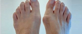
Most often, stages 2 and 3 are visible to the naked eye. Signs of hip dysplasia in children:
- femoral, gluteal or inguinal folds are asymmetrical, located at different levels;
- legs of different lengths;
- the presence of additional femoral folds;
- small amplitude when spreading the legs to the sides, bent at the knees;
- knees of different heights;
- crunching when bending and straightening the legs.
If the newborn has at least one sign, it is necessary to urgently go to the hospital.
There is an erroneous assumption that a baby with hip dysplasia cannot walk. He walks and runs no worse than his peers.
Pain and stiffness during movement occurs at a later age, more often after 5 years, as the load on the joints increases.
Prevention of dysplasia in children
For early detection and prevention of displacement of the femur, all newborns undergo an ultrasound of the hip joints in the maternity hospital or at 1 month.
When immaturity of the hip joint is detected, wide swaddling is used. One or two folded diapers are placed between the baby’s legs, giving the legs a position of extension and flexion.
The third diaper secures the baby's legs. It is quite possible to place a diaper on top of a disposable diaper. It is only important to ensure that the baby’s legs are not pressed against each other.
It is recommended to carry the child in a sling in a position on the side and on the stomach.
First of all, doctors recommend wide swaddling. You will need three diapers. The first diaper must be folded several times. So that you get a rectangle twenty centimeters wide. Place it between the baby's legs, spread apart.
Fold the second diaper into a scarf. Wrap the corners around the baby's hips. This way the legs are fixed at an angle of 90°. Wrap the third diaper around the baby's lower body. At the same time, with the help of a diaper, the legs are pulled up. This will prevent the baby from connecting his feet.
Gymnastics is especially good for prevention. In this case, focus on the abductor-adduction movements performed in the hip joints. Of course, this must be done carefully, without using force.
With a mild degree of dysplasia, this will be enough for the hip joint to develop as expected.
Exercises to prevent the development of the disease in children
The baby lies on his back. Starting position - the child lies on his back, abduct the legs bent at the knees to the sides, like a book, 150-200 times a day (but not at one time). It is necessary to place “free” fingers along the hips to control abduction.
There is no need to try your best to force your legs apart so that they touch the surface of the changing table. Movements shouldn't be painful! The main thing is not the force with which the legs are abducted, but the number of abductions. It is advisable to avoid strong rotational movements in the hip joints.
Baby on the tummy. Starting position - the baby lies on his stomach. You grab the baby's feet and try to bring the heels towards the buttocks. It should look something like a frog. In this case, you can lightly press your buttocks against the table. The number of times a day is about 100-150.
Stroking and rubbing. A light massage of the buttocks and thighs in the form of stroking and rubbing for 10 minutes a day can be done by the mother, but a more intense massage with kneading is best left to a professional children's massage therapist when the baby is already more than 2 months old. Naturally, an orthopedist must be seen at the ages of 1, 3 and 6 months.
Sources: spine5.com, deti.health-ua.org, orthoped.in.ua, www.mif-ua.com, doctorignatyev.com, asclepion.ru, www.medplus24.ru, 5gdp.by, www.moirebenok. ua
Diagnostics
In case of congenital pathology, doctors can make a diagnosis already in the maternity hospital after examining the child. Since the hip joint continues to form from the 3-4th month to the 8th, the pathology can be diagnosed during this period. This is why a routine examination by an orthopedist is so important.
The basis of diagnosis is ultrasound examination or x-ray.
How to independently determine hip dysplasia in newborns? There are a few simple tests you can do. The recommendations are:
- Check the folds on the hips, under the butt and knees , they should be at the same level. Place the baby on his tummy, straighten his legs.
- Check flexibility. Lie on your back, bend your knees and spread your hips to the sides. You can't use enormous force. In babies under one year of age, flexibility is very developed; if the amplitude is different or the angle is less than 90˚, then this is a pathology.
- Check the height of the bent legs. They must be on the same level. To check, place the baby on his back, legs bent at the knees, feet resting on the surface.
- Compare legs by length. When lying on your back, they should be the same length.
If you have even the slightest suspicion, you should go to an orthopedist.
Prognosis and complications
In the absence of medical intervention, hip dysplasia becomes the cause of the development of numerous complications. The functional activity of one or two hip joints is reduced, which leads to disruption of the entire musculoskeletal system.
Hip dysplasia provokes impaired motor skills of the spine, large and small joints of the legs. As the child grows up, gait becomes impaired due to developed flat feet, and scoliosis occurs—a persistent sideways curvature of the spinal column relative to its axis. This leads to an uneven distribution of loads on the vertebral structures during movement and the appearance of characteristic signs of osteochondrosis.
This is the name of severe degenerative-dystrophic pathology of the hip joint, which occurs due to the destruction of cartilage tissue with further deformation of the bones. In patients with dysplasia, the hip joints are formed incorrectly; under the influence of certain factors, the cartilage lining begins to thin out. After 25 years, dysplastic coxarthrosis can be triggered by low physical activity, excessive stress on the hip joint, changes in hormonal levels, and even taking drugs of certain clinical and pharmacological groups, for example, glucocorticosteroids.
Neoarthrosis
Neoarthrosis is a condition characterized by the formation of a false hip joint. With long-term dislocation, the femoral head flattens and the size of the acetabulum decreases. Where the head rests on the femur bone, a new joint gradually begins to form. Some doctors even consider this as self-healing, since the formed hip joint is capable of performing certain functions.
This pathology develops as a result of damage to the vessels supplying the head of the femur with nutrients. Aseptic necrosis in most cases occurs after surgery on the hip joint, including for the treatment of dysplasia. The femoral head begins to collapse, making independent movement impossible.
If treatment is not started immediately after the baby is diagnosed, then as the child grows older, when the child begins to stand on his feet, the load on the joint will be distributed incorrectly. In severe cases, joint dislocation occurs, even if the baby initially had an initial degree of dysplasia.
In this case, surgical intervention will be required to improve the patient's condition and regain mobility of the joint. The joint will no longer be completely healthy.
Lack of treatment for hip dysplasia in infants is dangerous; the following complications may occur:
- Scoliosis;
- Flat feet;
- Necrosis of the femoral head tissue;
- Osteochondrosis;
- Pathologies of the spine, legs and pelvis that interfere with the normal functioning of the musculoskeletal system.
Symptoms of untreated dysplasia
Treatment of hip dysplasia in babies under 3 months is the most effective, with more than 90% of cases of complete restoration of joint function. If therapy is started after six months, the chances of recovery become much lower.
Untimely treatment of the disease can lead to various serious consequences. Advanced forms of the described anomaly of the musculoskeletal system often provoke the development of the following complications:
- osteoarthritis of the hip joint in adulthood;
- decreased mobility of the spine, lower extremities and pelvic bones;
- scoliosis;
- flat feet;
- neoarthrosis;
- poor posture;
- osteochondrosis;
- necrosis of the femoral head tissue.
Treatment of THD is not particularly difficult and does not pose a threat to children's health. This disease can only be dangerous if its symptoms are completely ignored.
Treatment
Treatment is prescribed immediately after the examination. Every day is important, because as the child grows, the likelihood of negative consequences of hip dysplasia in children increases and the degree increases.
The therapy is long-term, ranging from 1 month to a year, and cannot be interrupted. During treatment, parents should be patient, as babies do not approve of therapeutic manipulations.
At an early stage, gymnastics and massage are enough; wide swaddling is important. Its essence is to keep the legs spread apart. To do this, place a diaper folded in the shape of a rectangle (15-17 cm wide) between them; it should reach the knees. The legs, bent at the knees, need to be spread apart so that the angle is 60-80˚. Then you can move on to swaddling. The baby will quickly get used to the abduction position and will not be capricious.
The massage is carried out by a specialist; if you do not apply the correct pressure on the hip, the dislocation can worsen. Therapeutic gymnastics is selected individually for each child; exercises during bathing are useful. Dysplasia in a one-year-old child is treated with active exercises, and in infants - with passive exercises. At the same time, a course of physiotherapeutic procedures is prescribed; electrophoresis is most effective.
Dislocations are treated using orthopedic devices:
- Pavlik stirrups;
- Gnevkovsky apparatus;
- spacer tires;
- plaster retainers;
- plastic corsets, for example, the Freyka pillow.
They hold the legs in a spread position.
Treatment of hip dysplasia in children after one year of age is often surgical. Using surgical manipulations, the head is set, then the limb is cast. In severe cases, multiple surgeries may be required.
Immaturity of the hip joints
Separately, orthopedists consider the immaturity of the hip joints. This is a normal condition that occurs in children in the first three months of life. This diagnosis is made in the maternity hospital or by ultrasound during the first medical examination in a children's clinic.
There is no treatment. Monitoring of the child and control ultrasound at 3 months are indicated. It is recommended to do therapeutic exercises and massage (at home on your own). Within 2–3 months, the immaturity of the tissue components of the hip joint disappears. If this does not happen, consultation with an orthopedist and treatment taking into account the severity of the child’s condition is indicated. After 3 months, untreated joint immaturity transforms into dysplasia.
Consequences
Negative consequences can be easily avoided if hip dysplasia in newborns is treated before 6 months. The treatment will be most effective and after a year you won’t have to remember about this disease. If therapy was not carried out in the first year, then severe complications arise that worsen the baby’s quality of life. How dangerous is the disease?
Consequences of hip dysplasia in newborns:
- hip dislocation, which is accompanied by severe pain;
- lameness;
- inflammatory diseases of the joints;
- arthrosis;
- dysplastic coxarthrosis.
The earlier treatment for hip dysplasia in children is started, the more effective it is. If diagnosed after one year of age, then recovery may take years. The success of therapy is questionable; sometimes chronic joint diseases occur.
With the development of arthrosis, pain interferes with the child’s movement. The symptoms that arise force one to resort to endoprosthetics.
How does hip dysplasia manifest?
To understand what DTS is, it is necessary to study the description of the disease, the list of symptoms, its variations and stages. In fact, dysplasia is not a disease; it is the name given to pathological changes in the structure of the hip joint, the anatomy of which normally guarantees mobility, providing movement in all planes.
The dysplastic type of development of the hip joints is the extreme limit of normal. During it, the anatomical and histological state of the joint changes, and the functionality of the limb is disrupted. In newborns with such deviations, different stages of development of the pathological process and, accordingly, the severity of the limitation of the body’s capabilities may be observed.
A severe form is considered to be a condition characterized by a discrepancy between the sizes of the head of the femoral joint and the acetabulum, an integral part of the massive pelvic bone. When contact is lost, the hip dislocates and when the head moves completely beyond the cup-shaped socket, hip dislocation occurs.
In the absence of adequate treatment, the consequences are serious: the cavity of the empty acetabulum becomes filled with connective and loose adipose tissue over time, and as a result, it becomes difficult to correct the dislocation.
There are unilateral and bilateral dysplasia (pathology on the right and left). It has been noticed that the left joint is more often affected, this is explained by the peculiarity of the intrauterine position of the fetus, in which the child’s left leg is compressed more strongly. Right-sided processes and bilateral dysplasia are recorded less frequently.
Prevention
Prevention of congenital dislocation should be carried out during pregnancy. It is necessary to eat properly and treat infectious diseases in advance. If the baby is overweight, you should give preference to a caesarean section.
We need to give up tight swaddling. Instead of diapers, it is better to dress the child in clothes so that his movements are not hampered. It is necessary to do exercises, massage, and also visit an orthopedist.
You should take care of your baby's health in the first year of life. Find out everything about dysplasia, what it looks like, and don’t skip routine examinations.
Author: Oksana Belokur, doctor, especially for Ortopediya.pro
What types and degrees of the disease exist?
In infants, the ligaments are overly elastic and are not always able to hold the femoral head in the glenoid cavity. Under unfavorable circumstances, she takes an unnatural position. Depending on this, four main types of hip joints are determined in a child with several subtypes:
- Normal joint
- There are minor violations.
- Subluxation of the hip.
- Severe dislocation.
Most babies have type 2a. This is a mild degree of the disease, pre-luxation. The muscles and ligaments have not yet changed, but if treatment is not started, the disease will progress to more serious stages. With subluxation, the ligaments lose tension and the head begins to move upward. A dislocation will cause it to come out of the cavity, and the treatment will be lengthy, possibly even surgical.
The form of the disease also influences the therapeutic course:
- Acetabular, when due to the irregular structure of the cotyloid cavity, inversion of the joint, cartilaginous ossification and displacement of the femoral head occurs.
- Epiphyseal, characterized by poor joint mobility and severe pain;
- Rotational – with incorrect placement of bones in the plane, leading to clubfoot.
Each form can appear on either joint or both.
Useful video about hip dysplasia in children
List of sources:
- Levanova I.V. Gribova I.V. Principles of treatment of congenital hip dislocation in children. //Matter. XXII Scientific and Practical Conference on the results of the work of the Medical Council of the Moscow Health Committee on the examination of long-term results of treatment of injuries and orthopedic diseases in children, Moscow, 1999.
- Malakhov O.A., Gribova I.V., Kralina S.E. Treatment of congenital hip dislocation in young children using functional methods. //Collection of scientific papers of the conference of young scientists New in solving current problems of traumatology and orthopedics, Moscow, 2000.
There are no similar articles.
Recovery period
Even if the treatment was successful, a child diagnosed with hip dysplasia remains under the care of an orthopedic doctor for a long time - in some cases until growth stops completely. Experts recommend performing a control X-ray examination of the hip joints once every 2 years. The child is subject to restrictions on physical activity, and it is recommended to attend special orthopedic groups in preschool and school institutions.
Hip dysplasia is a rather complex disease; many parents literally panic when they hear such a verdict from doctors. But there is no reason to be hysterical - modern medicine copes well with the pathology, timely treatment and the patience of parents make the prognosis quite favorable.
Comprehensive information about the signs of hip dysplasia, methods of diagnosis and treatment of hip dysplasia in children - in the video review of the pediatrician, Dr. Komarovsky:
Tsygankova Yana Aleksandrovna, medical observer, therapist of the highest qualification category.
44, total, today
( 167 votes, average: 4.69 out of 5)
Bruise on the face: how to get rid of it quickly?
Normal temperature in a baby: variants of the norm and causes of low-grade fever
Related Posts
Symptoms of pathology
When making a diagnosis of “hip dislocation,” certain difficulties arise in babies, because in newborns, pre-dislocation, the initial stage of the process, is more often observed.
To properly examine the child, you need a warm room. Before the examination, it is better to feed him. Under such conditions, it is easier to identify the symptoms of dysplasia.
The main symptoms of hip dysplasia:
- symptom of slipping;
- limitation of abduction in the hip joint;
- shortening of the limb;
- asymmetry of skin folds.
The most important symptom of preluxation is the symptom of slipping. It is explained by a fairly easy reduction and reverse dislocation of the femoral head from the joint cavity due to the stretched capsule and ligaments of the joint. The symptom of slippage cannot be heard during examination; it is felt with the hands as if the head of the bone is moving.
To identify it, the baby’s legs must be bent at the knee and hip joints, forming a right angle. At this moment, the doctor’s thumbs rest on the inner side, and the remaining fingers on the outer side of the thigh. Slowly begin to spread your hips to the sides. At this time, the femoral head slides into the acetabulum, and a push is felt.
As changes in the joint increase, other symptoms appear.
Limitation of abduction is mainly observed with increased tone of the muscles responsible for hip adduction. It manifests itself during neurological diseases, so when abduction is limited, an examination by a neurologist is necessary. When determining the abduction in the hip joints, the baby is placed on the back with the legs bent at the hip and knee joints.
To do everything correctly and identify this symptom, you need to achieve relaxation of the newborn’s legs, so it is better to examine the sleeping child or wait until the baby gets used to the doctor’s hands and completely relax.
Healthy joints allow you to spread your legs so that they touch the surface of the table with the outer sides of your thighs. The child grows, and the symptom loses its significance; it is detected inconsistently.
Leg shortening
Leg shortening in children is difficult to reliably determine. Shortening is determined by the kneecaps. For a baby lying on his back, the legs are bent at the hip joints and maximally at the knee joints, placing the feet side by side on the table. In this position, it is clear that the kneecap on the side of the dislocation is lower.
Also, when examining a child, the symmetry of the folds of the skin of the thigh is taken into account.
On the side of the dislocation, the inguinal and gluteal-femoral folds are deeper, and their asymmetry is visible.
If there is dislocation on both sides, this sign may not be present. And in newborns, asymmetry of the folds is often observed in healthy joints.
In newly born babies, the symptoms of congenital dislocation are mild and are not always detected. Therefore, relying only on the clinic, it is quite difficult to make a diagnosis. Doubting, the doctor sends the child for an ultrasound to clarify.
Depending on the type of THD, the disease will manifest itself differently in children at different periods of life. Severe symptoms of deviations from normal development can be noticed by attentive parents or a pediatrician during the next examination. If a diagnosis is suspected or made, the doctor prescribes a consultation with an orthopedist, who will subsequently see the child.
In newborns
Congenital hip dysplasia can be detected in newborns while still in the maternity hospital. This disease is difficult to visually recognize in grades 1 and 2, because a baby under 2 months does not feel any abnormalities, but if the problem is not eliminated in time, a feeling of discomfort and pain will begin to be felt as bones grow and cartilaginous tissue thickens.
With early dysplasia in newborns, parents may be alarmed by the following symptoms:
- asymmetry of skin folds in the area of the popliteal cavities and buttocks;
- the child reacts by crying when his legs try to separate;
- it is difficult to separate your legs bent at the knees.
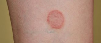
Asymmetrical skin folds on the buttocks and hips of a child with dysplasia
However, dysplasia in newborns of the 3rd degree is more pronounced, so it is difficult not to notice. In this case, the following symptoms are observed:
- Click syndrome. Occurs when the legs are spread and brought together. Always present during dislocation.
- Shortening one leg. This symptom is determined in case of severe dislocation of the limb. To do this, the child is placed on his back, and his legs are bent at the knees, placing his feet on the table. If asymmetry is noticeable at the knee level, then dysplasia is clearly present.
- Hip abduction is limited. Indicates muscle dystrophy with disruption of bone formations.
- Visible abnormal placement of the hip.
These symptoms are accompanied by additional signs:
- asymmetry of skin folds on the legs (but in infants under 2 months of age, this symptom is a variant of the norm);
- click syndrome;
- muscle atrophy;
- weak pulsation of the femoral artery;
- disturbance of the sucking reflex.
Undiagnosed and untreated hip dysplasia will cause many problems for the baby and his parents. As soon as the child begins to walk, he will feel pain and discomfort. Obvious signs of the disease will be:
- limping;
- pain when walking followed by inflammation of the joints;
- duck gait, which occurs with bilateral dislocation.
The first signs of hip dysplasia in infants may appear when they reach the age of 2–3 months, but they need to be diagnosed in the maternity hospital.
Main symptoms:
- Restriction during abduction of the unhealthy hip is typical for grades II and III dysplasia. In healthy children, the legs are bent at the knees and easily spread apart at an angle of 80–90 degrees. Pathological changes prevent this, and they can be separated by no more than 60 degrees.
- Asymmetry of folds under the knees, buttocks and groin. Normally they are symmetrical and of the same depth. Attention should be paid if, when lying on your stomach, the folds on one side are deeper and located higher. This sign is not considered objective, since it cannot indicate a problem with bilateral dysplasia. For many children, the pattern of folds evens out by three months.
- Symptom of sliding, or clicking. The head of the femur slips during movement, this is accompanied by a characteristic click when the legs are extended or adducted. This sign is a reliable symptom of abnormalities 2-3 weeks after the birth of the child. When examining children of other ages, this method is not informative.
- Shortening of one leg is a reliable sign of dysplasia and is detected when the kneecaps are aligned in the supine position. This symptom may indicate a mature hip dislocation.
- Late standing on your feet and improper walking can be observed already in the last stages of hip dysplasia.
Identification of at least one of the listed signs is a reason to contact a pediatric orthopedist.
The main symptoms of hip dysplasia in newborns can be identified simultaneously with associated symptoms.
- violation of the searching and sucking reflex;
- Muscle atrophy in the affected area;
- reduced pulsation of the femoral artery from the side of the changed joint;
- signs of torticollis.
The characteristic symptoms of hip dysplasia are also the main diagnostic criteria. The pathology can be detected by a pediatric orthopedist when examining a child in the maternity hospital. The doctor assesses the condition of the hip joints by the position and size of the legs, the mutual correspondence of skin folds in the thigh area, muscle tone, and the range of active and passive movements.
In newborns
The most informative symptom in newborns is shortening of the femur. The doctor places the child on his back, bends his hip and knee joints. Dysplastic changes are indicated by the location of one knee lower than the other. It is imperative to identify the Marx-Ortolani symptom with the child lying on his back with his legs bent. Dysplasia is indicated by a characteristic click, clearly audible when the hips are evenly and gradually abducted to the sides.
Signs of subluxation or dislocation of the hip in infants are visible upon visual examination. This:
- asymmetrically located skin folds in the groin area, buttocks and knees;
- shortened thigh;
- limited hip mobility.
Signs of the disease are best seen in children aged 3 months and older. In any case, if you notice symptoms of the disease, there is no need to panic, consult a doctor and start timely therapy. When examining the baby, the doctor pays special attention to the folds of skin in the groin, gluteal area and under the knees - in the affected areas they are larger and deeper than usual. However, with bilateral dysplasia, the asymmetry may not be so clearly visible.
However, the folds on the hips can be asymmetrical even in healthy children, so you need to pay attention to a few more indicators. Lay the baby on his back and gently bend his legs at the knees and hips. If you notice that the knees are at different levels, this is already a serious enough reason to show the newborn to the doctor.
During the examination, the orthopedist should check for symptoms of slipping and limited mobility of the hip joints. The presence of the last sign is most important when making the correct diagnosis.
- hormonal disorders during pregnancy;
- heredity: if the mother’s female relatives had or have dysplasia, the likelihood of this defect appearing in a newborn increases 4 times;
- problems during gestation: oligohydramnios, breech presentation, high weight of the child, all this contributes to a decrease in the motor activity of the baby in the womb and interferes with the normal development of the musculoskeletal system;
- birth and intrauterine injuries.
We invite you to familiarize yourself with: Arthrosis of the knee joint, stage 1, symptoms, diagnosis and treatment
Often the defect is noticed already in the maternity hospital or during the first ultrasound screening in the clinic, which is carried out at the age of 1-3 months. However, parents themselves can see the characteristic symptoms of underdevelopment of the hip joints in a baby: the folds on the surface of the thighs and buttocks are asymmetrical, the legs vary in length, and when spread in different directions, the knees do not touch the surface on which the baby is located.
A characteristic click may be heard when parents try to spread the baby's bent legs in different directions. All these symptoms should alert mom and dad and be a reason to visit an orthopedist in the near future. X-rays of the joints may be required to make an accurate diagnosis. After determining the degree of dysplasia, the orthopedist will prescribe appropriate treatment and recommend adhering to certain rules in the daily care of the baby.
Reasons for the development of DTS in children
Since DTS is a congenital pathology, it often develops during fetal development, and the following factors contribute to this:
- heredity: at risk are children whose maternal relatives had problems with joints;
- violation of early embryogenesis: pathology of the development of tissues from which the joint subsequently develops;
- hormonal imbalance in a pregnant woman;
- large fetus: the movements of such children in the womb are very limited;
- breech presentation of the fetus: babies who are born with their buttocks forward are more likely to injure their hip joints, since they are very weak and flexible;
- poor environmental situation where the pregnant woman lives: toxins entering the fetus’s body can negatively affect the development and formation of its bones;
- severe toxicosis during pregnancy: as a result of this condition, the pregnant woman loses her appetite, which means that the body does not receive the required amount of beneficial vitamins and microelements necessary for the full development of the fetus;
- oligohydramnios, multiple pregnancy, threat of pregnancy failure, taking medications;
- gynecological diseases, infectious, viral, bacterial, congenital pathologies of the uterus, heart and other organs, anemia;
- unhealthy diet, bad habits;
- tight swaddling of the baby;
- childbirth with complications.
It is possible to prevent the development of dysplasia while the baby is in the womb. To do this, you should eat right, avoid stress, give up bad habits, carefully monitor your health, and follow all the instructions of the gynecologist.
Topics you may be interested in:
Webinar on the topic of acute respiratory viral infections and acute respiratory infections
Constipation in infants with natural breastfeeding
Reviews of topics related to children's health issues
How to determine dysplasia?
To accurately identify leg dysplasia in a newborn or infant, you need to show it to a pediatrician, consult with an orthopedist, and undergo a series of diagnostic procedures. Parents can suspect the presence of pathology on their own. Dysplasia of the knee joints is easier to detect than dysplasia of the hip joints.
Signs of dysplasia
To identify it, you need to place the baby on its stomach with its legs bent. If the disease is present, asymmetry will be visible. To identify hip dysplasia, the baby should be placed on his back, legs bent at the knees spread to the side and made several rotations, holding the child’s hips. If there is pathology, it will not be possible to completely separate your legs.
In addition, the joints will move differently. The disease is also manifested by unequal length of the lower limbs. After discharge from the maternity hospital, the baby will be visited by a visiting nurse. It is important that the doctor carefully examine the baby for signs of dysplasia. At a later age, the disease manifests itself as lameness, X- or O-shaped curvature of the legs.
Other symptoms of pathology of the musculoskeletal system:
- pain when moving;
- dislocations, subluxations;
- curvature of the spinal column.
In order to diagnose pathology at an early stage, doctors prescribe a number of examinations:
- Ultrasound;
- CT scan;
- radiography;
- Magnetic resonance imaging.
Newborns are not usually subjected to any testing. But from the age of one month, when the adaptation period passes and the baby becomes stronger, ultrasound diagnostics are prescribed.
How to properly treat a child
The sooner treatment begins, the faster the correct formation of the hip joint will be achieved. To do this, various methods and means are used to help fix the child’s legs in a position of flexion and abduction. This is a special wide swaddling blanket, stirrups, splints and other devices. The younger the child, the softer and more elastic the orthopedic products that support the legs should be.
“Doctors are hiding the truth!”
Even “advanced” joint problems can be cured at home! Just remember to apply this once a day...
We suggest you read: Damage to the anterior cruciate ligament of the knee joint: treatment
{amp}gt;
Wide swaddling
This is more of a preventative measure rather than a curative one. Wide swaddling is recommended for parents of children who are at risk or who have been diagnosed with underdevelopment of the hip joint, which has not yet become the cause of pre-luxations, subluxations and dislocations. For the treatment of dysplasia, it is carried out only if it is impossible to use other, more effective methods of treatment.
To perform a wide swaddling, the baby is laid on his back, and two diapers are placed between his legs. They wrap loosely around each leg when flexed in abduction. The diapers are secured with a third, attached to the belt. This method of swaddling helps keep the legs apart at 60-80°.
In the treatment of hip dysplasia in very young children, the Freik pillow is often used. Outwardly, it looks like a dense cushion located between the knees. And to secure the legs in a physiological position for the “ripening” of the hip joint, the design provides fixing straps.
Another frequently used device is Pavlik stirrups. This is the name of an orthopedic product that resembles a chest bandage. To securely fasten the legs, it is equipped with straps located on the child’s shoulders and behind the knees, ankle locks, and straps. Less commonly used is the Vilensky splint - two leather cuffs with a metal telescopic spacer between them.
Massage treatment
Restorative massage is an important component of therapy. The pediatric orthopedist writes out a referral for sessions. The massage is performed approximately an hour after the last feeding, in a calm, relaxing environment. It begins with stroking, light kneading and rubbing. Then the massage therapist begins more intense, energetic movements. This is necessary to strengthen the muscles of the thighs and legs and improve blood circulation. At the final stage, stroking is performed again.
Daily physical therapy exercises are necessarily indicated for dysplasia. A set of exercises is compiled by a pediatric orthopedist, taking into account the severity of the disease, the age of the child, and his general health. He shows parents how to perform the movements correctly to avoid excessive stress on the hip joint. What exercises are most effective:
- the legs are retracted to the sides, and then they perform circular movements with a small amplitude;
- when lying on your stomach, the legs are smoothly moved to the sides and then brought together;
- lying on the back, the legs are raised and the child's feet are brought together.
Physiotherapy
To accelerate the “ripening” of the hip joint in its anatomical position, physiotherapy is used. Electrophoresis is prescribed with solutions of calcium, phosphorus, iodine - elements necessary for the proper formation of bone and cartilage structures. In total, about 10 sessions are performed, but if necessary, the course of treatment is extended.
UV irradiation of joints is also practiced according to a scheme determined individually. Thanks to the penetration of ultraviolet rays into the skin to a depth of 1 mm, local immunity is strengthened, metabolic and regenerative processes are accelerated.
Indications for surgical intervention include severe hip dysplasia, detected at the age of 24 months, and the presence of anatomical defects in which it is impossible to reduce the dislocation. Surgeries are performed when the joint capsule is pinched, underdevelopment of the pelvic bones or hip bones. If it is impossible to reduce the head of the femur using a closed method, then surgical intervention is also used. What operations are performed for dysplasia:
- open reduction of dislocation - reduction of the femoral head into the acetabulum after dissection of the articular capsule, followed by casting for 3 weeks;
- surgery on the femoral bone - giving the proximal end of the femur the correct configuration using osteotomy;
- surgery on the pelvic bones - creating a support for the head of the femur, preventing it from slipping out of the socket.
CLASSIFICATION
The classification of dysplasia is based on a violation of the development of a certain part of the hip joint. With acetabular pathology, the acetabulum is formed incorrectly - its size is reduced, and the cartilaginous rim is underdeveloped. Femoral dysplasia is characterized by a violation of the angle of articulation of the femoral neck with its body. And rotational pathology is manifested by changes in bone geometry in the horizontal plane.
Pediatric orthopedists also classify newborn dysplasia depending on the severity of hip joint underdevelopment. The degree of pathology always becomes the determining factor when choosing treatment methods.
| Severity of hip dysplasia | Characteristic features of the pathology |
| First (pre-dislocation) | The osteochondral elements are underdeveloped, the muscles and ligaments are not pathologically changed, the head of the femur is not deviated |
| Second (subluxation) | Underdevelopment of the bone and cartilaginous structures of the hip joint caused displacement (dislocation) of the femoral head upward and outward |
| Third (dislocation) | The articular surfaces do not contact each other, which leads to the femoral head slipping out of the acetabulum |
- Acetabular dysplasia is a deviation in the structure of the acetabulum. The limbus cartilage, located along its edges, is affected. Pressure from the femoral head causes its deformation, displacement and inversion into the joint. The capsule is stretched, cartilage ossifies, and the femoral head moves.
- Epiphyseal. Such dysplasia of the hip joints in newborns is determined by stiffness of the joints, deformation of the limbs and the occurrence of pain. It is possible to change the diaphyseal angle towards increasing or decreasing.
- Rotational dysplasia. The placement of the bones when viewed in the horizontal plane is incorrect, resulting in clubfoot.
- I degree – pre-dislocation. A developmental deviation in which the muscles and ligaments are not changed, the head is located inside the beveled cavity of the joint.
- II degree – subluxation. Only part of the femoral head is located inside the articulation cavity, as it moves upward. The ligaments are stretched and lose tension.
- III degree – dislocation. The head of the femur comes completely out of the socket and is located higher. The ligaments are tense and stretched, and the cartilaginous rim fits inside the joint.
Recommendations for parents
Hip dysplasia in children involves constant wearing of orthopedic devices, which sometimes negatively affects the mental development of the child:
- causeless tearfulness and anxiety;
- on the contrary, inhibition and lack of reaction to the environment;
- retardation in physical development.
Mothers need to be very careful when diagnosing joint dysplasia in children: even if the doctor plays it safe by insisting on constantly wearing a splint, first of all make sure that the diagnosis is valid using ultrasound and x-ray examination .
When dysplasia in children is at the stage where it is no longer possible to do without orthopedic devices, then you should not make a problem out of it: the disease is congenital, the parents have nothing to do with it. Discuss your concerns with your doctor, and consult with the parents of other sick children. Caring for children with hip dysplasia is difficult, but loving parents will go to great lengths to ensure their baby is healthy! If your child needs to correct the position of the hip joint using orthopedic devices, the following recommendations will help you:
- Study in detail the rules for handling stirrups or splints.
- Treat your child as normal without drawing his attention to the device he is wearing.
- Keep your baby busy with new toys, play fun games with him, and go for walks more often.
- Check the child’s skin in areas adjacent to the splint for abrasions, inflammation, and allergic reactions.
- Do not put any rough or irritating materials between your body and the splint.
- No matter how difficult it may be, do not give free rein to your emotions, learn to deal with stress, fatigue, take care of your health - in the future you will need a lot more strength and energy.
Possible consequences and prognosis
If therapy was started in a timely manner and selected correctly, then the prognosis for the treatment of leg dysplasia in children will be favorable. Ignoring the signs of the disease and an illiterate treatment plan can lead to the development of a number of complications.
Possible consequences of leg dysplasia in a child:
- gait disturbance, lameness . Its severity depends on the degree of damage to the joints of the limbs;
- complete dislocation . This leads to the fact that the baby will no longer be able to step on his foot. Walking will cause increased pain in him;
- severe shortening of the limbs . Observed at the age of 3-4 years. If the process is bilateral, then a slight delay in leg growth may be noticeable.
If there is severe leg dysplasia, the child is assigned a disability group. The decision to issue it is made by the medical commission. Experts assess the severity of the violations and the nature of the damage. In cases of moderate dysplasia and the appearance of persistent complications, a third disability group is usually assigned, and in more severe cases, a second disability group.
If a newborn has been diagnosed with leg dysplasia, then parents should try to help the child overcome this pathology. It is necessary to do wide swaddling, perform gymnastics, and take the baby to physiotherapeutic procedures.
By following the treatment plan and the recommendations of the orthopedist, in 90% of cases, before the age of six months, it is possible to achieve complete reduction of the hip joint. A timely course of therapy helps the child grow up healthy and lead a normal life.
Main forms of dysplasia
In infants, the ligaments are very soft and elastic, which is why they cannot always hold the hip joint in place; then they take an incorrect position, which is dysplasia.
There are several forms of the disease, the treatment of which differs from each other:
- Acetabular. The structure of the main elements of the hip joint is disrupted, most often the marginal surface and limbus are affected, due to which the position of the joint changes significantly. This form of dysplasia limits the usual movements of the joint;
- Epiphyseal. This disorder is characterized by impaired mobility in the joint and severe pain;
- Rotary. The anatomical structure of the joint turns out to be incorrect, which is why gait is disrupted and flat feet appear.
Also, the pathology can be of varying severity, the severity of the signs of dysplasia and, accordingly, its treatment depend on this. X-ray indicators of hip dysplasia, which are visible in the image:
- Pre-luxation is characterized by underdevelopment of the ligamentous apparatus, the femoral head is held in the acetabulum. This form of deformity rarely leads to disability;
- Subluxation is a position in which the head of the femur is located in the socket, but partially protrudes from it. This is a borderline condition that develops against the background of signs of pre-dislocation;
- Dislocation is the last degree of deformation in which the head of the femur comes out of the socket, which leads to gradual curvature of the joint and negatively affects the mobility of the leg.
There are unilateral and bilateral hip dysplasia; in newborns, the latter option usually manifests itself - simultaneous damage to both femoral joints.
Interesting!
Hip dysplasia has an ICD-10 code - M24.8.
Various forms of pathology in infants
Forecast
With timely detection of pathology and treatment from the first month of a baby’s life, the prognosis is quite favorable: conservative treatment is effective, and in 95% of children the diagnosis is removed by the age of one year.
If the treatment is ineffective or the diagnosis of DTS is late, the consequences can be very serious, and there is a high probability of the child becoming disabled.
In the case of undiagnosed or untreated DTS, the following complications may develop:
- necrosis of the femoral head without circulatory impairment (aseptic necrosis);
- coxarthrosis: degenerative process in the hip joint;
- neoarthrosis, or pseudo-joint, forms independently and cannot function normally: the femur completely comes out of the socket, the socket gradually heals, and the femoral head forms a new cavity in the pelvic bone; gradually the femoral head is destroyed, this leads to lameness, pain and disability.