Causes
Ulcers form against the background of tissue hypoxia, caused by disorders of microcirculation, metabolism and tissue innervation. The skin becomes very vulnerable, and even superficial microdamages lead to the formation of a poorly healing deep defect.
Secondary infection and blood stagnation contribute to the accumulation of toxins in the affected area, which accelerates the development of necrosis and the progression of pathology.
What causes trophic ulcers on the legs? These disorders can develop due to the adverse effects of various causes.
There are external factors that can provoke the formation of an ulcer. These include the following skin damage:
- wound (scratches, cuts, bruises);
- frostbite;
- thermal or chemical burns;
- chronic inflammatory and allergic skin diseases (furunculosis, eczema, dermatitis);
- radioactive skin damage;
- bedsores with prolonged immobility of the patient.
The causes of a trophic ulcer on the leg can be complications of the following diseases:
- chronic vascular insufficiency - varicose veins, thrombophlebitis, arterial circulation disorders due to stenotic processes;
- endocrinological pathology - diabetes mellitus, obesity;
- arterial hypertension;
- lymphostasis of any etiology;
- autoimmune diseases;
- immunodeficiency conditions, including HIV infection;
- CNS lesions - brain or spinal cord injuries, strokes, degenerative diseases of the nervous system;
- infectious pathology (tuberculosis, syphilis);
- blood diseases;
- poisoning with arsenic, chromium and heavy metals.
Experts often state a combination of external and internal factors that led to the formation of a trophic ulcer.
Preventing leg ulcers
The main means of prevention is timely treatment of diseases that can provoke the appearance of trophic ulcers. After surgery, patients should:
- avoid damaging the skin on the operated leg;
- refrain from excessive physical activity;
- use elastic bandaging or special orthopedic bandages;
- eat a lot of fresh vegetables and fruits;
- lead a healthy lifestyle, do not smoke;
- control blood sugar levels;
- undergo training at the “School of Diabetes” and learn all the information about diabetic feet.
Trophic ulcers are associated with impaired arterial or venous blood flow, as well as with impaired nervous regulation of metabolic processes. Such defects occur due to chronic tissue malnutrition, and therefore are resistant to treatment and take a long time to heal. After relieving acute inflammation, the only effective method of getting rid of a trophic ulcer is surgery.
Symptoms
How does a trophic ulcer begin? A person complains of rapid fatigue when walking, a feeling of heaviness in the legs with minor loads, which persists at rest. There is itching of the skin, a burning sensation, crawling in the area of the foot or lower leg, cramps in the calf muscles, especially at night.
Upon examination, the doctor discovers the following initial signs of a trophic ulcer:
- pastosity of the soft tissues of the lower extremities;
- thickening, roughening of the skin of the affected area;
- change in the color of the skin in the affected area - a growing spot of grayish or blue-violet color, reminiscent of a hematoma with a glossy tint;
- increased sensitivity at the site of ulcer formation, acute pain when lightly touching it;
- There may be serous-mucous discharge on the skin in the area of the spot.
With minor trauma, overload, or nervous fatigue, the disease progresses quickly. In the center of the spot, an area of atrophy of the epidermis of a whitish color forms, and a bloody discharge appears.
Symptoms of a trophic ulcer at the height of the disease are as follows:
- severe swelling of the leg or foot; more about leg swelling→
- the presence of an ulcerative lesion, which, if left untreated, tends to quickly increase and deepen;
- bloody purulent discharge with an unpleasant odor;
- excruciating pain in the legs, worsening with exercise and at night, limiting physical activity;
- general condition disorders (fever, chills, fatigue, poor appetite).
Prevention
As a rule, trophic ulcers are formed as a result of poorly treated varicose veins. Therefore, their prevention includes the prevention of varicose veins and other diseases that provoke the occurrence of ulcers. Give your legs a rest; if symptoms appear (spider veins, swelling), start using ointments, gels, bandages. Wear compression garments, do gymnastics, stick to a diet.
When the first symptoms of a trophic disease occur, special attention should be paid to the hygiene of the affected skin area.
Prevention of trophic ulcers caused by a sedentary lifestyle is based on improving its quality, style, and performing physical exercises that help normalize blood circulation and strengthen the immune system. Disruption of the endocrine system and metabolism can provoke an increase in a person’s body weight and negatively affect the restorative function of the dermis. As a result, the legs will experience excessive stress every day, and the skin may crack and collapse under the pressure of heavy weight.
Prevention of trophic ulcers includes giving up bad habits and a balanced, proper diet. If you notice the first signs of venous insufficiency (swelling, increased leg fatigue, small blue vessels), use elastic bandages. This will help improve skin microcirculation. Timely comprehensive treatment of the disease allows you to avoid the formation of ulcers.
Kinds
Ulcers are classified depending on the cause of their formation.
Venous trophic ulcer occurs in 8 out of 10 patients. The cause is stagnation due to impaired venous circulation. A trophic ulcer of the leg most often forms in the lower third on the inner surface of the leg.
Arterial
The background for the development of pathology in 20% of patients is obliterating diseases of the arteries of the lower extremities. Arterial stenosis leads to ischemia and necrosis of the soft tissues of the legs. Provoking moments are most often injuries or hypothermia of the extremities, the use of uncomfortable shoes.
Features of this type of trophic ulcers: most often occurs in old age, a person experiences difficulty walking up stairs and a feeling of coldness in the leg.
On examination, the leg is cold. Most often, small ulcers with symptoms of suppuration are found in the heel, big toe and on the outer surface of the foot. They have an oval shape, dense edges, and the skin around them acquires a yellow tint.
Diabetic
Trophic foot ulcer is a common complication of diabetes mellitus. Typical localization is the big toe or injured corns in the sole area. Develops in diabetic angiopathy against the background of sharp fluctuations in blood glucose levels.
Characteristic signs: decreased sensitivity in the legs, extinction of tendon reflexes, rapid increase in size of the defect, tendency to develop necrosis, resistance to therapy.
Neurotrophic
When the central nervous system is affected, the ulcers are located in the heels and soles.
Features of neurotrophic ulcers: small size and significant depth of wounds (down to the bone), abundant purulent discharge with an unpleasant odor in the absence of pain.
Hypertensive
They are rare, usually in elderly women against the background of long-term arterial hypertension.
They are characterized by slow formation, and on both legs at once, severe pain syndrome, and a high frequency of purulent complications.
Infectious
They form on the legs with advanced skin diseases against the background of unsanitary conditions and the accumulation of secondary purulent infection. Features: semicircular shape, small size and depth.
Mechanism of lesion formation
This process is quite lengthy, so it is quite difficult not to pay attention to it. Exceptions may be those cases when active and serious pathogens of various infectious diseases have entered the affected areas through the bloodstream. Then the first stage will be in all its glory in just a few hours.
The veins of the lower extremities cease to cope with their function normally. They overstretch, and the phenomenon of reflux occurs - the reflux of blood against its natural course.
In this condition, all trophic processes and innervation mechanisms are disrupted. Nutrients and oxygen are not fully supplied, local immune forces are practically zero. The skin ceases to perform barrier and protective functions. Thus, the gates for infection and bacteria to enter directly into the blood are open.
Negligent attitude towards oneself, non-compliance with doctor’s recommendations or arbitrary, uncontrolled self-medication entail huge problems that will not only reduce the ability to work, but also lead to disability or death.
Diagnostics
A trophic ulcer on the leg is accompanied by specific complaints and has a characteristic appearance upon examination. The main task of specialists is to find out the cause of the disease. Only by establishing the etiology of the ulcerative lesion can adequate therapy be prescribed.
The following diagnostic measures are carried out:
- A thorough history taking and examination by a therapist, phlebologist, angiosurgeon.
- Laboratory methods: general blood and urine tests, including determination of glucose levels, biochemical blood test, immunogram, bacteriological analysis of ulcer discharge and biopsy of biomaterials taken.
- Instrumental methods: rheovasography, angiography with the introduction of contrast agents, ultrasound of the vessels of the lower extremities (Dopplerography, duplex study).
How can you get infected?
A trophic ulcer develops not only as a result of diseases, but can also be diagnosed in the case of infection of the soft tissues of the legs, when the cause is anthrax or syphilis.
In the photo below you can see what a trophic ulcer of the lower extremities looks like.
Syphilis is transmitted through contact and household contact. Infection of the foot is quite possible if simple hygiene conditions, timely treatment of the wound, or a cut on the sole of the foot are not observed.
Infection can occur in children during intrauterine development, passage through the birth canal, or through mother's breast milk. Syphilis can be contracted through blood during a transfusion.
The source of anthrax infection can be contact with animals or consumption of contaminated milk or meat.
Treatment
The main directions of therapy are the impact on the underlying disease, the fight against secondary infection, and the stimulation of healing of the ulcerative surface.
Treatment of trophic ulcers depends on the following factors:
- the cause of the appearance and duration of existence of the skin lesion;
- patient’s age and presence of concomitant diseases;
- size and depth of the skin defect, growth rate;
- condition of the skin around the ulcer.
How to cure a trophic ulcer? Therapy is extremely complex, complex and lengthy. It is more often regarded as preparation for surgery, since conservative methods are not very effective.
If surgery is contraindicated for a patient for some reason, the goal of therapy is to prevent the skin defect from enlarging and deepening.
In a hospital, a patient with a trophic ulcer is provided with bed rest; the affected limb should be in an elevated position to improve blood and lymph circulation.
Conservative
A universal cure for trophic ulcers, equally effective for all types of disease, has not been developed. Treatment of trophic ulcers on the leg includes taking drugs of various pharmacological groups.
How to treat trophic ulcers with varicose veins? Experts prescribe complex therapy, the purpose of which is to stop inflammation and tissue necrosis and stimulate healing processes.
Medicines from the following groups are used:
- Vasoactive drugs , including phlebotonics (Troxevasin, Detralex) and antiplatelet agents (Thrombo-ass). Helps dilate blood vessels, improve microcirculation, strengthen vein walls and reduce venous stagnation. Read more about Trombo Ass and its analogues→
- Infusion therapy - solutions of hemodez, rheopolyglucin for symptoms of intoxication.
- Decongestants . Used according to strict indications, periodically, under the supervision of a doctor. When taking them, the content of electrolytes in the blood is monitored.
- Antibiotics - for infectious etiology of the ulcer or the accumulation of a secondary infection. Prescribed after isolating a pathogenic agent from ulcer secretions and determining its sensitivity to antibiotics of various groups.
- Anti-inflammatory drugs - in the presence of purulent discharge, inflammatory reaction of surrounding tissues and severe pain.
- Hyposensitizing drugs reduce the secretion of secretions from the ulcer surface.
The same drugs in different combinations are used in the treatment of trophic ulcers of any etiology.
During the regeneration period, antioxidant, metabolic and immunostimulating therapy is prescribed - injections of Mexidol, Actovegin, ascorbic acid, B vitamins.
Physiotherapy is used to heal trophic ulcers. UHF is used to enhance the anti-inflammatory effects of drugs, electrophoresis with solutions of wound-healing, anti-inflammatory and vascular drugs. Laser treatment is used to reduce pain and inflammation.
During the regeneration and postoperative period, ultraviolet irradiation, ozone therapy, mud therapy, hyperbaric oxygenation, and plasmapheresis are recommended.
Treatment of trophic ulcers of the lower extremities complements proper nutrition. To avoid increasing swelling in the legs, a person should eat more vegetables and fruits, control the amount of fluid consumed, and exclude salty, spicy, pickled foods. With diabetes, patients eat in accordance with the recommendations of the endocrinologist and regularly monitor blood glucose levels.
Local therapy
To cleanse the wound of pus and dead tissue, it is treated with disinfectants.
The following antiseptics are used:
- aqueous solution of chlorhexidine;
- 3% boric acid solution;
- potassium permanganate solution of a certain concentration;
- Rivanol;
- enzymes.
How to treat trophic ulcers on the legs after cleansing them of pus? For healing and accelerated scarring of skin defects, medicinal ointments are used: Solcoseryl, Ebermin, Actovegin. It is effective to use special dressings and sponges with antibacterial, antisecretory and wound-healing effects (Allevin, Algipor, Geshispon).
It is mandatory to use an elastic bandage, which is replaced as necessary several times a day. It not only protects the affected area from secondary infection and injury, but also helps reduce swelling.
Surgical intervention
A trophic ulcer on the leg can be completely cured through surgery. In order for the operation to be successful, it is necessary to competently prepare the patient for the upcoming intervention, achieve improved well-being, stabilize the general condition and begin healing of the wound surface.
The following methods have been developed in angiosurgery:
- bypass surgery for vascular diseases;
- removal of a section of a vein with varicose nodes (phlebectomy).
If there is a large and deep ulcer, a skin flap is transplanted.
Traditional methods
The question of the possibility of outpatient treatment of trophic ulcers is decided only by the treating specialist. With a short duration of the disease, small single ulcers that are at the stage of regeneration, the patient can use folk recipes as a supplement to the main therapy.
How to treat a trophic ulcer at home? Clean the wound surface from any remaining pus, as it slows down healing. For this purpose, decoctions of chamomile, celandine, string, and calendula are used.
After washing the ulcer, with the permission of the doctor, apply pharmaceutical ointments that help relieve inflammation and healing, including Vishnevsky ointment and ichthyol ointment.
You can treat the ulcerative defect with golden mustache juice, make a bandage with an ointment prepared on the basis of propolis, comfrey, and arnica. Apply compresses with powdered leaves of tartar, golden mustache, willow or oak bark, which can be left overnight.
You cannot rely only on traditional medicine; refusal of comprehensive hospital treatment can lead to life-threatening consequences.
Treatment of trophic ulcers at home
Herbal medicine
Traditional medicine offers the following recipes based on the use of medicinal herbs:
- Pour two tablespoons of dried lavender inflorescences into 500 ml of boiling water and let it brew for several hours. After which the broth should be filtered and used as a local antiseptic, treating the wound daily.
- Pour three tablespoons of eucalyptus and the same amount of calendula into 500 ml of water and boil for several minutes. Use as lotions. Eucalyptus has an antimicrobial effect, and calendula dries and accelerates the healing of ulcers.
- Take one tablespoon of chamomile, oregano, fireweed, mix with 250 ml of boiling water and let it brew for 6 hours. Next, one tablespoon of propolis tincture is added to the decoction and used as a lotion.
Compresses
The following compresses are often used to treat trophic ulcers:
- Mix egg yolk with natural, liquid honey, in a ratio of 1 to 1. The mixture is applied to a previously cleaned wound, covered with a sterile bandage and left overnight. It is recommended to carry out at least 5 such procedures.
- Several streptocide tablets are ground to a powder, mixed with a teaspoon of 3% hydrogen peroxide, applied to the wound and left overnight. This compress should be done daily for 5-7 days.
- Take 100 ml of olive oil, 50 grams of natural wax and 4 quail yolks, pre-cooked (the yolk must be firm). All ingredients are mixed to form an ointment, then the product is applied in a thick layer to the wound, covered with a sterile bandage and left for 10-12 hours. A course of 10 procedures is recommended.
How dangerous is the disease?
Ulcers are dangerous due to rapid progression in severe cases of the underlying disease. At the same time, they not only increase in size, but also become numerous and can merge into a large wound surface.
This pathology is also distinguished by the following features:
- tendency to relapsing course;
- resistance to all methods of therapy, except surgical treatment, which may have contraindications;
- the possibility of developing sepsis - with ulcers of venous origin, gangrene - with diabetic ulcers, osteomyelitis.
Stages of the disease
Doctors classify several degrees of trophic ulcers, photos of which can be easily found on the Internet:
- initial . At this stage, the disruption of the trophic processes of the skin just begins. The color may be slightly changed: pallor, bluishness or redness. The main symptom is the phenomenon of “patent leather”: it is so taut that it has an almost artificial shine. It is difficult to pinch because of the swelling. Sometimes moisture starts to appear. This stage can last from several hours to 1-1.5 months. It all depends on the general condition of the patient, the presence of additional microbial insemination or concomitant diseases. Sometimes the patient may experience fever, mild chills, general weakness,
- second . Intensive rejection of affected and necrotic areas. This phenomenon is accompanied by an unpleasant odor, fever, loss of strength, and bleeding. The duration of this stage is also purely individual. The big problem is the itching sensation. The patient scratches the wound, thereby disrupting the process of its cleansing, introducing pathogens,
- the third stage is characterized by the beginning of epithelization of the lesion . Slowly the wound decreases in size. It can simultaneously have several foci of healing and islands of young tissue. There is a fairly high risk of re-starting the first stage. To prevent this, ensure the process is aseptic as possible,
- fourth : Complete healing or scarring. It sometimes drags on for years, so the task of doctors is to restore regenerative abilities and local immunity as much as possible.
During the initial stage, trophic processes in the skin are disrupted. A color change occurs. There is pallor, cyanosis or redness. The main feature is patent leather. She's stretching a lot. An almost artificial shine appears. It is impossible to pinch it because it is swollen. Sometimes there is moisture. The period lasts from several hours to one month. Everything depends on the general condition of the patient, how additional microbial insemination occurs or concomitant diseases. Occasionally, patients experience an increase in temperature, mild chills, and general weakness.
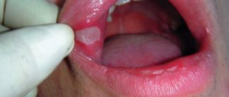
Mechanical ulcers: mouth ulcers, etc.
The symptoms of all ulcers depend on their location and origin, especially when it comes to defects of the mucous membranes. Mechanical type defects appear mainly after pressure, friction or stretching of certain areas of the mucous membranes or skin. In this case, we can distinguish ulcers in the mouth, which, according to statistics, most often appear due to uncomfortable dental prosthetic structures and teeth that scratch the mucous membrane of the mouth. Such problems are easily eliminated and the defect, as a rule, disappears after eliminating traumatic moments. The same effect applies to ulcers of the stump of an arm or leg, which appear due to a poorly fitted prosthesis.
Therapy aimed at eliminating ulcers that appear due to sprains is difficult. Often, anal fissures even require surgical intervention, and cracked nipples and lips take a long time to heal.
Mechanical ulcers are often encountered in military field surgery. They appear mainly on the legs, in particular after injuries to the calf muscle or popliteal fossa.
Ulcer: features, symptoms of peptic ulcer, treatment of ulcers
An ulcer is a granulating defect of the mucous membranes or skin that is not prone to rapid healing. These defects are quite diverse in their morphology. Based on criteria such as shape, type of edges and depth, several types of ulcers are distinguished: callous, slit-like, round, etc. Experts also note internal ulcers, which are located in the internal organs, and external ones, located on the surface of the body (often visible to the naked eye) .
Chemical ulcers
Chemical ulcers are often diagnosed in people who work in the fishing industry and are involved in salting. Defects that appear on the skin of the hands are sometimes accompanied by swelling and redness of the surrounding skin, which indicates the addition of a purulent infection. The ulcers are usually multiple and can be easily treated if you stop working for a while and use antiseptic dressings on the advice of your doctor.
Skin ulcers that appear due to the negative effects of digestive juices are difficult to treat, for example, when intestinal fistulas or pancreatic fistulas are diagnosed.
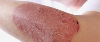
Traumatic ulcers
Traumatic ulcers often appear on the elbow and under the knee, that is, in those places where the defect in the skin is constantly subject to stretching/friction. Treatment of ulcers can be effective if the patient adheres to bed rest and reduces the “use” of the affected limb to a minimum.
It is worth considering that both traumatic and other types of ulcers become trophic after a certain period of time. This occurs due to inflammatory and cicatricial changes that affect small nerve branches around the ulcer.
Complications
The ulcerative process involves not only the skin, but also muscle tissue, the Achilles tendon and bones.
Ulcers must be treated promptly, otherwise the following complications develop:
- bleeding;
- pyoderma;
- eczema, dermatitis;
- fungal infections;
- osteomyelitis, arthritis, arthrosis;
- phlegmon;
- tetanus;
- erysipelas;
- inguinal lymphadenitis;
- lymphangitis;
- thrombophlebitis;
- purulent varicothrombophlebitis;
- wound myiasis;
- sepsis.
The most dangerous complication is death due to blood poisoning.
Trophic diabetic ulcers on the toes most often cause gangrene and amputation.
If the infectious process is not stopped in time, vascular obliteration (lymphedema) develops. With ulceration of the heel area, such a complication can lead to disability.
anthrax
This is an infectious disease characterized by a severe course, affecting both people and animals. The disease is accompanied by acute intoxication and is characterized by damage to the skin and lymph nodes. There are several forms of anthrax manifestation:
- cutaneous;
- pulmonary;
- gastrointestinal;
- septicemic;
- other forms (meningitis);
- unspecified form.
Anthrax can be localized or generalized. The localized, that is, cutaneous form of the disease is most common.
The causative agent of the disease is Bacillus anthracis, a non-motile bacterium of rather large size in a transparent capsule shell, resembling a bamboo cane in shape. These bacteria are vulnerable and die from disinfectants and even not too high temperatures. But the greatest danger is posed by the spores they produce, which can remain in the soil and aquatic environment for decades, in wool for months, and in skins for years.
The virulence of bacilli lies in their ability to create a shell and produce an exotoxin - a protein substance that includes protective, lethal and swelling-provoking components. The capsule shell helps the pathogen to gain a foothold in the tissues.
The main source of infection is infected cattle, which produce anthrax bacillus into the external environment throughout the entire duration of the disease. However, an infected person is not dangerous to healthy people, unlike animals.
Animals usually become infected by eating, for example, grass or as a result of a horsefly bite. As a rule, the disease occurs in them in the form of sepsis or in the intestinal, less often cutaneous (carbunculous) form.
Humans can become infected through infected animals and the bites of insects such as horseflies, burner flies, and mosquitoes. Anthrax primarily affects children and adolescents, most often boys. The pathogen enters the human body through any damage to the skin, in rare cases through the mucous membranes of the gastrointestinal tract, mouth, nose and eyes.
Having penetrated and fixed itself, the rod begins to multiply, simultaneously producing products of its vital activity, which is nothing more than an exotoxin and a specific shell. A carbuncle appears on the infected skin, which is a necrotic hemorrhagic inflammation of the skin, as well as subcutaneous tissue.
With mobile macrophages, the bacillus from the site of its penetration begins to spread to nearby lymph nodes, resulting in the development of acute lymphangitis, sepsis and lymphadenitis.
All people are susceptible to anthrax. After the disease is cured, a person receives stable post-infectious immunity, however, cases of re-infection after several years have been recorded in medical practice.
Stomach and duodenal ulcers
This is a chronic disease, which is characterized by a violation of the integrity of the mucous layer of the stomach and/or duodenum. According to statistical data, such an ulcer is diagnosed mainly in men.
Gastritis and stomach ulcer: what are the differences
Gastritis and ulcers are among the most serious diseases of the gastrointestinal tract. These diseases can be distinguished both by symptoms and by the results of diagnostic studies.
Gastritis is characterized by an inflammatory process that develops on the mucous membrane of the body of the stomach. Both acute and chronic gastritis can develop as a result of exposure to irritating factors on the mucous membrane. It can be:
- chemical substances;
- food (hard to digest, spicy, too cold or hot, smoked, fatty);
- drinks containing alcohol;
- improper, irregular nutrition;
- medicines (sulfonamides, biomycin and others);
- microbes (staphylococci, salmonella);
- toxic substances;
- products produced due to metabolic disorders;
- infection with the bacterium Helicobacter pylori, which causes inflammation of the mucous membrane;
- regular stress, neuroses, depression;
- food allergies;
- vitamin deficiency;
- diseases of endocrine nature;
- genetic predisposition (heredity);
- malfunction of the immune system.
The gastric mucosa can become inflamed either entirely (with diffuse gastritis) or in patches (with focal gastritis). Gastritis is also classified depending on the nature of the abnormalities of the mucous membrane and can be:
- catarrhal (simple);
- fibrinous;
- purulent;
- necrotic.
Each form of gastritis has its own clinical characteristics.
The outcome of an acute disease depends on how deeply the mucous membrane is affected. In a simple form, it is able to fully recover, but relapses of the disease ultimately lead to its chronic course. If the changes in the mucosa are significant, the membrane begins to atrophy.
Chronic gastritis is also classified in terms of the severity of the disease and can be superficial or atrophic. In the first case, dystrophy affects the surface epithelium, in the second, atrophy develops, not only of the mucous membrane, but also of its glands. Chronic disease can be mild, moderate or severe.
Thus, the basis of the disease becomes inflammatory processes in the gastric mucosa, which can subsequently result in serious complications.
Stomach ulcers, unlike gastritis, are chronic. This is a cyclical disease, since its main clinical manifestation is a periodically recurrent ulcerative defect. Both gastritis and ulcers have generally similar symptoms, such as belching and heartburn, problems with appetite and bowel movements, nausea and vomiting.
Like an ulcer, gastritis causes pain, including on an empty stomach. However, the frequency of pain and the location of pain in gastritis and ulcers differ. With a stomach ulcer, pain mainly occurs at night. They do not decrease until the patient eats. But they can also appear after eating. Gastritis bothers the patient mainly during the day.
The most common complication of peptic ulcer disease is bleeding. This symptom usually occurs during an exacerbation of the disease and becomes a threat to the patient’s life. Clinically, ulcerative bleeding appears as hematemesis, vascular collapse, and tarry stools. Bleeding can also be hidden, and then it can only be detected by analyzing stool using the Weber reaction method.
The reasons for the development of peptic ulcer disease, which, unlike gastritis, more often affects men, are largely related to stress. As a result of severe stressful situations, excessive mental and emotional stress, and neuroses, the areas of the cerebral cortex that regulate the functioning of the gastroduodenal system are disintegrated. Meanwhile, the neurogenic nature of the ulcer does not always occur. Other reasons for its occurrence include:
- poor nutrition;
- smoking and alcohol abuse, drugs;
- taking a number of medications, such as acetylsalicylic acid;
- heredity.
The ulcerative defect forms gradually: at the beginning it is erosion, then an acute ulcer. Erosion is predominantly superficial and appears as a consequence of necrosis of the membrane area, on which hemorrhage is then observed, and later, rejection of necrotic tissue. Usually, erosions are quickly covered with epithelium, but there are cases when they do not heal. During the development phase of necrosis, not only the mucous membrane dies. The deeper layers of the gastric wall undergo necrosis, resulting in peptic ulcers.
There is usually only one chronic ulcer inside the stomach. Multiple defects are rare. Microscopically, gastritis and ulcers look different, so they are easy to distinguish during a specific examination. One way or another, only a doctor can make an accurate diagnosis.
Symptoms of gastrointestinal ulcers
Not only Helicobacter pylori, but also some medications, as well as neuropsychic factors can provoke the development of ulcers in the stomach or duodenum. Hereditary predisposition and diseases that contribute to ischemia and hypoxia of the mucous membrane also play an important role. The pathogenesis is not fully understood today.
Clinically, peptic ulcer disease manifests itself in a very diverse manner. Ulcer symptoms vary depending on gender, age, season, location and size of the ulcer. Moreover, the patient’s personality and profession matter.
Peptic ulcer disease, also called peptic ulcer, manifests itself in two clinical and morphological variants. This:
- duodenal ulcer;
- stomach ulcer.
These forms of the disease can be distinguished through research methods such as endoscopy, radiography and clinical examination of gastric ulcers, the symptoms of which may be different.
Traditionally, the main symptom of the disease (both gastric ulcers and duodenal ulcers) is pain. Based on the characteristics of the pain, the doctor can make a clinical guess about where exactly the ulcer is located. For example, if pain appears in the morning, there is a high probability that an ulcer has formed in the stomach. When pain occurs at night or accompanies a feeling of hunger, this indicates that the defect is in the duodenum.
Typically, pain is concentrated in the epigastric area: with a stomach ulcer - usually in the center or on the left side of the midline; in the case of a duodenal ulcer - on the right side of the midline. The pain is sometimes not too intense.
The pain can be different: dull and aching, sharp and cutting, cramping, boring. If pain occurs as contractions, it is necessary to differentiate it from the so-called acute abdomen syndrome. The time intervals between attacks of pain depend on the exacerbation of the disease, which often occurs in the autumn and spring. Pain usually decreases when the patient eats, drinks milk, or takes antacids. The pain usually decreases after vomiting.
Symptoms of ulcers in dyspeptic syndrome are belching and heartburn, problems with appetite and stool, nausea and vomiting. Meanwhile, this symptomatology is not typical for all patients. In particular, from 60 to 80 percent of patients with peptic ulcers suffer from heartburn, and not necessarily during an exacerbation. Heartburn may well last for years and appear from time to time (depending on the season) and after eating. There are cases when heartburn is the only symptom of an ulcer, its obvious manifestation.
As for belching, it appears more often with a gastric ulcer than with a duodenal ulcer. The occurrence of belching is a consequence of dysfunction of the cardia and antiperistalsis of the stomach.
Vomiting with peptic ulcers is a relatively rare symptom. It often appears at the peak of pain and brings relief to the person. For this reason, patients often induce vomiting themselves in order to alleviate their condition. If vomiting is repeated repeatedly, pyloric stenosis can be assumed. This symptom is also very typical for unbalanced patients who experience autonomic instability.
Nausea is typical mainly for the chronic form of gastritis and gastric ulcers, but almost never appears with a duodenal ulcer.
Appetite disturbances in peptic ulcer disease can sometimes occur, but in general patients' appetite is normal and even slightly increased.
Stool disorders are manifested by constipation, which usually occurs with a chronic form of duodenal ulcer (mainly in young people).
Generally speaking, the clinical manifestation of peptic ulcers in each specific case has its own characteristics, which, among other things, depend on the location of the ulcers and their number.
Cardiac and subcardial ulcerative defects are manifested by characteristic symptoms, namely:
- Mild pain radiating to the heart area. The pain occurs after eating and is quickly relieved with antacids.
- Heartburn, belching, and vomiting, which often accompany pain and appear due to disruption of the cardia sphincter and the development of reflux.
- Hernia in the area of the esophageal opening of the diaphragm.
The most common complication of peptic ulcer disease is bleeding. As a rule, gastric ulcers, the symptoms of which are characteristic of this form of the disease, are observed in the area of the lesser curvature. And if the ulcer is located exactly there, clinically it looks like this:
- Painful sensations as symptoms of an ulcer occur an hour or an hour and a half after a meal and subside when food is evacuated from the stomach.
- It happens that patients experience late dull hunger pains localized on the left side of the midline. This most often affects patients over 40 years of age.
- Heartburn and nausea appear. Much less often - the urge to vomit.
- Gastric secretions are usually normal.
Gastric ulcers that form in the area of the greater curvature are observed quite rarely. In half of such cases, they portend the development of stomach cancer.
Prepyloric ulcers most often occur in young people. Their clinical manifestation is similar to the picture observed in duodenal ulcers.
Ulcerative defects of the pyloric canal also have their own clinical characteristics. This disease is persistent and is accompanied by characteristic attacks of pain, which are almost never associated with food intake. Clinical symptoms include constant heartburn, increased salivation, and a feeling of fullness after eating.
When relapses of gastric pyloric ulcers occur over several years, dangerous complications may develop, in particular:
- pyloric stenosis;
- bleeding;
- ulcer perforation;
- penetration;
- malignancy.
If the ulcers are localized both in the stomach and in the duodenum, the peptic ulcer regularly recurs. Such cases are also characterized by sluggish scarring of ulcers and developing complications.
The classic complication of the disease is bleeding, observed in 20 percent of cases. This symptom usually occurs during exacerbation, neglect of the disease and becomes a threat to the patient’s life. Clinically, ulcerative bleeding appears as bloody vomiting, vascular collapse, and tarry stools. Bleeding can also be hidden, and then it can only be detected by analyzing stool using the Weber reaction method.
If perforation develops with a stomach ulcer or duodenal ulcer, a picture characteristic of an acute abdomen is observed: the Shchetkin-Blumberg symptom is positive, percussion reveals the absence of hepatic dullness, and muscle defence occurs.
Diagnosis of duodenal and gastric ulcers
A gastroenterologist treats and diagnoses stomach and duodenal ulcers. To confirm or refute the diagnosis, the attending physician prescribes the patient an esophagogastroduodenoscopy, which is most often simply called gastroscopy.
This diagnostic method is performed on an empty stomach in an outpatient clinic. A thin endoscope with a special optical device at the end is inserted through the oral cavity. Thanks to this modern device, a medical specialist can examine the duodenum, stomach and esophagus.
In order to clarify the diagnosis, the attending physician may recommend additional ultrasound diagnostics and radiography.
Treatment of stomach and duodenal ulcers
Today, a stomach and duodenal ulcer is no longer perceived as a lifelong sentence. The discovery of Helicobacter pylori, which plays a role in the appearance and development of defects in the mucous membrane, helped to facilitate the approach to the effectiveness of therapy.
There are several treatment regimens for ulcers. One of the main components of such regimens are antibiotics. The patient is also prescribed gastric juice neutralizers (this could be Ranitidine or, for example, Omeprazole) and agents that form a special film coating on the mucous membrane (for example, De-nol). Treating an ulcer can take a long time. It is important for the patient to adhere to a special diet, eliminate stress and not drink alcohol.
Surgery for peptic ulcers may be necessary only in rare cases. Especially when it comes to ulcers that do not heal for a long period of time or complications have appeared.
Diet for ulcers
Proper nutrition plays a special role in the treatment of stomach and/or duodenal ulcers. A diet for ulcers is primarily aimed at muting the symptoms of the disease and restoring the functioning of the gastrointestinal tract.
- Doctors strongly recommend excluding fried, salted and canned foods from the daily menu. All food must be chopped. Dishes should not be scalding or icy.
- It is advisable to reduce heavy foods to a minimum. Those foods that require heat treatment should be cooked using steam or baking.
- It is important to eat food in small portions. The break between snacks should not be more than 3 hours. And in the evening, dinner is allowed no later than a few hours before bedtime.
- The amount of salt should be kept to a minimum. No more than 8 g per day is allowed. Salt should also be taken into account in ready-made products, and not just that which is added directly during cooking. When the disease worsens, it is advisable to remove salt from the diet altogether.
A diet for duodenal ulcers may include the following foods:
- porridge (buckwheat, oatmeal, rice, semolina);
- poultry and lean fish;
- dried white bread;
- juices from non-acidic fruits and vegetables in diluted form;
- honey;
- jelly, various rosehip decoctions, weak teas;
- boiled chicken and quail eggs;
- milk;
- soups prepared according to vegetarian recipes.
A diet for ulcers requires the exclusion of certain foods that can negatively affect the health of a person suffering from peptic ulcer disease. Here it is worth highlighting:
- bakery products made from bran and rye flour;
- baked goods;
- sour fruits and vegetables;
- legumes;
- coffee;
- alcoholic drinks;
- any mushrooms;
- wheat and pearl barley porridge;
- fatty fish and meat soups;
- fermented milk products, etc.
It is advisable to adhere to a diet for ulcers for at least several months. If the disease is chronic, then the diet should be lifelong. Outside the acute stage, the daily menu for stomach or duodenal ulcers may look like this:
- The first breakfast is jelly from non-acidic fruits, steamed omelette, semolina porridge.
- Second breakfast – pumpkin and carrot casserole.
- Lunch – vegetable stew, rosehip decoction, steamed meatballs and buckwheat soup.
- Afternoon snack – weak tea, preferably green. You can supplement your afternoon snack with dietary marmalade.
- Dinner – compote of non-acidic fruits, rice porridge, lean fish, steamed.
The attending physician will tell you in detail about the features of the diet. In addition, this specificity depends on numerous factors, including the age of the patient and the severity of the disease.