A cerebral aneurysm (also called an intracranial aneurysm) appears as a small abnormal formation in the blood vessels of the brain. This compaction can actively increase due to filling with blood. Until it ruptures, such a bulge is not dangerous or harmful. It only exerts slight pressure on the organ tissue.
Online consultation on the disease “Cerebral aneurysm”. Ask a question to the specialists for free: Neurologist, Surgeon.
- Etiology
- Varieties
- Symptoms
- Complications
- Diagnostics
- Treatment
- Prevention
When an aneurysm ruptures, blood enters the brain tissue. This process is called hemorrhage. Not all aneurysms can be complicated by hemorrhage, but only some types. In addition, if the pathological bulge is quite small in size, then it usually does not cause any harm.
Aneurysms can occur anywhere in the blood vessels that supply the brain. The age of a person does not matter. But it is still worth noting that middle-aged and older people are most often susceptible to the disease; children are diagnosed very rarely. Doctors note that neoplasms in the brain vessels appear less often in men than in the fair sex. People between thirty and sixty years of age are often at risk.
The rupture of a cerebral aneurysm becomes “fertile ground” for strokes, central nervous system damage, or more dire consequences. It is noteworthy that after one rupture such a pathological formation can appear and burst again.
General information
An aneurysm is a condition in which there is a protrusion of the wall of an artery , or in more rare cases, a vein . This occurs as a result of stretching or thinning of the artery. Due to this process, an aneurysmal sac appears, sometimes compressing tissue located nearby. As a rule, an aneurysm is a congenital phenomenon. At birth, such a pathology is not detected, the child develops normally. An aneurysm manifests itself as a result of diseases in which the blood vessels gradually become thinner. The disease can also be a consequence of trauma or injury to blood vessels and the appearance of infected blood clots . Quite often, an aneurysm is discovered accidentally during an X-ray or ultrasound examination. Immediately after such a diagnosis is made, it is necessary to take action, because when an aneurysm ruptures, bleeding , which can be fatal.
When an aneurysm ruptures, a person feels pain and his blood pressure drops sharply. Acquired aneurysm also occurs, but its manifestation is more typical for people at an older age - after fifty years. In people at a younger age, acquired aneurysm occurs as a consequence of injury. There are several types of aneurysm.
First aid
If an aneurysm rupture is suspected, urgent hospitalization is necessary. But in some situations, when characteristic symptoms appear, a person needs help immediately, otherwise the risk of death is high. What needs to be done before the doctors arrive:
- The patient is laid horizontally, the head should be in an elevated position. This is necessary to ensure the outflow of venous blood and reduces the risk of developing severe cerebral edema.
- A person needs to ensure a flow of oxygen; to do this, unbutton the top buttons on his clothes and untie his tie. This will help improve blood supply to the brain, reduce hypoxia, and delay the death of neurons.
- In case of loss of consciousness, the airways should be cleared: dentures are removed, the head is turned to the side to prevent inhalation of vomit.
- To reduce the spread of swelling and hemorrhage in the head, cold objects are applied. Whatever you have on hand, any bag from the refrigerator will do. Cold promotes vasoconstriction and accelerates the process of blood clotting.
Manipulations do not always help with extensive hemorrhages; often the patient dies in the first minutes of an attack. But you should fight for a person’s life until the ambulance arrives. Urgent measures will help reduce the amount of irreversible changes and save his life.
Brain aneurysm
A cerebral aneurysm , also called an intracranial aneurysm , is a formation that occurs on a cerebral blood vessel. Gradually increasing, it fills with blood. Often there is pressure from the convex section of the aneurysm on the brain tissue, on the nerve. But still, the most dangerous condition for a person is a rupture of a brain aneurysm, which causes hemorrhage in the brain tissue.
If the size of the aneurysm is small, then it cannot lead to hemorrhage. Such pathology occurs in almost any area of the brain. However, most often it appears in the place where branches arise from the artery, that is, between the base of the skull and the lower surface of the brain.
Often an aneurysm manifests itself as a consequence of the presence of a congenital pathology of the vessel walls. Sometimes a cerebral aneurysm occurs in people who have certain genetic disorders. These are connective tissue diseases , circulatory disorders , polycystic kidney disease .
In addition, the cause of an aneurysm in the vessels of the brain can be a previous head injury, constant high blood pressure, tumors, infectious diseases, atherosclerosis and a number of other ailments of the vascular system. Heavy smoking and drug addiction lead to the occurrence of an aneurysm.
Today, experts distinguish three types of cerebral aneurysms. A saccular aneurysm is a roundish sac filled with blood that is attached to where blood vessels branch off. This type of aneurysm, also called a “berry” aneurysm due to its structure, is the most widespread. This pathology is typical for adults.
With a lateral aneurysm, a kind of tumor of the wall of the blood vessel occurs. The formation of a spindle-shaped aneurysm occurs as a consequence of the expansion of the vessel wall in a certain area.
There is also a classification of aneurysms according to their size. If the size of the aneurysm is less than 11 millimeters in diameter, then it is a small aneurysm; a medium aneurysm is usually called an aneurysm with a diameter of 11-25 millimeters; a giant aneurysm is more than 25 mm.
This disease can strike a person at any age. This pathology is found a little more often in women.
It is important to consider that aneurysm rupture and, accordingly, hemorrhage can occur with each type of cerebral aneurysm. Various factors can provoke the rupture of a cerebral aneurysm: high blood pressure, alcoholism, cocaine use, etc.
As a result of a cerebral hemorrhage, a person can experience a hemorrhagic stroke , serious damage to the nervous system, and death. It is also possible for the aneurysm to rupture again or for the subsequent development of new aneurysms in the vessels of the brain. The most common cause of aneurysm rupture is subarachnoid hemorrhage, which in turn leads to hydrocephalus . In this condition, cerebrospinal fluid accumulates in the ventricles of the brain, which later put pressure on the brain tissue.
As a complication of bleeding, vasospasm , which is a narrowing of the blood vessels, can also occur. In this case, blood flow to certain areas of the brain is impaired, leading to tissue damage or a stroke.
Classifications
A complete diagram of the types of aneurysms makes it clear that the disease is quite extensive in scope
Based on the type of vessels affected, aneurysms are divided into:
- Arterial, when the protrusion resembles a sphere or sac. Such swellings are usually located in the area of the circle of Willis, at the base of the skull, where there are the most vascular branches. They can be single or multiple, small or giant.
- Arteriovenous. Bulges of this type represent a tangle of varicose vessels of unequal diameter and are a consequence of a genetic defect. This type of aneurysm is formed due to the difference in pressure in the arteries and veins that communicate with each other. As a result, the walls become wider and take on an abnormal shape, which leads to compression of adjacent tissues and disruption of the blood supply to brain structures.
Based on their shape, cerebral aneurysms are divided into:
- Saccular, which, as the name suggests, is a round cavity filled with blood. The connection with the feeding vessel occurs through the neck.
- Fusiform, representing an area of uniform stretching of the walls of the vessel.
- Fusiform, which is similar to the saccular but smaller in size and does not have a neck.
A clear example of the types of aneurysms
All these formations can consist of one or several chambers, which is determined by the number of protrusions that are separated from each other by a layer of the vessel.
Based on location, cerebral aneurysms are classified as located on:
- anterior cerebral artery;
- middle cerebral artery;
- internal carotid artery and in the vertebrobasilar system.
In size they can be:
- miliary (less than 3 mm);
- small (from 3 to 10 mm);
- medium (10–15 mm);
- large (15–25 mm);
- gigantic (more than 25 mm)
There are also aneurysms such as:
- False, representing a cavity near the arteries, but not being part of it. It receives blood from a hole in the wall of a vessel located nearby;
- True - a swelling located directly on an artery or vein;
- Dissecting, which is formed in the thickness of the vascular wall and is connected to its lumen through an opening in it.
Types of anurysms: a) False b) True c) Dissecting
Aneurysms occur where the choroid is partially or completely devoid of submucosal membrane and muscle cells. Microtraumas often occur in these same areas, which contribute to the formation of blood clots.
Symptoms of a cerebral aneurysm
In general, with a brain aneurysm, severe symptoms of the disease do not appear until the aneurysm ruptures or the formation becomes very large. With a large aneurysm, pressure occurs on tissues and nerves. As a result, pain appears in the eye area, periodic spasms of the face, and paralysis of one side are possible. A person's vision may become blurred and pupils may dilate. If an aneurysm ruptures, symptoms include severe and sudden headache , vomiting, and double vision . The patient may lose consciousness. It should be noted that the nature of the headache in this case is especially acute and intense. Sometimes a person feels a “warning” headache a few days before the aneurysm ruptures. When an aneurysm ruptures, seizures may also occur, and in rare cases, the patient may fall into a coma . If you have such symptoms, you should contact your doctor immediately.
Treatment
Nowadays, the most effective method of treating an aneurysm is surgical intervention. Drug therapy is carried out only for prevention and stabilization of the patient, because pharmaceutical drugs will not destroy the aneurysm, but will only reduce the risk of its rupture.
In modern medicine, there are several operations aimed at eliminating an aneurysm from the brain.
Surgical treatment methods:
- craniotomy and clipping of cerebral aneurysm. The intervention consists of opening the skull and installing a clamp on the neck of the formation, which will keep the formation intact and prevent it from bursting. After applying the clamp, the aneurysm dies and is replaced with repair tissue;
- endovascular intervention. It is carried out in the middle of the vessels, so that you can get to the aneurysm from the inside. The operation is carried out under observation using an X-ray machine. When the doctor reaches the place with the aneurysm with a catheter, he inserts a spiral there, which will lead to its death. This method can also be used after aneurysm rupture.
Clipping of a cerebral aneurysm
Before the aneurysm ruptures and when it is small, only the patient decides how to treat, whether to undergo surgery or not. The decision should be based only on consultations with a doctor, who will provide detailed information about the possible outcomes of the operation or refusal of it.
Self-medication for cerebral aneurysm is prohibited.
Diagnosis of cerebral aneurysm
A cerebral aneurysm is often detected during examinations related to the diagnosis of other diseases. In case of an aneurysm, examination is usually carried out after the subarachnoid hemorrhage has occurred in order to confirm the diagnosis. The study of blood vessels using an x-ray method is called angiography . With an intracerebral angiogram, you can see changes that take place in an artery or vein, and find out whether the arteries are narrowed or destroyed.
Computed tomography is used to detect a cerebral aneurysm or hemorrhage after the aneurysm has burst.
Magnetic resonance imaging allows you to obtain an informative image of the brain. Magnetic resonance angiography provides detailed images of the blood vessels of the brain.
If the doctor suspects a ruptured aneurysm, a cerebrospinal fluid test may be ordered for the patient. Using a surgical needle, cerebrospinal fluid is extracted from the subarachnoid space for analysis.
Causes and risk factors
To date, there is no single theory explaining the formation of this vascular pathology. Most researchers believe that cerebral aneurysm is a multifactorial pathology. Changes in the structure of the walls of blood vessels can lead to:
- atherosclerosis;
- hyalinosis;
- exposure to ionizing radiation;
- hereditary predisposition;
- inflammation of the vascular wall of a bacterial or mycotic nature;
- traumatic vascular injuries.
In addition to those listed, there are factors that directly influence the development of an aneurysm, and then provoke the rupture of its sac. These include:
- arterial hypertension;
- uneven blood flow, in which the movement of blood through the vessel becomes turbulent rather than laminar.
Heart aneurysm
Cardiac aneurysm is one of the most serious complications after myocarditis , myocardial infarction , and also after injuries. With a cardiac aneurysm, there is a limited bulging of the heart wall, in which certain changes previously occurred. Most often, cardiac aneurysm occurs in people who have suffered a myocardial infarction, because the development of such a pathology is directly related to a violation of the nutrition or integrity of the heart muscle.
If coronary circulation is disrupted for a long time, then necrosis occurs in a certain area of the myocardium. Later, such an area is replaced by fibroplastic masses, and scarring occurs. There is a classification of cardiac aneurysms: they are usually divided into acute , subacute and chronic . If we consider the shape of the aneurysm, then we distinguish saccular , diffuse , and mushroom-shaped aneurysms.
The manifestation of an acute aneurysm occurs during myocardial infarction in the first weeks. Then the non-contracting necrotic area of the heart is stretched due to the impact of intraventricular pressure on it. Eventually it bulges. This phenomenon occurs due to the presence of a number of factors - high blood pressure , an extensive focus of necrosis. However, the disruption of rest immediately after myocardial infarction becomes decisive.
After a few weeks, scarring of the necrotic muscle fibers occurs, and the aneurysm becomes chronic. After some time, its wall thickens.
Much less common are subacute aneurysms, which appear in fragile areas of scar tissue.
With an aneurysm of the heart, its activity is disrupted. A person’s condition sharply worsens, acute left ventricular failure develops, which later turns into chronic total failure. Blood stagnates in the left atrium, pulmonary arterial pressure increases. The walls of the ventricles gradually hypertrophy, and the heart enlarges.
Often with this condition, heart pain , which can last for several hours or several days. With physical exertion, the pain becomes more intense; it is not relieved by analgesics and Nitroglycerin . Sharp pains are replaced by dull ones. Sometimes a person periodically feels suffocated , lack of air. pulmonary edema gradually appears , which is characterized by periodic coughing and noisy breathing. As the swelling increases, severe wheezing, copious sputum production appears, and the cough becomes stronger. Often an aneurysm is accompanied by thromboendocarditis , low-grade fever , and tachycardia .
There is also a risk of heart rupture in the area of the aneurysm. This happens suddenly, the patient develops severe pallor and cold sweat. The skin on the face quickly becomes cyanotic, and blood overflow is observed in the neck veins. The limbs become cold and consciousness quickly disappears. Death comes very quickly. As a rule, this phenomenon occurs between the 2nd and 9th day of illness.
Also, as a result of an aneurysm, the heart rhythm may change and fibrous pericarditis .
When the aneurysm becomes chronic, the patient experiences other complaints. From time to time, the heart feels hot or sinking, the person suffers from shortness of breath and weakness, and becomes dizzy . At first, with a chronic aneurysm, tachycardia , later the walls of the ventricles expand. The heart increases in size, and a little later there are signs of right ventricular failure.
Diagnosis of a cardiac aneurysm is carried out using an electrocardiographic examination and an X-ray examination of the chest organs.
Treating a cardiac aneurysm is a very difficult task. It is carried out exclusively in a hospital setting. The main method of treatment is surgery to excision and suturing the defect in the heart wall. But this operation is performed only in the presence of complications of the disease.
To prevent heart aneurysm, it is important to diagnose myocardial infarction in time and provide a competent approach to the treatment and recovery of the patient.
Symptoms
When there is a threat of rupture, some (up to 15%) patients develop nonspecific symptoms within 1–5 days: widespread headache, focal neurological manifestations associated with the location of the aneurysm, and sometimes seizures. Therefore, when a person knows about the pathology, if the condition changes, it is better to immediately consult a doctor.
But more often, a hemorrhagic attack begins unexpectedly. The clinical picture of what is happening depends on the amount and speed of the blood being shed and the area where the damage occurred:
- Among the first manifestations, an intense headache stands out; it comes suddenly and is compared by patients with a sharp blow to the head. More often it involves the entire head, sometimes it is local in nature.
- After a few seconds, dizziness occurs and vomiting occurs.
- Often, the pain syndrome may be replaced by confusion or loss of consciousness. The condition can last 20 minutes, sometimes several hours, and sometimes a coma develops.
- Upon returning to consciousness, the patient is weakened, dizzy, and has poor orientation.
- Autonomic disorders are accompanied by rapid breathing (up to 20 times per minute) and an increase in heart rate.
- Neurological manifestations are expressed in severe stiffness of the neck muscles, impaired oculomotor functions, tremors, paresis, loss of speech functions and paralysis. Generalized seizures are observed in 10% of patients.
- With a hematoma in the area of the thermoregulatory center, persistent hyperthermia develops.
- In some cases, mental disorder and disorientation in space are observed.
The general condition is serious and requires immediate medical attention.
With minor hemorrhages, when a tear occurs or microcracks form in the wall of the aneurysm, a small amount of blood enters the brain. In this case, the symptoms are blurred, pass without loss of consciousness and vomiting with a slight increase in temperature.
Aortic aneurysm
Most often, an aortic aneurysm develops in the abdominal region, and in more rare cases - in the thoracic region. Aneurysms of other arteries are also sometimes diagnosed - the popliteal artery, carotid , femoral , cerebral , coronary arteries. Most often, an aneurysm develops in places where arteries branch, where the vessel wall is subjected to more pronounced stress and, accordingly, is more often injured. The cause of arterial aneurysm is most often determined to be vascular atherosclerosis; in more rare cases, its occurrence is associated with trauma. Blood flow in the artery is disrupted, turbulent blood flows may occur, which contribute to the formation of blood clots and their separation. Renal failure often occurs as a complication of an aortic aneurysm .
If the diameter of the aneurysm does not exceed 5 cm, then such an aneurysm ruptures infrequently. Consequently, medications that lower blood pressure are used to treat pain. They are used to reduce the likelihood of rupture. It is important to undergo regular examinations to see the dynamics of the development of the aneurysm. If it increases too quickly, the patient may be prescribed surgery. Surgery is also prescribed if the diameter of the aneurysm is more than 5 centimeters.
There are two methods of surgical treatment of abdominal aortic aneurysms. The first consists of making an incision in the abdomen and suturing a graft into the aorta. When using the second method, a catheter with a stent is inserted through the femoral artery. It is installed in the aorta. Both operations are technically complex. The same treatment methods are used for thoracic aortic aneurysm.
Treatment prognosis and possible complications. Consequences of the operation
Those who have undergone treatment for an unruptured intracranial aneurysm will require less time and effort to recover than people who have experienced a ruptured vascular bulge. The general recovery period lasts from several weeks to several months.
Postoperative complications can manifest as:
- oxygen deficiency of the brain, which occurs when there is obstruction of a deformed vessel, complete or fragmentary. Modern medicine solves this problem by artificially expanding the lumen and strengthening the walls of veins and arteries;
- spasm of veins and arteries, especially in cases where the intervention took place in the acute period;
- damage to the walls of the aneurysmal formation, which occurs when they are pierced with a microspiral.
The likelihood of an adverse outcome increases when:
- The aneurysm is at a late stage of development and is gigantic in size. To avoid this, it is important to carry out surgical treatment at an earlier stage, when it is less dangerous;
- There are concomitant pathologies that may not be related to the vascular problem, but cause other serious dysfunctions of the body.
The mechanism of aneurysm formation is that the inner lining of the wall bulges when the veins and arteries are unable to contain the pressure caused by hemodynamic stress. This means that even if the protrusion is successfully eliminated, there is a possibility of re-formation of similar cavities, as has already been mentioned. It's all about influencing factors. Until they are eliminated, the danger exists.
Diet, nutrition for aneurysm
Diet for vascular atherosclerosis
- Efficacy: therapeutic effect after 2 months
- Dates: no data
- Cost of products: 1700-1800 rubles. in Week
Diet for cleansing blood vessels for diseases of the cardiovascular system
- Efficacy: therapeutic effect after 3 months
- Timing: constantly
- Cost of products: 1700-1800 rubles. in Week
Diet after a heart attack
- Efficacy: therapeutic effect after 2-6 months
- Terms: 2-12 months
- Cost of products: 1800-1900 rubles. in Week
Symptoms and characteristic signs
Rupture of the aneurysmal sac is accompanied by a sudden and unbearable headache of both a localized and diffuse nature. This is the most typical symptom of a vascular accident.
At the same time, dizziness occurs, severe weakness in the limbs (the person cannot stand on his feet), disorientation, confusion or complete loss of consciousness occurs, sometimes with a transition to a coma.
The condition is aggravated by nausea and repeated vomiting, tachycardia with transition to bradycardia. Additionally, depending on the specific location of the ruptured aneurysm, hyperthermia (increased temperature), problems with speech or color vision (the patient sees everything in a red hue) may occur.
List of sources
- Medvedev Yu.A., Matsko D.E. Aneurysms and malformations of cerebral vessels. Etiology, pathogenesis, classification, pathological anatomy. - St. Petersburg: Publishing house RNHI im. prof. A.L. Polenova, 1993;
- Essays on angioneurology / ed. BEHIND. Suslina. — M.: Atmosphere. 2005;
- Troshin V.D., Gustov A.V., Troshin O.V. Acute cerebrovascular accidents - N. Novgorod: Publishing House of NGMA, 2000;
- Krylov V.V., Tkachev V.V., Dobrovolsky G.F. Microsurgery of aneurysms of the Willis polygon. - M.: Antidor, 2004;
- Surgery of cerebral aneurysms; edited by V.V. Krylova: in 3 volumes. - M.: Medicine, 2012.
Prevention
Prevention of cerebral aneurysms should be based on eliminating risk factors that contribute to damage to the vascular wall. Here's what it consists of:
- cessation of smoking and alcohol abuse;
- normalization of body weight;
- blood pressure control;
- proper nutrition with the obligatory inclusion in the diet of foods rich in polyunsaturated fatty acids;
- moderate exercise;
- timely detection and treatment of diseases.
Video from YouTube on the topic of the article:
Actions of doctors to save lives
In case of a ruptured aneurysm, we can only talk about radical surgical intervention : the aneurysm must be completely excluded from the bloodstream. This can be done in two ways - the clipping method and endovascular embolization.
The choice of the most optimal method always depends on the size of the formation, the amount of time that has passed since the rupture, the age and general condition of the patient, and the location of the cerebral aneurysm.
Clipping
The operation takes place on the open brain under endotracheal anesthesia by craniotomy.
After identifying the necessary vessels, the aneurysm is switched off from the bloodstream using a transverse installation of a titanium clip on the neck of the aneurysm. The operation lasts from 3 to 4 hours. Rehabilitation involves a period of up to 2 months with partial restriction of activity. You will find all the details about the aneurysm clipping procedure in a separate material.
Endovascular surgery
A minimally invasive technique in which the aneurysm is blocked by introducing a catheter with a microspiral into the femoral artery. The coil approaches the site of the aneurysm and forms an artificial thrombus, filling the expanded space. Blood flow stops.
The recovery time in this case is significantly shorter than after clipping, and complications are quite rare.
Sometimes the wall of the vessel is strengthened with surgical gauze, and the affected area is wrapped with it. Over time, this area is encapsulated by connective tissue. However, the method is used less and less due to the high risk of possible re-bleeding.
The operation is relevant within the next 72 hours from the moment of rupture . After this time, irreversible destructive processes lead to ischemia and vasospasm, which entails the death of the patient.
Complications after surgery
Clipping disrupts the proper circulation of cerebrospinal fluid. The trepanation site swells, and the nerve centers in the meninges are severely irritated.
As a result, the patient may experience problems with coordination of movements, hearing and vision . Rehabilitation is often accompanied by severe headaches. But such manifestations are temporary.
During endovascular intervention, there is a risk of perforation of the aneurysm by the coil, and during the operation it ruptures again. If the IUD is installed incorrectly, further filling with blood may occur. Blockage of a neighboring vessel and the formation of a blood clot cannot be ruled out.
Symptoms indicating the need for examination
The examination is recommended for people who experience the following symptoms:
- Persistent headaches that are not relieved by taking analgesics;
- Unsteady gait;
- Lagging of one leg when walking;
- Decreased vision and hearing for no apparent reason;
- drooping eyelids;
- Tendency to faint;
- High hypertension not controlled by taking antihypertensive drugs;
- Feeling of “fullness” in the head;
- Convulsions;
- Transient numbness of the limbs, facial muscles, tongue.
For cerebral aneurysms, there are no symptoms, the identification of which clearly indicates this disease. Some patients are asymptomatic.
Non-surgical treatment methods
Despite the fact that the main and radical method to combat the disease is surgery, conservative treatment is also carried out. First of all, you must constantly be under the supervision of a doctor. Each patient needs an individual approach, it is necessary to take into account his condition as a whole, all the characteristics of the body. This approach is also important when choosing surgical treatment. Various drugs are used to prevent aneurysm rupture and improve general condition.
- Antiemetic and painkillers. They are necessary to alleviate the patient's condition.
- Medicines to stabilize blood pressure. The most important thing is to provide a certain fixed threshold above which the pressure will not rise. An increase in blood pressure can lead to aneurysm rupture and hemorrhage.
- Anticonvulsant medications. These drugs are also usually prescribed because seizures are likely to occur.
- Calcium channel blockers. The drugs prevent cerebral spasm and stabilize blood vessels. It is necessary to use medications so that blood access to those parts of the brain that are damaged due to the development of an aneurysm is not interrupted.
It is optimal to combine conservative and surgical treatment, since a cerebral aneurysm requires surgical intervention to reduce the risk of its rupture and prevent death.
Prevention of aneurysm and its consequences
The first step is to take into account genetic predisposition. If a person has relatives with a similar disease, he should be regularly observed by a doctor and undergo appropriate examinations.
Secondly, it is important to minimize the influence of negative factors on the condition of blood vessels. You need to lead a healthy lifestyle, eat healthy food, don’t expose yourself to stress, don’t overwork, play sports, and give up bad habits.
Thirdly, you need to pay special attention to your blood pressure level. Strive to reduce it in every possible way; if necessary, regularly take blood pressure-normalizing medications.
The fight against excess weight will relieve many diseases, including helping to prevent the development of an aneurysm.
Likely consequences
All probable consequences of aneurysm rupture always directly depend on the affected area of the brain.
- Hemorrhage in the left hemisphere results in speech incomprehensibility and problems with reading and writing.
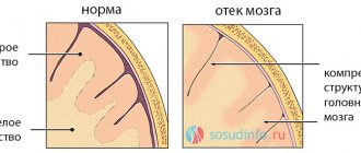
- The leakage of blood into the subarachnoid space leads to cerebral edema due to disturbances in the circulation of the cerebrospinal fluid and its stagnation.
- Penetration of blood into internal tissues is fraught with the formation of hematomas. Fabric saturated with decay products becomes necrotic.
- Hemorrhage into the ventricular area leads to immediate death or coma.
Each area of the brain into which blood is shed undergoes irreversible changes in one way or another, which does not go unnoticed for the functioning of individual systems and organs. A person may experience:
- unusual emotional instability;
- problems with the swallowing reflex;
- throbbing headaches;
- violation of orientation in space;
- episyndrome;
- Difficulty with independent bowel movements and emptying the bladder.
It is not always possible to fully restore lost brain functions. As a rule, we are talking about partial restoration. But systematic observation by specialists and high-quality rehabilitation make it possible to achieve at least complete self-care.
Treatment of aneurysm
In the early stages, acupuncture and preparations based on natural ingredients are used. Surgery is the most reliable method. But it is used in extreme cases, when other treatment is not effective. There is always an alternative. Endovascular embolization is a new method that appeared relatively recently. An embolus is injected into the vessels, the blood supply is blocked in the deformed areas. In this case, craniotomy is not necessary, which means that the rehabilitation period will be less difficult and protracted. Still, this method is not ideal. The body’s reaction can also be negative: the procedure will lead to a major stroke, instead of complete remission. In some cases, it cannot be carried out due to the anatomical features of this part of the circulatory system. Despite all these shortcomings, treatment with this method is popular, it gives hope for the best, motivates not to stop working, to continue to look for ideal methods of treatment.
Symptoms and signs
Signs of a cerebral aneurysm are sometimes completely absent and this is what often leads to a vascular catastrophe. In the event that the aneurysm initially did not manifest itself in any way, and then either began to grow or ruptured, then either a course similar to this (tumor-like symptoms) or apoplectic symptoms are possible. When a large aneurysm ruptures, the clinical picture of a large, hemispheric stroke occurs, and it is enough to read any description of this vascular catastrophe to understand that a large defect in the vascular wall is a “time bomb.”
The clinical course of each aneurysm has several stages:
the period before rupture, or pre-hemorrhagic.
In the event that there are no signs of a growing mass formation, and the patient is not bothered by any symptoms, then the only thing left is either screening MRI or even angiography, but it is not easy to convince a healthy person to “check” whether he has such disorders or not. In rare cases, a thorough history can help to suspect an aneurysm. Patients may have atypical attacks of migraine, throbbing, asymmetrical, weather-related, or miscellaneous headaches (TIAs).
If this happens, regardless of the typical migraine “reasons” and risk factors for ischemic stroke, you need to look for an aneurysm.
Quite rarely, symptoms of a space-occupying formation occur - when a vessel protrudes in an area, the compression of which leads to obvious complaints, for example, loss of sensitivity, paralysis, strabismus, or decreased visual fields.
Rupture of a cerebral aneurysm, or hemorrhagic period.
Here it is necessary to clarify that previously this incident was classified as a complication. Then they realized that this was a fundamentally wrong approach: it lulled doctors’ vigilance and inspired hope that one could live one’s entire life, and consequences in the form of a vascular accident would arise if one “behaved incorrectly.”
However, numerous studies have shown that the consequences of rupture have almost no correlation with lifestyle modification and attempts at conservative, protective treatment. This vascular pathology is insidious because the aneurysm “lives” according to its own laws, and its rupture is a matter of time, so it must be included in the course of the disease.
The risk of rupture is 1% each year. For example, an aneurysm was accidentally discovered in a 40-year-old patient. The doctor must explain that in 10 years he will have a 50% chance of getting a gap, since it is necessary to count not from the moment of discovery, but from the moment of birth, in order to include the maximum risk in the prognosis.
It occurs suddenly, in the midst of complete health, and often against the background of physical exertion or nervous stress. The main factor is an increase in blood pressure, which exceeds the rupture limit of the defective protrusion wall.
A sharp headache, nausea and vomiting occurs, and progressive depression of consciousness quickly sets in. This is what distinguishes the rupture of an aneurysm from SAH, or subarachnoid hemorrhage: there is no initial “blow to the head”, and there is no “light interval” during which there is imaginary well-being. As a rule, a rupture is accompanied by general cerebral symptoms, but focal symptoms are mild, and in 50% of cases they are not present at all.
Posthemorrhagic period. It is here that persistent and focal signs begin to appear, which are associated with the developed necrosis of brain tissue. These are paralysis, paresis, speech disorders, ataxia, vestibular disorders, aphasia and other signs.
About complications
We indicated that aneurysm rupture and bleeding are not a complication. What then is considered a complication? There are several of them, and all of them further aggravate the patient’s condition after a rupture. Here they are:
- repeated hemorrhage. If the aneurysm ruptures, then re-bleeding may resume from it, and the risk of its occurrence is much higher: 1% not in a year, but in 3 days;
- pronounced vasospasm, which occurs first in the artery of the aneurysm, and then in all vessels of the brain, as a reaction to blood breakdown products. Its peak occurs on the 10th day, and it leads to the occurrence of repeated ischemic foci and a significant deterioration in the patient’s condition;
- the most severe symptoms with rapid descent into coma occur when a large amount of blood breaks into the ventricles of the brain. Hyperthermia develops and most often death occurs within the first or second day;
- occlusive hydrocephalus;
- ischemia of the brain (essentially an ischemic stroke).
Share the article on social media. networks:
About diagnostics
It is sad when the diagnosis of “ruptured aneurysm” is established only at autopsy. Currently, as research results show, aneurysms are often detected as “surprises” on CT and MRI scans performed for other reasons (multiple sclerosis, sinusitis). And the number of such finds in absolute numbers exceeds the aneurysms found during a targeted search.
The “gold standard” for diagnosis is MR angiography, which allows, even without the introduction of a contrast agent, to evaluate the most significant formations that are intact and unruptured. To do this, you need a tomograph with a magnetic field voltage of at least 0.4 T (screening of large vessels), and for a full study you need a tomograph of at least 1.5 T on magnets, using a contrast agent based on the paramagnetic gadolinium (“Omniscan”, which is injected intravenously). The examination takes a little time, but the result is complete information about the condition of the aneurysm. This method completely replaced routine cerebral angiography, when patients were injected with a radiopaque substance containing iodine and X-rays were taken.
In some cases, patients with subarachnoid rupture undergo a lumbar puncture, but this is now recognized as dangerous.
If the aneurysm ruptures, then it is necessary to do not an MRI, but a CT scan, since CT “sees” free blood well. An entire aneurysm may be completely invisible on a CT scan.
Other research methods are also used, for example, EEG (to clarify the timing of surgery for subarachnoid hemorrhages, multiple aneurysms), as well as ultrasound. Dopplerography allows you to identify the onset of vasospasm and take action.
Intravital diagnosis of a completely “silent” aneurysm can be either the result of a conscious search (for example, with strokes in relatives), or an accidental discovery
How it manifests itself
Cerebral aneurysm has minor manifestations; symptoms of cerebral aneurysms can only be recognized with appropriate examination. An unruptured aortic aneurysm can manifest itself symptomatically only if it is large (giant) in size and puts pressure on the nerve endings.
Symptoms of an aneurysm that maintains its integrity:
- visual impairment if the pathological formation compresses the optic nerves;
- headache in the area where the affected vessel is located;
- convulsions if the vessels near the motor parts of the brain are greatly dilated;
- pain in the face with pressure on the facial nerve;
- strabismus;
- drooping lower eyelid;
- noise in ears.
Important information: How to treat an aneurysm of the apex of the left ventricle of the heart and what is the prognosis for life
Signs of an aneurysm that may rupture become stronger. Added to them:
- dizziness;
- aphasia;
- hemiparesis or hemiplegia;
- lack of coordination.
A ruptured aneurysm of the middle cerebral artery will provoke an attack of severe headache caused by the effect of hemorrhage on the lining of the brain.
The presence of brain aneurysms that require immediate medical intervention can be determined by the following signs:
- the appearance of photophobia;
- difficulty swallowing;
- dilated pupils;
- neck muscle tone;
- pain and paresis of the muscles of the neck and legs;
- loss of consciousness.
In women over 40 years of age, signs of menopause (hot flashes), which are accompanied by symptoms of aneurysmal pathology (dilated pupils, speech impairment, convulsive or pain syndrome), will help identify damage.
Kinds
According to their origin, vascular aneurysm is often of the following types: congenital and acquired. In turn, acquired disorders arise due to:
- injuries;
- severe cerebral atherosclerosis;
- infectious diseases (for example, when purulent emboli are introduced into the brain). This aneurysm is called mycotic (not to be confused with a fungal infection).
Naturally, aneurysms, due to the special structure of the vessel walls, belong to arteries. After all, an absolutely rigid and rigid vessel simply cannot begin to bulge under blood pressure. Therefore, when examining mature and ruptured aneurysms, the muscular membrane is absent, and the elastic fibers are fragmented.
As serious clinical studies show, aneurysmal dilatation of cerebral vessels in most cases is localized in the subarachnoid space, that is, under the arachnoid membrane, and they are located at the base of the brain. About 70% of all cases of diagnoses of diseases associated with rupture of these formations were identified within the circle of Willis, and in its anterior sections. In the medulla (for example, deep in the white matter), aneurysms appear much less frequently.
Since the localization site is the subarachnoid space, this is why this vascular pathology most often manifests itself as a dangerous complication - namely SAH, or spontaneous subarachnoid hemorrhage, which neurosurgeons and neurologists have to treat. Sometimes chronic aneurysms occur, which can rupture repeatedly. In this case, when there are many adhesions in the structure of the protrusion, the blood breaks through either into the ventricles or into the brain.
Epidemiology and classification
According to modern data, aneurysms are often asymptomatic. When conducting a section of corpses that died from a variety of causes (that is, in the population), the frequency of detection of aneurysms was 5%, that is, it occurred in every 20th healthy person. The vast majority of spontaneous subarachnoid hemorrhages (more than 80%) are caused by this particular pathology.
The classification of aneurysms varies, but from the point of view of the final event and morphological changes, ruptured aneurysms occur in 80% of all cases. Unruptured (10%) – manifested by focal signs of nerve compression or other symptoms. The remaining 10% are “silent” aneurysms that do not manifest themselves in any way during the patient’s lifetime. This is international statistics data.
The shape of the aneurysm is similar to either a sac or a spindle. That's what they are called, but saccular forms are much more common. By size, miliary aneurysms are distinguished (up to 3 mm), small aneurysms (or small) - up to 10 mm, the size of medium aneurysms reaches 15 mm, large ones - 25 mm. However, there are also giant aneurysms, whose size may exceed the indicated values.
Share the article on social media. networks:
Surgery
The insidious feature of this disease is that surgery is in many cases the only rational solution that can save a person’s life, but it does not guarantee the patient’s survival.
However, neurosurgery is offering new methods for eliminating such formations:
- Clipping is the most complex intervention, requiring microscopic equipment and, accordingly, highly qualified neurosurgeons. During the operation, the doctor opens the skull and, by applying a metal clip, separates the dilated part of the artery from the general blood flow, while simultaneously removing the consequences of the rupture - spilled blood from the hematoma and the space between the membranes.
- Strengthening the membranes of blood vessels with special surgical gauze. The risk of surgery is the possible development of bleeding.
- Endovascular intervention with strengthening of the vascular bed with microspirals. In this case, the opening of the skull is not provided, and the progress of the process is monitored using angiography. A very complex type of treatment, like clipping.
Complications of such attempts at surgical intervention may include the occurrence of spasms, bleeding when adjacent arteries or veins are damaged, or the development of hypoxia. On the other hand, any of these techniques can prevent death or severe complications of the disease itself.
After the destruction of the membranes of the bloodstream and the occurrence of bleeding, there can be no questions about the need for intervention - this is the only solution. The choice between conservative and surgical treatment methods is faced only by patients who have not had hemorrhage.
The diversity of this pathology in structure, location, probable consequences, and clinical features determines the uniqueness of each case of the disease. It is impossible to draw conclusions about certain likely consequences in this case from the experience of another patient.
The person himself, naturally, is unable to in any way exclude the development of congenital varieties of this disease. The likelihood of intrauterine defects can only be reduced by a mother who, during pregnancy, tries to avoid negative influences on the body of her unborn baby.
There is also no special targeted prophylaxis to prevent acquired cases of this disease. You can only strengthen your body so that it is less vulnerable to such diseases. Increasing your own physical activity and resistance to stress, rational nutrition, living without dependence on alcohol and drugs are the main principles of reducing the risk of such ailments.
If, nevertheless, a diagnosis is discovered, you need to be more attentive to your well-being and not neglect medical recommendations. If hemorrhage is suspected, the life and future of the patient greatly depend on how correctly and quickly he receives adequate assistance.
Rehabilitation
The need for repeat clipping is 35% within the first 14 days after the first bleeding. Therefore, neurosurgeons prefer to perform open surgical or endovascular intervention immediately after diagnosing an aneurysm to reduce risks.
The recovery process (especially after clipping) is lengthy and may take several months or years to fully restore all brain functions.
The incisions heal within 6 weeks. After the operation, the patient is provided with a clear set of instructions and prescriptions. After endovascular surgery, the patient may be prescribed blood thinners, such as aspirin, and other medications to relieve pain symptoms (pain medications are indicated for 1-3 weeks). Return to full activity after aneurysm clipping or endovascular intervention depends on the patient's condition before and immediately after treatment. A physical therapy program is tailored to each individual patient. Moderate activity after discharge is encouraged.
Symptoms of the disease
In the acute type of the painful condition, the patient becomes weak, has a fever, sweating increases, shortness of breath occurs, which is replaced by asthmatic manifestations and swelling of the lungs, tachycardia is observed and heart rhythm dysfunction occurs.
If the disease occurs in a subacute form, then the symptoms of insufficient blood circulation progress.
In the chronic form of the disease, the symptoms are similar to heart failure. You feel shortness of breath, syncope, angina pectoris, your heart is working intermittently. In the last stages of the development of the disease, the veins of the cervical region swell, swelling is observed, as well as phenomena that indicate that adhesions are developing in the chest.
The chronic form of the disease is represented by thromboembolic syndrome near the cardiac region. After a chronic aneurysm, there can be a variety of complications such as gangrene of the extremities, stroke, renal infarction, pulmonary embolism, occlusion in the mesenteric vascular region, and recurrent infarction can also occur.
But a chronic aneurysm is almost impossible to rupture. But acute, unlike chronic, usually ruptures from 2 to 9 days after the onset of a heart attack in the myocardial region and usually leads to death.
With an aneurysmal rupture, there is a sharp pallor of the skin with a quick change to a red tint of the skin, cold sweat flows, and the veins in the neck become oversaturated with blood. The patient is unconscious, that is, he is not at all aware of what is happening to him.
The lower extremities become cold. A person breathes noisily, hoarsely, shallowly, rarely. The patient often dies quickly, within a day or two.
Correction of the situation
Treatment is selected by the doctor individually. So, for example, if the defect is small, the doctor may recommend observation and risk monitoring. In addition, those diagnosed with an aneurysm are prescribed a gentle diet, excluding fatty foods, and also have to give up alcohol and smoking.
Another option for correcting the situation is clipping. In this case, the patient has a metal clip applied to the aneurysm stalk, which limits the growth of the defect and prevents rupture.
A replacement for surgical intervention is endovascular occlusion. This option is considered new and involves the use of a special catheter. Metal spirals are placed in it, which fill the aneurysm, as a result of which the blood does not press on the walls of the vessels.
This pathology should not be ignored; it is better to undergo examinations on time. Then there is a chance to save health and life.
What causes aneurysms?
The main reason for the development of an aneurysm is a defect in the walls of the arteries. The following factors lead to it:
- Genetic predisposition to thinning of the muscle layer (collagen deficiency).
- Developmental anomaly: pathology in the structure of the vascular wall.
- Renal polycystic disease.
- Tuberous sclerosis.
- Systemic lupus erythematosus.
- Coarctation of the aorta.
- Arteriovenous malformation.
Neoplasms can be acquired. With this type of pathology, rupture of a cerebral aneurysm occurs in the same percentage as with congenital aneurysm. Causes of thinning of the vascular wall, narrowing of the vascular bed:
- Atherosclerosis of the vessels of the head and neck.
- Arterial hypertension.
- Head injuries, skull fractures, concussion, accompanied by a violation of the integrity of the vascular wall.
- Radioactive exposure.
- Hyalinosis of vascular walls.
- Bad habits: alcoholism, smoking, use of psychotropic and narcotic substances.
If infectious emboli (cells containing fungal organisms, bacteria, cancerous elements) enter the cerebral arteries, a mycotic aneurysm may develop.
A combination of factors, when there is a genetically determined predisposition to thinning of the arterial wall and atherosclerosis has already been diagnosed, increases the chances of the occurrence and rupture of an aneurysm sac. The deterioration of brain function is more pronounced in older people.