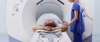
Computed tomography is a procedure that produces a layer-by-layer image of the area under study or the entire body. Unlike an X-ray machine, a tomograph gives less radiation exposure due to a narrow beam of X-ray radiation. After scanning, the data collected by the tomograph is sent to a central computer, where the processes of decoding and improving image quality take place.
Computed tomography room
Features of preparation
Three days before the examination, it is recommended to visit a specialist who will tell you in detail how to prepare for an abdominal CT scan, based on the individual characteristics of the body. Preliminary preparation will help to obtain the most accurate examination result.
Typically this period is viewed from the following angles:
- A consultation with a doctor, during which the patient’s medical history is clarified and the medications he is taking are examined.
- Assessment of the absence of contraindications to the study.
- Carrying out bowel cleansing activities.
- Compliance with the recommended dietary intake.
How to take orally before an abdominal CT scan
In preparation for the study, it is proposed to take Urografin orally. It takes place in two stages:
- In the evening, stir 2 ampoules of 75% composition in cold boiling water (1.5 l), take 500 ml immediately.
- Divide the remainder equally. Drink the first part after waking up. The second – 30 minutes before the CT scan.
The development of side effects is possible:
- Mild nausea.
- Attacks of vomiting.
- Skin itching and rashes.
Pain does not occur while taking Urografin.
https://youtu.be/O6kvPJN0V6o
Diet food
The main goal in the preparatory period for abdominal tomography is to improve the visualization of the organs as much as possible. The most serious obstacle is caused by intestinal loops, which swell from excess gases. To get the right results, you need to pay attention to a diet that is aimed at reducing gas formation. The recommended diet is shown both for CT scanning and for ultrasound and MRI of the abdominal cavity.
While preparing for a CT scan, you should not eat a flour diet.
During the 3 days before the examination, the following is prohibited:
- fresh fruit;
- vegetables;
- cabbage;
- legumes;
- dried fruits;
- yeast-containing products;
- rice, semolina porridge;
- any alcoholic beverages;
- chocolate;
- juices;
- carbonated drinks;
- coffee;
- strong tea;
- whole milk.
During the preparatory period, it is recommended to include in the diet:
How many times can an MRI be done?
- food made from lean fish;
- steamed meat;
- light vegetable soups;
- low-fat varieties of cheese, cottage cheese;
- steamed vegetables;
- croutons from white bread.
If the CT scan is scheduled for the morning, then the last meal is at 7 pm. The procedure, carried out in the afternoon, requires a 6-hour break from eating. For breakfast, you can eat a little low-fat broth or liquid porridge. You can drink a small mug of tea.
Instructions for use
The method of use and dosage depend on the type of procedure.
Intravenous urography
Injections . The composition is administered slowly - 20 ml/min. To obtain a large volume - over 100 ml - the procedure lasts at least half an hour. The dosage is determined by the patient's age group.
The rate of contrast solution for adult patients (in ml): 76% - 20, 60% - 50.
In children, 76% Urografin is used in the following doses:
| Age, months/years | Volume, ml |
| 0–12 | 7,0–10,0 |
| 1–2 | 10,0–12,0 |
| 2–6 | 12,0–15,0 |
| 6–12 | 15,0–20,0 |
| Over 12 | As for adults |
An image of the kidney parenchyma is taken immediately after receiving contrast. To visualize the pelvis, ureter and bladder, the first image is obtained after 3–5 minutes, the second:
In infants and children of primary preschool age, the study is carried out after two minutes. If the picture turns out to be uninformative, then the pictures are taken a little later.
Drip infusion . When using the system, the time for administering 100 ml of a solution of any concentration falls within the range of 5–10 minutes.
If the patient is diagnosed with heart failure, it increases to half an hour. The first image is taken immediately after the infusion is completed. Subsequent ones - after 20 minutes.
Retrograde urography
The procedure is prescribed to visualize the bladder. The drug is injected directly into its cavity using a catheter.
The duration of the procedure is half an hour, since the main reactions occur during this period. A 30% composition is used, obtained by diluting 60% Urografin with cold boiling water in a 1:1 ratio.
The dosage is determined by the following factors:
- Age.
- Weight.
- Myocardial contraction frequency.
- General health.
Angiography of the abdominal cavity
Preparation for the procedure involves complete cleansing of the human intestinal tract. For these purposes, two days before the expected date of the study, foods that increase the formation of gases are excluded from the menu:
Dinner should take place the day before the procedure no later than 18:00. Before going to bed, take a laxative.
To reduce anxiety, you are allowed to drink a sedative. If a person has myeloma, decompensated diabetes, or gout, he can consume any amount of liquid; drinks are unlimited. This also applies to infants and very young children.
For confirmed pheochromocytoma, alpha-adrenergic blockers are recommended. This measure helps prevent the formation of a hypertensive crisis.
Purgation
Often, preparation for an examination requires cleansing enemas or taking mild laxatives, which will be prescribed by the specialist conducting the diagnosis. This measure will help cleanse the intestines of feces and food debris. If a patient suffers from irregular bowel movements, he may be prescribed laxative medications for 3 days, which are familiar to him.
When a patient has a tendency to increased gas formation, then during the preparation period he is recommended to take activated charcoal and Espumisan. If there are problems with the digestive system, enzyme preparations, for example, Mezim, Festal, are often prescribed. In case of increased motor function of the gastrointestinal tract, the patient is recommended to take No-shpa once immediately before the diagnosis.
Important! It is prohibited to take medications on your own. All prescriptions must be made by a doctor.
Contraindications
There are complete and conditional bans on the use of Urografin.
Strict restrictions are:
- Heart failure in the stage of decompensation.
- Hyperthyroidism.
- Pancreatitis during an exacerbation - if the patient is indicated for endoscopy of RCP.
- Gestation, inflammation in the pelvic cavity - when prescribing hysterosalpingography.
Possible contraindications for the use of contrast include:
- Decompensated myocardial dysfunction.
- Deviations in the functioning of the kidneys and liver.
- Finding a person in serious condition.
- Emphysema.
- Diabetes in the stage of decompensation.
- Subclinical form of hyperthyroidism.
- Generalized myeloma.
- Individual reaction of the body to iodine.
With these diseases and conditions, the administration of Urografin is carried out under the supervision of physicians.
Preparation before contrast injection
Quite often, a computed tomography scan of the abdominal cavity is done with contrast. Barium and iodine-containing substances are introduced into the human body, which during diagnosis will contrast the organs located in the abdominal cavity. This will allow you to most accurately visualize pathological foci. These components are completely safe for the patient; they accumulate in areas of increased blood supply.
Following the preparation rules allows you to get results the first time
Before undergoing a CT scan with contrast, you should consult with the doctor conducting the study. He will collect information about concomitant diseases and medications taken. It may be necessary to cancel some of them during the preparatory period. In addition, during preparation you should find out whether there is an allergy to the administered substances.
To do this, you need to arrive half an hour earlier than the appointed time. The nurse will perform a contrast test. If the reaction is not determined, then you can safely proceed to the examination. You should definitely inform your doctor if you have hypersensitivity to iodine or seafood.
special instructions
Against the background of the administration of Urografin, the development of turnip hypersensitivity is possible. Signs of the condition are:
- The appearance of shortness of breath, a feeling of lack of air in the chest.
- Severe redness of the skin as a result of dilated capillaries.
- Hives.
- Itching.
In exceptional cases, there is a possibility of the formation of angioedema - the vocal apparatus may be affected - anaphylactic shock and bronchospasm.
Reactions to the administration of a contrast agent occur and fully develop no later than 60 minutes after receiving it. Delayed symptoms are also likely: side effects occur after several hours or days.
The possibility of forming a response to iodine-containing drugs is significantly increased if a person has already had similar reactions in the past. In case of predisposition, it is permissible to use antihistamines and glucocorticosteroids for prevention purposes.
With bronchial asthma, there is also a possibility of symptoms of a hypersensitivity reaction or bronchospasm.
When signs of an allergic response of the body develop, the administration of Urografin is stopped immediately. If necessary, symptomatic therapy is performed.
Additional measures
Immediately before performing MSCT of the abdomen, preparation is carried out in the clinic. First of all, the patient or the person responsible for him signs the necessary document. Then the patient goes into a room where he takes off his clothes containing metallic inclusions, watches, and all jewelry. If you have a hearing aid, it must be removed.
If the patient’s body contains devices or prostheses made of metal, then diagnostics cannot be carried out. Since metal objects can disrupt the operation of the tomograph, which will lead to an incorrect diagnosis. Preparation before an abdominal CT scan is no less important than the examination itself. Because only with the right approach to diagnosis can you get accurate results that influence subsequent treatment.
By-effect
The development of adverse symptoms depends on the method of administration of the substance.
Intravenous
The drug is generally well tolerated. Unpleasant sensations are mild and pass quickly. The most common concerns are nausea, vomiting, hot flashes, and slight pain in the injection area.
Other side effects include:
- Violation of myocardial contractile function and blood pressure indicators.
- Thromboembolic complications.
- Shortness of breath, cough, swelling of the lung tissue.
- Pain in the intestines.
- Deviations in kidney function.
- Tremor of the limbs, photophobia, cephalalgia, dizziness.
- Drowsiness.
- Skin itching, rashes.
- Conjunctivitis, urticaria, runny nose.
Intracavity
Unpleasant reactions do not form immediately, but several hours after receiving the contrast. This is explained by the slow absorption and redistribution of Urografin.
An increase in amylase during ERCP cannot be ruled out. Visualization of acini in rare situations becomes an impetus for the development of pancreatitis.
Systemic allergic reactions are uncommon and are expressed in the form of skin rashes and itching.
pharmachologic effect
Iodine-containing radiocontrast agent. The iodine contained in the preparation increases the contrast of the image due to the absorption of X-rays.
Fever, dizziness, headache, nausea, vomiting, tachycardia, cyanosis, lacrimation, erythematous rashes, urticaria, anaphylactoid reactions, anaphylactic shock, pulmonary edema, neurotic disorders, diarrhea, exacerbation of enteritis or colitis, acute renal failure.
Local: pain at the injection site, phlebitis, thrombosis.
From the heart: abnormal heart rate, cardiac arrhythmia; tachycardia, including reflex; bradycardia cyanosis impaired cardiac function; transient ECG changes; ventricular fibrillation; asystole; heart failure; severe thromboembolic events leading to myocardial infarction.
From the side of blood vessels: circulatory disorders, which is manifested by dilation of peripheral vessels and a subsequent decrease in blood pressure, collapse, hot flashes, possible increase in blood pressure, vasculitis.
From the blood and lymphatic system: inhibition of blood coagulation and platelet aggregation, disseminated intravascular coagulation, thrombocytopenia, hemolysis, platelet microangiopathy.
From the endocrine system: hyperthyroidism.
From the nervous system: dizziness, headache, agitation, amnesia, subchordal edema, increased symptoms of myasthenia gravis, speech impairment, visual impairment, hearing impairment, photophobia, temporary loss of vision, tremor/shaking, convulsions, epileptic seizures, coma, disorientation, drowsiness , paresis/paralysis, confusion, loss of consciousness, severe thromboembolic events leading to stroke.
From the urinary system: impaired renal function, reversible renal failure (including acute).
From the immune system: anaphylactic reactions / hypersensitivity reactions, including anaphylactic shock, angioedema, conjunctivitis, increased lacrimation, cough, rhinitis, sneezing.
From the skin and subcutaneous tissue: skin rashes, itching, redness/erythema with vasodilation, pallor of the skin, urticaria, skin reactions, including Stevens-Johnson syndrome, Lyell's syndrome.
From the respiratory system: transient disturbance of respiratory rate, shortness of breath, respiratory distress, cough, bronchospasm, spasm/swelling of the larynx, respiratory arrest, pulmonary edema.
From the digestive tract: nausea, vomiting, abdominal pain, metallic taste in the mouth.
General disorders: feeling of heat, weakness, malaise, fever, vasovagal reactions, sweating, increased body temperature, swelling of the salivary glands.
Local reactions: changes at the injection site, including pain, swelling, inflammation, possible development of phlebitis and thrombosis; when the drug flows paravasally, increased pain, swelling, tissue necrosis.
With intravascular administration
Adverse effects associated with intravascular administration of iodinated contrast agents are usually mild to moderate in severity and transient in nature. However, severe, life-threatening and fatal reactions have been reported. The prevalence of side effects in patients treated with ionic contrast agents was found to be more than 12% compared to more than 3% in the case of non-ionic contrast agents.
Most often, with intravascular use, reactions such as nausea, vomiting, pain and a general feeling of heat are observed.
When introduced into other body cavities
After administration of a contrast agent into body cavities, adverse reactions rarely occur. Most of them develop within a few hours after administration due to slow absorption from the injection site and distribution throughout the body, mainly through a controlled process of diffusion.
After ERCP (endoscopic retrograde cholangiopancreatography), a slight increase in amylase levels is common. Acinar contrast imaging has been shown to be associated with an increased risk of developing pancreatitis after ERCP. Isolated cases of necrotizing pancreatitis have been described.
Cases of vasovagal reactions associated with hysterosalpingosis are rare.
Anaphylactic reactions/hypersensitivity
Systemic hypersensitivity is rare, mostly mild, and usually occurs in the form of reactions.
However, the possibility of developing a severe hypersensitivity reaction cannot be completely excluded. For a complete description of anaphylactic reactions, see the relevant information in the section "Adverse reactions", "
After administration of the drug, in rare cases, a slowdown in psychomotor reactions is possible. You should avoid driving vehicles and machinery for 24 hours after administration of the drug.
In patients suffering from diabetic nephropathy and taking biguanides, lactic acidosis may develop when a contrast agent is administered. In order to prevent such a complication, it is necessary to stop taking biguanides 48 hours before the procedure. Before resuming the use of biguanides, ensure that there is no impairment of renal function.
Hypersensitivity reactions may be more severe in patients taking beta-blockers.
Patients taking antipsychotics are more likely to experience delayed reactions (fever, rash, itching, joint pain) and flu-like symptoms.
The incidence of delayed reactions (eg, fever, rash, flu-like symptoms, joint pain and itching) is higher in patients taking interleukin.
Interaction with diagnostic tests
After intravascular administration of iodinated contrast agents, the ability of thyroid tissue to absorb radioisotopes for the diagnosis of thyroid diseases is reduced for a period of up to 2 weeks, and in some cases even for a longer period.
When is MSCT prescribed?
MSCT of the abdominal cavity and retroperitoneal space is prescribed in cases where other diagnostic methods cannot provide clear, unambiguous results. Direct indications for diagnostics using a spiral tomograph are:
- jaundice, especially asymptomatic;
- sudden weight loss for no apparent reason;
- chronic digestive disorders;
- chronic forms of dysfunction of the intestines, stomach and other organs;
- acute injuries of the anterior abdominal wall and penetrating wounds of the abdominal cavity.
Also, patients who are preparing for surgery on the abdominal organs or undergoing a complex course of therapy that requires monitoring even minimal changes are referred to MSCT.
Decoding the results
Research results are provided in several formats. The paper reflects a description of the main organs, vascular elements and neoplasms, if any. If the description is qualitative, then the topography, or the relative position of the organs relative to each other, will be reflected.
In addition, the entire shooting is recorded on digital media in Dicom format. It is impossible for a person without a medical education to independently understand these graphic images, sometimes animations or video files. Therefore, you need to rely on the conclusion given by the doctor who interpreted the MSCT results.
First of all, you should pay attention to the size of the organ. The identified dimensions and normal indicators are described.
When the liver becomes enlarged, one should think about steatohepatosis (fatty degeneration), and when its size decreases, on the contrary, about cirrhosis. If the spleen is enlarged, blood diseases should first be excluded, and then infectious diseases. In the second case, the liver enlarges in parallel.
Normal bile should be visible in the gallbladder. Its walls are not thickened. Otherwise, we are talking about a diagnosis such as cholesterosis or biliary dyskinesia. If a stone is described in the lumen of the gallbladder, then this is a manifestation of cholelithiasis.
Neoplasms in any organ can be of any shape and size. Only an oncologist can determine their nature. They are differentiated from cysts, malignant and benign tumors, metastatic, purulent (abscesses) foci. It is impossible to identify them independently.
An ordinary cyst looks like a round formation with clear contours. Within one cystic cavity there may be several daughter ones. Cysts of parasitic origin pose a danger.
Blood flow in cystic cavities is not pronounced. Malignant tumors have a clearly defined intensive blood supply. They are characterized by uneven contours and heterogeneous contents. Purulent foci (abscesses) also look like rounded formations with a horizontal level of liquid.
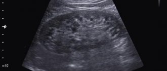
MSCT of the abdominal cavity with contrast immediately after injection and after 10 minutes
Adenomas are considered benign neoplasms. They are also visible on MSCT. They often develop in the liver. When contrasted, an adenoma looks clearer because its tissues capture the contrast compound better and retain it longer. A fibrous field is visualized around it, the contours of which usually follow the contours of the tumor.
MSCT of the abdominal cavity with contrast allows you to determine the presence of hepatocellular cancer. The sizes can be absolutely any. At the same time, the structure of the intrahepatic formation is extremely heterogeneous; multiple collaterals and vascular shunts are visible. In pancreatic cancer, on the contrary, the vessels feeding the tumor are very rarely visible.
Composition of Urografin
The drug Urografin is available in the form of an injection solution. Composition of the drug:
| Description | Transparent, slightly colored liquid |
| Concentration of meglumine amidotrizoate, mg per ml | 520 or 660 |
| Concentration of sodium amidotrizoate, mg per ml | 80 or 100 |
| Iodine content, mg per ml | 292 or 370 |
| Auxiliary components | Water, sodium hydroxide, sodium calcium edetate |
| Package | Ampoules of 20 ml, packs of 5 ampoules with instructions for use |
Analogs
There are substitutes for radiopaque contrast agents, but with slightly different compositions (contain sodium amidotriosate):
- Angiographin.
- Visitor trust.
- Urovizin.
- Urotrast.
- Verografin.
- Trazograph.
- Triombrine.
Triombrast solution is also used. In addition to sodium amidotrizoate, the drug also contains meglumine.
The cost of the medicine is high, which is explained by the peculiarities of its composition and scope of application. But at the same time, it is fully justified by the effectiveness of Urografin. The price of 1 ampoule largely depends on the region of sales. A dose of the product sells for 200–260 rubles.
Price Urografin
You can buy the drug via the Internet or pharmacies at prices depending on the concentration of the solution and the trade markup. Approximate cost of Urografin and its analogues in Moscow:
| Name of the drug, volume | Internet price tag, rubles | Pharmacy cost, rubles |
| Urografin 76% 10 ampoules of 20 ml | 2200 | 2250 |
| Urografin 60% 20 ml 10 pcs. | 1950 | 2000 |
| Trazograph 76% 5 ampoules of 20 ml | 1400 | 1450 |
Conditions of release and storage
The medicine is a prescription drug. You can buy Urografin in Moscow in large pharmacies.
To prevent the solution from losing its original properties, it is stored at room temperature. Choose a dark place out of direct sunlight.
Urografin belongs to the group of radiopaque iodine-containing drugs and is not a drug in the usual sense of the term. Its independent use is unacceptable. Prescription of the solution is possible only according to indications.