Congenital ailments and bad habits can affect the physiological structure of the liver.
What it is?
Conventionally, the liver is divided into 4 lobes, which consist of very small functional lobes, similar to grains, interconnected by connective tissue. Such “grains” surround the central vein, from which the hepatic beams branch, which consist of hepatocytes - cells that are capable of recovery. The liver is penetrated by blood vessels, capillaries, and bile ducts. Its normal texture is slightly granular. Based on the “graininess”, to a greater or lesser extent, the condition of the organ and the presence of diseases are determined.
What is echostructure and echogenicity, indicators are normal
On ultrasound, it is the dense or parenchymal organs that are clearly visualized: liver, spleen, glands, kidneys. Liquid media have the best ultrasonic conductivity, namely the bladder with urine, the gall bladder filled with bile, and blood vessels.
Echostructure is a concept that defines the structure of an organ and the state of its parenchyma at the moment. It can be homogeneous, focally or diffusely heterogeneous. Echogenicity refers to the degree of density of the tissues that make up the gland or other organ being examined. It happens:
- average , or normal (the main landmark is the echogenicity of the healthy liver parenchyma);
- reduced - hypoechoic, when the organ looks “darker” than the liver;
- low (anechoicity), which is typical for the bladder, cysts;
- increased (“lighter” than normal);
- high – hyperechogenic (such echogenicity is comparable to the color of fatty tissue and bones on ultrasound).
A normal (healthy) pancreas may be either homogeneous or somewhat heterogeneous when scanned. Visualization of small or large “grains” in the parenchyma of this organ depends on the level of equipment and the doctor’s sensitivity. If, in the absence of complaints, the structure of the pancreas is diffusely heterogeneous, and the dimensions are within normal limits, then there is no pathology of the organ.
The echogenicity of the pancreas is usually slightly higher or equal to that of the liver. High echogenicity of the gland is established when it is on the same level with the “color” of the fatty tissue. Hypoechogenicity is recorded if the gland is darker than the liver or kidney parenchyma. Thus, normally the pancreas has a fine-grained, somewhat heterogeneous echostructure, medium or increased echogenicity.
Diffuse changes
Such changes imply an increase in the liver parenchyma and indicate its transformation. This disorder is diagnosed even in young children. Diffuse changes are not considered diseases. Enlarged liver “grains” are most often the result of bad eating habits. The parenchyma can change with the slightest negative influence.
The change is manifested by discomfort in the right side, mild, quickly passing pain. Sometimes yellowing of the eyes is observed. A modified liver entails changes in the pancreas and vice versa if one or another organ is sick or does not function properly. If diffuse changes appear due to external factors, bringing the liver back to normal will not be difficult. The course of treatment and diet prescribed by your doctor will bring positive results.
What pathologies does the heterogeneous structure of the pancreas indicate on ultrasound?
If the doctor simply indicates that the structure of the pancreas is heterogeneous, this means that its fatty transformation has occurred (that is, the amount of fat deposits in the parenchyma has increased).
To make a more serious diagnosis, “diffuse heterogeneity” alone is not enough. The doctor must take into account other ultrasound criteria for various pathologies, and also describe why the structure of the organ has changed.
Causes and consequences
Changes in liver tissue occur from bad habits, obesity, taking pills, and unhealthy eating.
The causes of diffuse changes can be caused by poor nutrition, when the diet consists of fatty and heavy foods, excessive consumption of unnatural foods and alcohol. Fashionable mono-diets for weight loss today are also not in vain. But there is nothing to say about a person who is obese: he definitely has structural changes, and often irreversible ones. More serious causes of changes are viral and bacterial diseases, genetic pathologies.
If the tissue is modified even slightly, you should take this seriously and make efforts to restore the liver. Otherwise, this is fraught with the development of cirrhosis and hepatitis. sclerosing cholangitis (disease of the bile ducts, which leads to stagnation of bile).
Classification
In medicine, there are rules according to which a diagnosis is formed - and this applies, in particular, to the division of pathologies into types and types according to causative factors, development mechanism, severity of symptoms and other criteria. The classification of diffuse liver changes is no exception.
By severity
Liver tissue may undergo changes that are persistent and severe or moderate, in some cases even reversible. This depends on the nature of the unfavorable process and the aggressiveness of the provoking factors, as well as the initial state of the organ.
Diffuse changes in the structure of the liver may correspond to the degree of:
- I – mild, there are signs of widespread damage, but this does not yet have a serious impact on the functionality of the organ;
- II – moderate, the affected areas are clearly registered on ultrasound, there is no obvious liver failure yet;
- III – severe, or severe, symptoms of cirrhosis are noted.
Thus, there is a direct relationship between the severity of the diffuse process and symptoms.
According to the specifics of the changes
Transformation of the liver structure may be associated with different types of pathologies:
- Inflammation.
- Hepatosis (lipid degeneration).
- Hemochromatosis (the liver suffers due to impaired iron metabolism).
- Cirrhosis (replacement of normal parenchyma with rough and dense connective tissue).
The basis of all unfavorable changes is hypoxia (oxygen starvation of cells), leading to dystrophy - a violation of metabolic reactions in the parenchyma. If the pathological process remains active, necrosis occurs - hepatocytes die, and in their place non-functional connective tissue is formed.
Fine-grained echostructure
The normal structure of the liver is fine-grained and homogeneous (homogeneous), the ideal dimensions of the portal vein are from 8 to 12 mm. A healthy organ has a clear, even contour with sharp edges. It is important to note that a variant of the norm in some people is a more granular surface of the organ.
The echogram shows a clearly visible network of vessels and bile ducts. If the portal vein is enlarged by at least 2 mm, the echogenicity increases slightly, then we can talk about incipient portal hypertension (increased blood pressure in the portal vein, the system of which collects blood from the stomach, spleen and intestines.). Viral diseases, poor nutrition, and bad habits contribute to this. If the liver is slightly changed, it is reversible.
Medium grain
Medium-grained heterogeneity of liver tissue, if not treated in a timely manner, enters an irreversible phase,
The medium-grained echostructure of the liver parenchyma appears when there is a clear disturbance of metabolic processes. These are excess weight, diabetes. This liver is enlarged and has a vague outline. It is impossible to determine the cause of medium grain size on ultrasound; additional tests are prescribed. Such an echostructure can be considered intermediate between the norm and the beginning of the development of the disease, when the damage becomes irreversible.
Coarse grain
Coarse-grained echostructure is diagnosed in hepatitis (echogenicity is slightly increased), obesity, diabetes, alcoholism, when studies show increased echogenicity. This is an advanced and dangerous stage of the disease. The structure of the liver, in this case, gives the impression of being weak and flabby, the surface is dull, covered with tubercles. The blood vessels are not visible, they seem to be “smoothed out”. In this condition, the liver loses its ability to recover. Chronic diseases can lead to this result. This condition is dangerous because cell death - necrosis - can occur.
Types of changes
All changes in the pancreas may have different ultrasound signs, depending on different reasons. Most often, doctors use ultrasound to detect differences that occur during dystrophic or metabolic changes in the organ. This happens when there is poor circulation, dysfunction of the bile ducts, etc.
A diffusely heterogeneous structure can also be detected with age-related changes in the pancreas. The organ decreases in size, and part of it is replaced by connective and fatty tissue. Let's consider the types of changes in the structure of the pancreas.
Echogenicity increased slightly and significantly
A change in the permeability of organ tissue may indicate the development of pathology. But often it can show the normal functioning of the organ.
If there are areas of high echogenicity, this is always a sign of a pathological condition of the gland. There are no hypoechoic foci.
Moderately increased echogenicity indicates that altered objects can be detected in the gland:
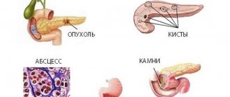
- Pseudocysts. These are formations that have liquid inside. Develop after acute inflammation. The outline becomes unclear and sometimes has jagged edges. There is a general increase in echogenicity.
- The presence of a cystic-solid structure is noted if there is no fluid in the detected formations.
- Areas of calcification. They are also called calcifications. They are formed as a result of an inflammatory disease, and also occur with chronic degenerative changes in the organ.
- Presence of areas with adipose tissue. Fatty degeneration of the structure is observed due to an increase in body weight as a result of obesity and if the patient consumes a lot of fatty foods. Often such changes occur when a person abuses alcoholic beverages.
- Fibrous areas appear where normal cells are no longer present as they are replaced by connective tissue. Most often, areas of fibrosis develop with pancreatic necrosis. The heterogeneity of the structure is characteristic.
- Presence of stones in the ducts.
- Degeneration of pancreatic tissue. It is the outcome of chronic pancreatitis in an advanced stage.
- Tumors that appeared as a result of the spread of metastases.
High echogenicity
Disturbance and a sharp increase in the echogenicity of the gland structure indicates that an acute inflammatory process is developing in it. Such a patient requires urgent treatment in a surgical hospital. Acute inflammation of the gland is a life-threatening condition.
A sharply increased hyperechogenicity may also indicate an active fibrotic process in the tissues. This means that normal pancreatic tissue is quickly replaced by connective tissue.
Severe hyperechogenicity is also diagnosed in diabetes, especially the insulin-dependent type, as well as in elderly patients. Portal hypertension leads to pronounced changes in ultrasound signs, i.e. increased pressure in the portal vein. This condition often results in significantly increased echogenicity.
Heterogeneous
Diffuse heterogeneity of liver tissue is a harbinger of cirrhosis.
A diffusely heterogeneous structure, in other words, degeneration of the liver tissue, is characteristic of cirrhosis. On ultrasound, the echogenicity is significantly increased, irregularities and tuberosity are clearly visible. In some areas it is noticeably greater, in others less. Changes lead to compaction of the organ and proliferation of connective tissue. This is a consequence of hepatosis (incorporation of fatty grains and its subsequent growth), chronic alcoholism, and hepatitis. In this case, an enlargement of the lymph nodes in the hepatic region occurs. A heterogeneous structure can provoke inflammation and dystrophy of the bile ducts and liver cells.
What can cause heterogeneity in the echostructure of the pancreas?
Increased echogenicity of the pancreatic structure is observed in diabetes.
Increased echogenicity of the pancreatic structure is observed in pathological conditions:
- Lipomatosis. This means that part of the organ is replaced by fatty tissue. The size of the pancreas is normal. In this state, a person practically does not feel any symptoms.
- Edema that develops with acute tissue inflammation. This state of the structure is always accompanied by severe pain, diarrhea, and vomiting.
- Tumors. A person experiences symptoms such as severe weight loss, weakness, lack of appetite, and stool disorders. The same signs occur when the organ is filled with cysts.
- Pancreatic necrosis is a disease accompanied by destruction and death of pancreatic tissue. On ultrasound, areas of pancreatic necrosis are visualized as having a hyperechoic structure. With pancreatic necrosis, pain in the abdominal region is severe, and patients often develop pain shock. In such cases, vomiting can be uncontrollable, and the person experiences severe diarrhea.
- Diabetes. If the causes of this disease lie in an autoimmune change in the organ, then it almost always decreases in size.
- Fibrosis is the formation of connective tissue. The structure of such tissues will be uneven. The condition develops due to inflammatory phenomena.
The disturbance in the echogenicity of the structure can also be temporary. It manifests itself due to the flu.
The disturbance in the echogenicity of the structure can also be temporary. It appears due to:
- reactive inflammation;
- a significant number of infectious pathologies - influenza, pneumonia, meningococcal infection;
- diet changes;
- sudden change in the patient’s lifestyle;
- development of gastritis, cholecystitis and other pathologies of the digestive tract;
- a hearty lunch.
A temporary increase in echogenicity is also observed with the change of seasons - spring, autumn.
Treatment
As a rule, diffuse changes are a consequence of exposure to external factors or liver diseases. Treatment begins with identifying the cause that needs to be eliminated. If the changes are weak or moderate, you should simply adjust your lifestyle, improve your diet, and most importantly, permanently eliminate fatty fried foods from your diet. Use the recommendations of diet No. 5.
For changes caused by viruses or bacteria, antiviral drugs or antibiotics are prescribed. For obesity, treatment is aimed at losing weight and normalizing metabolism. Diet is the main treatment. In case of severe illnesses, treatment is carried out under the supervision of a specialist. Medicines are prescribed to support organ function. The patient’s task is to make his work easier, for which it is necessary to follow a diet and not burden the organ.
Why does this condition develop?
Any factors affecting the liver can cause the development of the most severe degree of damage. Diffuse changes develop for various reasons, sometimes, at first glance, seemingly insignificant. The list of such reasons is extensive:
- viral pathogens;
- excessive alcohol consumption;
- long-term smoking;
- a diet rich in fatty fried foods;
- side effect from taking medications;
- hereditary predisposition;
- diabetes;
- some types of liver diseases;
- dangerous working conditions.
In fact, there are much more interrelated diseases that provoke the development of a violation of the integrity of the liver and deterioration of its functioning; it is impossible to list all the cases. All body systems are interconnected and any failure affects overall health.
As the density of the liver increases, it becomes more difficult for it to perform its filter function, and blood flow worsens.
Diseases in which the density of liver tissue increases:
We recommend reading:
Leukocyte formula
- fatty liver deformation as a result of severe diabetes, hepatitis or alcoholism;
- virus infection;
- hepatitis - in this case, the edges of the liver become rounded, the lymph nodes become enlarged;
- cirrhosis - the main reason is alcoholism, with cirrhosis the liver tissue is transformed into connective tissue, which means the activity of the liver changes, the most dangerous thing in this case is the irreversibility of the process, diffuse changes cannot be reversed.
The lists above are just some of the possible cases. Only a competent specialist can make an accurate diagnosis and determine the relationship.
How is liver heterogeneity dangerous for human life?
The presence of such a disease significantly worsens a person’s quality of life, this happens for the following reasons:
- Impaired liver function leads to intoxication of the body.
- An inflammatory process of the liver with complications is possible.
- Disturbance of metabolic processes of the whole body.
- Dystrophy and disruption of the normal functioning of the bile ducts.
Prevention
Preventive measures to avoid diffuse changes are simple and known to everyone:
natural food (use recipes from national cuisine, where the dishes are healthy, easy to prepare, uncomplicated); avoid alcohol (if you drink occasionally, take care of the quality of the drink, and give preference to red wine); timely visit to the doctor.
Gradually reduce the amount of beer per day, drink in moderation. It negatively affects the liver. However, you don’t have to give up a cup of black coffee - it stimulates the liver and, according to recent research, prevents many diseases. It would not be superfluous to periodically clean the organ using traditional methods.
Signs of diffuse changes and compaction of the liver parenchyma
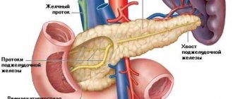
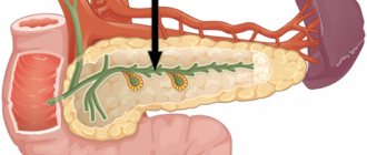
The liver is an unpaired parenchymal organ; it consists entirely of hepatic tissue. This organ is located in the abdominal cavity in the right hypochondrium. The basis of the parenchyma is the liver lobules, between which blood vessels and bile ducts pass. The bile ducts supply bile to the gallbladder, after which this fluid flows through the bile duct into the duodenum, where it connects with the pancreatic duct.
Diseases of the liver and gall bladder always affect the condition of the pancreas, and vice versa, the health of the pancreas speaks about the condition of the liver and gall bladder. The liver is the main organ of hematopoiesis and performs important functions for the body. An ultrasound examination can detect liver diseases, but laboratory and instrumental studies are necessary for an accurate diagnosis.
Normally, the liver parenchyma is a homogeneous, weakly echogenic structure, against which the vessels and bile ducts are clearly visible. Diffuse changes in the liver parenchyma mean that the entire tissue of this organ is changed. These can be severe lesions. Therefore, more research is needed to clarify the extent of these lesions.
In this case, all organs of the gastrointestinal tract are examined. Diffuse liver damage can be divided into acute hepatitis, chronic hepatitis, cirrhosis, fatty infiltration, diffuse changes in the liver with other concomitant diseases. With hepatitis, the liver enlarges, but the structure of the parenchyma may remain homogeneous. When the hydrophilicity of the parenchyma increases, its structure may be disrupted and thickening of the liver walls occurs.
The more pronounced the inflammatory process, the greater the swelling of the parenchyma, the echogenicity decreases, and the sound conductivity increases. The increase in echogenicity of the parenchyma during hepatitis can be uneven, low or high in individual areas in different ways. The structure of the parenchyma in liver cirrhosis usually becomes diffusely heterogeneous and has many areas of increased, average or decreased echogenicity.
Found an error in the text? Select it and a few more words, press Ctrl + Enter
The size of areas of heterogeneity can be from 0.5 to 2.0 cm or more. Violation of the homogeneity of the parenchyma is possible as a result of stagnation in the liver ducts and, in connection with this, an increase in bilirubin. The cause of diffuse processes in the parenchyma can be fatty degeneration of the liver.
The influence of viral and parasitic infections, malnutrition, the predominance of fatty, high-calorie foods and alcohol cannot be excluded. Clinical signs of parenchymal compaction may include general weakness, headaches, nausea, fatigue, a bitter taste in the mouth, irritability, and emotional instability in mood.
The liver is the only organ that detoxifies food and metabolism; it carries the maximum load, eliminates various foreign substances and excess hormones. Participating in the digestive processes, it provides the body with glucose and is a producer of protein and cholesterol.
Our liver is able to restore its shape thanks to complex treatment, which includes cleansing the body, normalizing nutrition, taking immunosuppressants, drugs from the group of hepatoprotectors and immunomodulators. With tumors, cysts and stones, tissue density will have local changes. Always diffuse changes are constant companions of fatty liver disease, hepatitis, cirrhosis, and various metabolic lesions.
Due to its size and density, the liver reflects ultrasound waves quite well and therefore is excellently scanned using an ultrasound machine. To assess the condition of the liver, its size, wall thickness, structure of its constituent elements and parenchyma are measured. In addition to the liver, the same manipulations are also performed with the spleen. An ultrasound examination of the liver is required to determine the diagnosis of hepatitis, cirrhosis and other serious diseases. After the ultrasound diagnostician gives a conclusion that sounds like “Diffuse changes in the liver parenchyma,” the origin of these changes should be determined using a blood test and other instrumental studies - x-rays, retrograde cholangiopancreatography, liver biopsy and laparoscopy.
Excellent prevention of compaction of the liver parenchyma is early diagnosis, timely hospitalization and productive, competent treatment, protection from various industrial, medicinal and household intoxications. If the liver functions are impaired, then harmful substances entering the bloodstream become toxic to the body. It is very important to maintain the health of one of the vital organs by eliminating problems that have arisen with the help of a highly qualified doctor.
So, today we will talk about diffuse changes in the liver. In my previous article “What are diffuse and focal changes?” I tried to talk in detail about what these terms, so often used by ultrasound doctors, mean. Today I propose to continue our series of lectures on the liver. And talk about its diffuse changes.
When a doctor writes in a conclusion that the liver is diffusely changed, this means that the entire liver tissue has undergone certain changes. The entire fabric, not individual sections.
Diffuse changes in liver tissue are of different types and occur in various diseases.
Let's talk about those liver diseases that are characterized by diffuse changes in its tissue (parenchyma).
What is echogenicity
Echogenicity is the degree of reflectivity of internal organs, based on which doctors can judge the density of living tissues. In this case, the reflectivity of the liver is taken as a standard. The echogenicity of other organs is compared with it, and based on the data obtained, conclusions are drawn about the condition of the tissues. For example, the echostructure of the pancreas should be identical to the corresponding structure of the liver. If the first structure is heterogeneous or differs from the structure of the liver, then this is a reason to contact a gastroenterologist.
Some healthy organs exhibit mixed echogenicity. What does it mean? This means that the organ has heterogeneous density or consists of several types of tissue.
On the monitor of the ultrasound diagnostic machine, all organs are displayed in the form of grainy silhouettes. The specialist conducting the examination can observe that the granularity of the organ is normal or abnormal. Based on this, he can draw conclusions about the condition of the patient’s organs. For example, the finer the image on the monitor, the more reduced the echogenicity of the tissue.
The liver, kidneys, thyroid gland, as well as the human skeleton are quite dense in structure. For this reason, ultrasound waves do not pass through them completely, but are reflected from them. The monitor will display a predominantly coarse-grained image. Sometimes a dense pathogenic formation may appear in the human body, which will reflect almost all ultrasonic radiation. This may be a calcified area of the organ or a stone inside it. In this case, doctors diagnose hyperechogenicity.
Important information: How long do people live with stage 4 pancreatic cancer?
Many organs have a homogeneous, loose structure, which is why ultrasonic waves pass through them without being distorted. These organs include some glands, as well as the urinary and gallbladder. In a healthy state, they are practically indistinguishable by ultrasound, even if
Acute hepatitis
Acute hepatitis is an acute inflammation of the liver tissue.
Dimensions
With this disease, an ultrasound examination may reveal an increase in the size of the liver, especially its right lobe.
Outlines
The contours of the organ remain smooth and clear.
Heterogeneity
But the liver parenchyma (tissue) becomes heterogeneous. This happens because different areas of the liver become inflamed to different degrees, and some of them remain unchanged.
More affected areas become darker or, to put it correctly, less echogenic. They reflect ultrasound rays less well, and therefore the doctor sees a “dark” liver on the screen. Or the liver, which has both dark and lighter areas of parenchyma.
Dark areas or areas of reduced echogenicity are those areas where inflammatory tissue swelling is most pronounced.
Vascular pattern
Against the background of such a dark liver, dense, light-colored walls of blood vessels are more clearly visible, which is also one of the signs of swelling of the liver tissue, and therefore its inflammation.
Of course, relying only on an ultrasound picture of the liver, it is impossible to make a diagnosis of acute hepatitis. To make a diagnosis, other studies are also necessary, starting with a survey and examination of the sick person and ending with blood tests, etc.
What is the value of liver ultrasound for hepatitis?
But still, ultrasound in this situation provides very valuable information for the doctor. After all, patients with acute hepatitis are usually jaundiced. And it is ultrasound examination that makes it possible to easily and simply determine the nature of jaundice. Namely: exclude obstructive jaundice.
Once an ultrasound doctor examines the liver of a person with icteric discoloration of the skin, he can confidently answer the main question in this situation: what causes jaundice? Does the patient have a mechanical block or blockage of the biliary tract? And the answer to this question completely determines how the patient will be treated further.
Symptoms of underlying diseases that are considered diffuse heterogeneity of the pancreas
Main symptoms: lack of appetite, persistent constipation or diarrhea, feeling of a full stomach regardless of food intake. But, there are symptoms that are characteristic of certain diseases:
- Acute pancreatitis. With this disease, necrotization of the inflamed tissues of the organ occurs, which leads to intoxication of the entire body and disruption of the function of gastric juice secretion. Severe pain begins in the left side of the hypochondrium, nausea occurs, which can be aggravated by profuse vomiting. All this is accompanied by a decrease in pressure and constant tachycardia. If the condition does not improve with drug therapy, then surgical intervention is indicated.
An ultrasound picture will show a diffusely heterogeneous structure, increased brightness of the organ and enlargement of the pancreas due to existing inflammation of this organ.
- Chronic pancreatitis. Has a long-lasting character. First, the gland is damaged and swells. After some time, it decreases and loses its elasticity, because of this, the production of enzymes is disrupted. In the acute stage, noticeable pain attacks appear on the left side.
Ultrasound picture: diffusely heterogeneous structure, normal size of the gland with reduced brightness in the monitor.
- Fibrosis. This is not a disease, but the consequences of exacerbations of chronic pancreatitis. With this disease, part of the gland tissue damaged by inflammation is gradually replaced by tissue from connective cells. Because of this, the incorrect production of enzymes and hormones necessary for metabolism and digestion occurs. A severe lack of enzymes is accompanied by nausea, vomiting, and diarrhea. Dramatic weight loss is also possible. If treatment is not started in time, there is a risk of diabetes.
The ultrasound picture will have a slightly reduced size of the organ and increased brightness on the screen, which will also indicate a diffusely heterogeneous structure.
- Lipomatosis. This is not a disease, but an irreversible age-related pathology. The size of the gland decreases, and the lack of volume is replaced by fat. It is asymptomatic and can only be detected by ultrasound. It is considered an age-related pathology and is often found in diabetics. Can be passed on genetically.
An ultrasound picture of the pancreas will show the normal structure of the organ with an increase in the brightness of the organ on the screen, but the doctor will write “diffusely heterogeneous structure.”
If you periodically experience discomfort in the gastrointestinal tract after eating, nausea, vomiting, or paroxysmal pain, do not delay, seek help from specialists.
After all, a disease is easier to prevent than to treat. Adequate and timely treatment will help avoid serious complications.
Source: pankreatitum.ru
Chronic hepatitis
With this liver disease, its ultrasound changes may be different. Ranging from quite diverse and pronounced to the complete absence of any changes. And this depends on the duration, stage and severity of the disease.
In the initial stages of the disease, the liver remains unchanged and normal during ultrasound examination. And only with further progression of the process do signs characteristic of this disease appear.
Signs of chronic hepatitis
The size of the liver increases. Moreover, not only the right, but also the left lobe increases. Its echogenicity (ability to reflect ultrasound rays) increases. And the liver becomes lighter. This indicates that its tissue has become denser. The granularity of the liver increases. In these cases, the doctor says that the liver parenchyma has become coarse-grained. Tissue heterogeneity appears. In the initial stages of the disease, the contour of the liver is clear and even. And only at the stage of transition of chronic hepatitis to cirrhosis of the liver, the contour becomes uneven and less clear. The liver vessels are clearly visible at the beginning of the disease, but gradually, with further progression of the process, the small liver vessels become invisible. And the doctor writes about “impoverishment of the vascular pattern of the liver.” At the same time, the large vessels of the portal vein become denser and more echogenic. The sound conductivity of the liver tissue decreases. This happens because the liver tissue becomes denser, it better reflects ultrasonic waves. And with the progression of tissue compaction, this reflection of ultrasonic rays becomes so good that they (the rays) do not penetrate far into the liver. And often its lower edge becomes invisible.
Methods for treating heterogeneous structure
The heterogeneity of the structure of the pancreatic parenchyma revealed by ultrasound examination is not a diagnosis, and therefore does not always require treatment. Basically, these changes develop asymptomatically and over a long period of time (even with existing latent diabetes mellitus), the patient may not be aware of them. Since they can be physiological in nature (with existing age-related tissue transformations), further examination is necessary in order to clarify the diagnosis. Only when a pathology is identified is treatment prescribed.
It is not possible to cure the tissue transformation that exists at the time of examination. But it is necessary to prevent their progression. Complex treatment is used, which includes:
- lifestyle modification;
- dietary nutrition;
- therapeutic methods;
- physiotherapy.
In order for the pathological process of cell death to slow down, you need to give up bad habits - smoking, drinking alcoholic beverages. It is recommended to take medications only as prescribed by a doctor and not to self-medicate. There are no medications that are completely harmless. Any medicine if taken uncontrolled leads to irreversible consequences.
If changes are detected in the pancreas, treatment is impossible without diet. This is an important part of therapy. Dietary nutrition is prescribed depending on the identified pathology that led to disorders in the pancreas tissues. For diabetes mellitus, table No. 9 according to Pevzner is used with the restriction or exclusion of carbohydrates. Pancreatitis requires avoiding fatty, fried, smoked, and salty foods. Spicy seasonings with a juice effect, canned foods, sausages, and packaged juices are prohibited. The basis of the diet is porridge and lean meat and fish. For diseases of other organs of the digestive tract (liver, gall bladder, stomach or duodenum), which have caused changes in the pancreas, a diet and drug therapy appropriate to the identified pathology are prescribed.
Treatment with medications is individual. Drug therapy is prescribed based on data from laboratory tests, functional diagnostics, the objective status of the patient, and the complaints he makes. Treatment and correction of the underlying disease is carried out under the supervision of a doctor. During the period of remission (if it is pancreatitis or pathology of other organs of the digestive system), physiotherapeutic methods are prescribed.
To restore quality of life and prevent further pathological changes in the pancreas, it is necessary to follow all doctor’s recommendations, both therapeutic and related to lifestyle.
Diffuse heterogeneity of the pancreas is not an independent disease. This is a sign of pathology that can be detected during an ultrasound examination of the organ. The diagnosis is made with further examination. The reasons for this phenomenon can be different: from a serious illness to a diet disorder. Treatment is prescribed only after identifying the causes.
Cirrhosis of the liver
In the initial stage of this disease, the ultrasound image of the liver is no different from that of severe chronic hepatitis.
Simply put, with ultrasound it is not always possible to determine where hepatitis ends and cirrhosis begins.
And only at later stages of the process do signs characteristic of this disease appear.
Ultrasound signs of cirrhosis
The size of the organ is initially significantly increased. Both the right lobe and the left. In the later stages of the disease, the size of the liver normalizes and even decreases. Particularly characteristic is a decrease in the size of the right lobe of the liver. The contours of the liver are uneven and even lumpy. The liver capsule is often not distinguishable. The liver parenchyma becomes markedly heterogeneous: with areas of tissue of increased, average or decreased echogenicity. Tissue compaction is noted in the area of the portal of the liver, along the branches of the portal vein and around the biliary tract. The overall echogenicity of the organ is significantly increased, and the conductivity of ultrasound is correspondingly reduced. The vascular pattern of the liver also changes significantly. Small hepatic veins become invisible. Only large branches of the hepatic vein are visible. The branches of the portal vein become dense. Its small branches also disappear from view. Further, expansion of the portal and splenic veins and enlargement of the spleen develop. Free fluid appears in the abdominal cavity, which is very clearly visible during ultrasound examination.
What changes occur in the pancreas
As already noted, various types of abnormalities on ultrasound indicate that pathological processes are occurring in the gland. With diffuse changes, the organ may increase or decrease.
Tissues can become denser, their structure becomes heterogeneous. Often the contours of the pancreas become unclear. Decoding the diagnostic results describes in detail all such phenomena.
This is what happens in the gland in the presence of certain pathologies:
- In acute pancreatitis, the pressure in the duct increases. The organ tissues are destroyed and the body is poisoned. Such processes signal themselves with terrible pain.
- In the first stages of chronic pancreatitis, the gland is swollen. Then it decreases and becomes sclerotized.
- With fibrosis, some areas of the organ are replaced by connective tissue.
- Replacement of organ areas with adipose tissue is an irreversible process. With a massive process, the pancreatic parenchyma is compressed.
- In pancreatitis or diabetes, ultrasound shows various signs of changes in the parenchyma, hyperechoic areas are noted in it.
- Structural changes affect the parenchyma, since it consists of many glands.
- The formation of cysts and tumors is possible.
- Reactive changes indicate that the patient has problems with the liver and gall bladder.
- Finally, due to cell death, ultrasound shows fatty degeneration.
It is possible that mild changes may occur that do not affect the functionality of the gland.
Fatty liver or fatty liver
There are three forms of this disease.
Diffuse form, when the entire liver parenchyma is affected; Local form, when individual large areas of damage to the organ are detected; and focal form, in which a single lesion of the liver parenchyma can be seen.
In this article we will talk about the diffuse form of fatty hepatosis
On ultrasound examination, the liver is usually enlarged. Its echogenicity is increased (from a slight increase to a very pronounced one). Sometimes the echogenicity is increased so much that the liver becomes “white”. Sound conductivity is reduced. With significant damage to the organ, sound conductivity is reduced so much that the lower edge of the liver becomes invisible because ultrasound rays do not reach it. The shape and contours of the organ usually do not change. The parenchyma is usually homogeneous, but coarse-grained. The vascular pattern does not undergo any significant changes.
Diffuse changes in the liver parenchyma are observed not only in diseases of the liver itself, but also in diseases of other organs. For example, for heart disease.
Predisposing factors
The liver is a damage-resistant organ. But in unfavorable conditions, if the influence of provoking factors is prolonged or the trigger that has a destructive effect is too aggressive, adaptation failure occurs - depletion of adaptive reserves.
Diseases
The cause of diffuse changes in the structure of the liver can be such pathologies as:
- Fatty hepatosis, otherwise lipid degeneration (may, in turn, be associated with oxygen deficiency, or hypoxia, metabolic dysfunction, intoxication, poor nutrition and excess body weight, exhaustion and starvation).
- Chronic hepatitis (this is an inflammatory process in the liver with progressive development, caused by infections, toxins, alcohol and other unfavorable provoking factors).
- Neoplasms of a widespread nature (lymphomas, hemangiomas) or metastases (secondary foci of tumor growth located outside the boundaries of the liver).
- Small (micronodular) abscesses (these are purulent foci that form during inflammatory processes of an infectious nature) and cysts (cavities with fluid that can be congenital or acquired).
- Sarcoidosis (a systemic disease of unknown etiology in which numerous granulomas are detected in the liver).
- Cirrhosis (characterized by a structural restructuring of the parenchyma due to necrosis of cells (hepatocytes) and the appearance in their place of coarse scar connective tissue; it can be a consequence of inflammation, vascular and metabolic unfavorable processes).
Pathologies of the gastrointestinal tract, nervous and endocrine systems become contributing or background. The patient’s condition can be aggravated by stressful situations, leading to a more rapid depletion of the liver’s reserve capacity, as well as abdominal injuries and disorders of the heart and blood vessels.
Nutrition and lifestyle
The nature of the diet inevitably affects the state of the hepatobiliary system. Individuals at risk:
- those who eat irregularly, with long breaks and habitual “hungry pauses”;
- eating a lot of fatty and fried foods;
- including little fiber and protein in the diet.
The likelihood of liver damage increases with frequent consumption of spicy seasonings and fast food.
Lifestyle can make patients feel worse if they:
- They smoke.
- They drink alcohol.
- They move little.
The risk is higher for healthcare workers and others who come into contact with blood and other potentially infected biological material.
Ecology
The state of the environment, especially in megacities, leaves much to be desired - the air is polluted by emissions from industrial facilities and highways, toxic substances can settle in the soil and even end up in food or water. Of course, this cannot but affect the condition of the liver. The likelihood of diffuse lesions is higher if a person:
- lives near industrial facilities;
- comes into contact with hazardous metals (lead, mercury);
- has chronic gastrointestinal pathologies.
The risk of diseases of the hepatobiliary system is increased by the treatment of fields where food crops grow with pesticides.
Medicines and toxic substances
It is known that some chemical compounds can provoke mild, severe and moderate diffuse changes in the liver parenchyma. This happens because upon contact with them, organ cells are damaged and an inflammatory process occurs. So, the following are considered dangerous:
- Mercury, lead.
- Ethyl alcohol, toluene, xylene, naphthalene.
- Phenol and its derivatives.
- Cyanides, pesticides.
- Picric acid, nitrobenzene.
- Trichloroethane, ethylene glycol.
- Components of household chemicals.
Potentially hepatotoxic drugs include:
- "Tetracycline";
- group of anabolic steroids;
- "Chlorpromazine";
- "Isoniazid";
- "Rifampicin";
- "Metronidazole";
- sulfonamides;
- antitumor drugs;
- antidepressants;
- medications used to fight fungal infections.
The hepatotoxicity of drugs is widely known using the example of the drug Paracetamol.
This is a medicine from the group of non-steroidal anti-inflammatory drugs that is used for pain and fever. It poses a threat to the liver if taken for a long time or if the dosage is exceeded.
For heart diseases
Blood flow is disrupted, and edema forms in many organs. Including in the liver. In this case, they talk about the so-called congestive liver.
Signs of congestive liver
a congestive liver is characterized by an increase in size, a decrease in echogenicity (it becomes darker), the shape and edges of the liver remain normal, the inferior vena cava and hepatic veins are enlarged
If you observe a patient before and after treatment, you can see how the size of the liver decreases under the influence of adequate treatment.
At first, the liver tissue is homogeneous.
With prolonged heart failure, liver fibrosis gradually develops and the ultrasound picture changes. The liver becomes denser, its echogenicity increases, and heterogeneity with foci of fibrosis appears. In severe cases, free fluid may be seen in the abdominal cavity - ascites.
Diagnosis and treatment
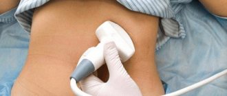
Diffuse heterogeneity can be diagnosed using ultrasound. This is a simple, non-invasive and painless procedure. The examination is carried out using ultrasound, which passes through tissues and organs and is reflected from them, displaying an image on the screen. The examination is carried out using an ultrasonic sensor. The patient lies on his back, the doctor lubricates the skin of the abdomen with gel, applies the sensor and moves it, pressing lightly. Painful sensations should not occur, but if the pancreas is inflamed, they may appear when pressed.
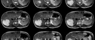
In addition to ultrasound, for diseases of the pancreas, an endoscopic examination, a blood test (biochemistry) is prescribed to check pancreatic indicators; if a tumor is suspected, a computed tomography or MRI may be prescribed.
Treatment directly depends on the diagnosis, but for any disease of the pancreas, diet is an essential part of treatment.
In case of acute pancreatitis, it is recommended to go on a strict diet for a couple of days and drink more to relieve the organ. It is necessary to give up alcoholic beverages, fatty and fried foods, smoked, spicy, coffee, chocolate, cream cakes, eggs (except soft-boiled).
For pancreatitis and other diseases of the pancreas, digestive enzymes are often prescribed, in particular Pancreatin (Creon, Mezim, Festal). They reduce the load on the organ and improve digestion. You can take them for a long time, if necessary (several months or even years with breaks).
In case of severe nausea, symptomatic drugs are prescribed that relieve the urge to vomit, for example, Cerucal or Motilak. They are taken as needed an hour before meals.
More information about acute pancreatitis can be found in the video:
If the disease is accompanied by severe abdominal pain, painkillers are prescribed (Spazmalgon, No-Shpa, Bral, etc.). These drugs are not taken in long courses, as they have a number of side effects.
During the period of remission, it is important not to break the diet, not to overeat, and to avoid taking medications for no reason, especially antibiotics. For people with diseases of the pancreas, alcohol is contraindicated, but if the patient decides to break this rule, then it is better to drink without fatty snacks (lard, meat, sausage, kebab, etc.)
Source: DiagnozLab.com