Oncological diseases are dangerous for long periods that are asymptomatic. Gastric adenocarcinoma is not an exception. What should a patient do when faced with a cancerous tumor?
- 2 Classification of the disease: papillary, cricoid, poorly differentiated, highly differentiated and other forms of pathology
2.1 Stages of development
2.1.1 Determination of stages of gastric adenocarcinoma using the TNM system - table
- 6.1 Traditional medicine
6.1.1 Gallery of traditional medicine
- 6.2.1 Prohibited products - gallery
What is gastric adenocarcinoma
Gastric adenocarcinoma is a common oncological pathology. The tumor consists of tissue from the mucous membrane of the organ. Under the influence of several reasons, the protective layer is destroyed, as a result of which healthy cells degenerate into cancerous formations.
Adenocarcinoma occurs in 95% of cases of gastric cancer.
Adenocarcinoma of the stomach is a dangerous condition that can spread to other organs.
The pathology occurs in males more often than in females. The tumor can form over a long period of time - from 1 year to 15–20 years. The neoplasm most often appears after 45 years.
Gastric adenocarcinoma is often called glandular cancer.
https://youtu.be/h5QI_4wFrJs
general description
Speaking about what gastric adenocarcinoma is, it should be noted: this is a form of cancer classified not by clinical manifestations, but based on which cells have changed under the influence of oncogenic factors, giving rise to the tumor. To do this, it is studied under a microscope, conducting a histological examination. Adenocarcinoma is a derivative of glandular epithelium. According to the WHO histological classification, there are:
- Tubular adenocarcinoma of the stomach, which can be highly differentiated (that is, the cells do not differ much from normal glandular tissue) and moderately differentiated (the cells are greatly changed, but you can “guess” their origin).
- Poorly differentiated gastric adenocarcinoma. Differentiation is the process of cell maturation, during which they acquire a structure and properties characteristic only of a certain tissue. In poorly differentiated carcinoma, they are so “immature” that they have practically nothing in common with normal glandular epithelium.
The lower the cell differentiation, the more malignant and aggressive the tumor behaves; therefore, other things being equal, well-differentiated gastric adenocarcinoma has a more favorable prognosis than poorly differentiated one.
The so-called mucinous adenocarcinoma of the stomach or “mucosal cancer” is distinguished separately, which is characterized by increased mucus formation and early metastasis.
Causes
The main reason for the formation of adenocarcinoma is the degeneration of glandular epithelial cells due to deterioration of nutrition of the stomach walls, failure of blood supply and inhibition of organ secretion. Numerous factors lead to this:
- unhealthy diet - consumption of salty, fatty and smoked products in large quantities;
- abuse of diets - leads to a decrease in dietary fiber intake, as well as a deficiency of vitamins and nutrients;
- bad habits - ethyl alcohol and tobacco tars negatively affect the condition of the organ, so adenocarcinoma is more often found in people who abuse alcohol or have a long history of smoking;
- Helicobacter pylori is a bacterium that often causes gastritis and ulcers. Long-term infection leads to an increased risk of developing adenocarcinoma;
- heredity - the likelihood of developing cancer increases if close relatives have adenocarcinoma;
- diseases: gastritis, gastric epithelial dysplasia, polyposis, chronic ulcer, duodenogastric reflux.
Risk factors include being over 50 years old, living in an area with poor environmental conditions, and working in hazardous enterprises.
Stomach ulcers often cause cancer
How does pathology manifest itself in the early stages and during tumor disintegration?
At the initial stage, the disease does not manifest itself - this is the main danger of adenocarcinoma. Minor changes in the body, characteristic of other diseases, do not make the patient wary. These symptoms are:
- gradual decrease in body weight;
- weakness;
- fast fatiguability.
As the tumor develops, other signs appear:
- decreased appetite;
- painful sensations in the abdomen, depending on food intake;
- belching and heartburn.
In the later stages, the patient's condition worsens. Symptoms appear:
- aversion to meat and other protein foods;
- anemia;
- tarry stools due to internal hemorrhages (indicates tumor disintegration);
- stagnation of food in the stomach;
- feeling of a full stomach;
- increased salivation;
- nausea and vomiting.
It must be taken into account that even in the later stages the pathology may be mild - it depends on the patient’s condition and the type of glandular cancer.
Clinical manifestations
At the initial stage, the disease does not manifest itself in any way. The first symptoms appear only when the tumor begins to increase in size. As the tumor grows, it affects nearby organs: kidneys, bladder, liver. The development of the disease is accompanied by the following symptoms:
- cramping pain in the abdominal area;
- decreased appetite;
- weight loss;
- increase in temperature indicators;
- general weakness and constant fatigue;
- paleness of the skin;
- the presence of blood and mucus in the stool;
- bloating;
- constipation gives way to diarrhea;
- pain during bowel movements.
After some time, the malignant formation begins to disintegrate. Decay products penetrate into the stool, and the stool acquires a foul odor.
The disease occurs in four main stages. At the last stage there is a high risk of intestinal obstruction.
Diagnostics
To identify adenocarcinoma, an examination is performed, during which typical symptoms are detected. After this, a set of diagnostic measures is prescribed, which includes the following methods:
- general blood test - the patient shows an increased number of leukocytes and a decrease in the number of red blood cells, which indicates an inflammatory process and anemia;
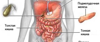
- computed tomography - the method is used to exclude metastases to other organs;
- gastroscopy - evaluate the nature of changes in the mucous membrane of the organ;
- blood test to detect tumor markers;
- laparoscopy - detect metastases in other organs and determine the degree of development of pathology;
- fluoroscopy with barium sulfate - reveals a filling defect, which indicates destruction of the stomach walls or the growth of adenocarcinoma;
- esophagogastroduodenoscopy with biopsy - determine the degree of differentiation;
- Ultrasound examination of the abdominal cavity - metastases are detected.
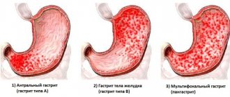
Gastroscopy helps assess the condition of the stomach and changes
Diagnostic tests
To detect a malignant neoplasm of the stomach, the doctor prescribes a number of laboratory and instrumental studies:
- The most informative picture of the disease is provided by esophagogastroduodenoscopy. The patient's throat is numbed and a probe with a camera at the end is inserted into the esophagus. A specialist examines the organ from the inside; if a tumor is detected, EGD allows you to take a piece of tissue for examination.
- Ultrasound diagnostics determines the presence and types of tumors in all organs of the gastrointestinal tract and lymph nodes.
- To determine the area of the lesion and the size of the tumor, an X-ray of the stomach is performed with contrast.
- Computed tomography and magnetic resonance imaging detect tissue changes in all parts of the body.
- A biochemical blood test evaluates the functioning of internal organs.
- In a general clinical blood test, ESR indicators are important. Their increase indicates a pathological process. When hemoglobin levels decrease, internal bleeding is likely.
- To confirm the nature of the neoplasm, histology of the biopsy specimen is performed.
- If the tumor has metastases, the cervical lymph nodes, genitals are checked, vascular angiography and MRI of the brain are performed.
Treatment of adenocarcinoma
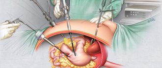
The main treatment method for adenocarcinoma is surgery. Based on the size of the formation and its location, the following methods of intervention are used:
- gastrectomy - removal of the stomach with surrounding tissues: lymph nodes, part of the small intestine and esophagus;
- subtotal resection - part of the surrounding tissue or stomach is removed.
Gastrectomy - complete or partial removal of the stomach
If it is impossible to eliminate adenocarcinoma using a surgical technique, and the patient has complications, then the following types of intervention are prescribed:
- endoluminal laser therapy - the use of a laser to remove cancer cells;
- Endoluminal stenting is the use of a stent that is inserted into the stomach so that the patient can feed himself.
In addition to surgery, additional methods can be used:
- Immunotherapy. To have a positive effect on the patient’s immunity, immunomodulatory drugs are prescribed - this will reduce the growth and spread of the tumor.
- Radiation therapy. The technique is aimed at destroying the cells remaining after surgery. Special radiation not only eliminates diseased areas, but also reduces pain and the likelihood of bleeding.
Ftorafur is used in chemotherapy to slow down the growth of pathological cells
- Chemotherapy (Cisplatin, Ftorafur, Bleomycin). Used to reduce discomfort and slow down the growth of pathological cells. As a result, the spread of metastases is reduced.
ethnoscience
Alternative medicine recipes can improve the patient’s well-being. They are used only after consultation with a doctor.
Natural juices are effective for stomach cancer. To prepare them you need:
- Pass the following components through a juicer:
- 1 potato;
- 100 g celery;
- 300 g beets;
- 30 g black radish;
- 100 carrots.
- Strain the drink and leave for a few minutes until sediment settles.
This drink is consumed 0.5 liters per day.
Herbal infusions are also useful for the patient. The following ingredients are needed:
- 50 g celandine;
- 50 g sage;
- 100 g wheatgrass rhizomes;
- 50 g eyebright;
- 50 g of Rhodiola rosea roots;
- 50 g hogweed.
- Mix the ingredients and take 1 tbsp of raw materials. l.
- Add the herbal mixture to the thermos and pour 1 liter of boiling water.
- Leave the container overnight and strain the next morning.
The product is taken before each meal, 1 glass (250 ml).
Gallery of traditional medicine
Celery is effective against stomach cancer
Using sage along with herbal remedies brings relief to the patient
Carrot juice is a source of vitamin A
Hogweed contains a large number of amino acids.
Beets neutralize the development of tumor cells
Black radish is rich in vitamins B and C
Celandine has an antitumor effect on the body
An invalid diet
There are 3 possible diet options for people with gastric adenocarcinoma:
- For patients without significant metabolic disorders and with normal body weight:
- energy value - no more than 2400 kcal per day;
- proteins - 90 g, half of which are of animal origin;
- fats - 120 g, including 30 g vegetable fats;
- carbohydrates - 330 g.
- Patients with impaired excretory function of the kidneys and liver, as well as those who are overweight:
- energy value - no more than 2650 kcal;
- fats - 90 g, of which 30 g are vegetable;
- carbohydrates - 400 g;
- proteins - 60 g, half - animal origin.
- For patients with low weight and obvious metabolic problems:
- energy value - 3600 kcal;
- proteins - 140 g;
- fats - 120 g, including 40 g vegetable fats;
- carbohydrates - 500 g.
The doctor will give specific nutritional recommendations after examining the patient. The calorie content of the diet and the BJU ratio may vary depending on the age of the sick person and the characteristics of the course of the disease.
Nutrition principles:
- cooking method - stew, boil or bake;
- number of meals - 4–6 times a day.
Prohibited products:
- pickles and marinades;
- smoked products;
- fatty and fried foods;
- alcohol;
- strong coffee;
- hot dishes.
Products useful for the patient:
- black currant;
- sea fish;
- carrot;
- citrus.
It is recommended to select foods for your diet together with your doctor.
Prohibited products - gallery
Salty foods are excluded from the patient’s menu
Smoked products are dangerous for the patient
Fried foods have a negative effect on the stomach, especially in the development of cancer
Alcohol increases stress on the gastrointestinal tract
If you have stomach adenocarcinoma, you should avoid drinking strong coffee
How are they treated?
Therapy is based on the patient's condition. Drug treatment is ineffective, so surgery is prescribed if the stage allows. In phase 4, surgery is unsuccessful. Folk remedies are ineffective, so official medicine does not use them. Surgery provides several options for the operation, the choice is made by the doctor:
An operation in which part or all of an organ is removed through surgery is called a gastrectomy.
- Gastrectomy is the total removal of an organ. Along with the stomach, part of the esophagus and the upper parts of the intestine are cut out.
- Resection is the excision of part of an organ and nearby tissues.
- Endoluminal stenting is the placement of a tube to improve the lumen in the organ. It is used if oncology blocks the pathways.
- Laser therapy. The beam removes cancer cells. The procedure is used in the first stages of development.
Radiation and chemotherapy are prescribed as auxiliary and are carried out before and after surgery. They are not able to completely stop the process; they are used to reduce the size of the tumor formation. In the postoperative period, beams are used to clear away any remaining malignant cells. During the treatment process, a special diet is important.
Treatment prognosis, survival and life expectancy of patients
The prognosis for recovery depends on the stage of development of adenocarcinoma:
- initial stage - high probability;
- Stage 1 - 60–80%;
- Stage 2 - 30–40%;
- Stage 3 - 12–20%;
- Stage 4 - less than 5%.
The stage of tumor development affects life expectancy. In the later stages, a sick person will be able to live for about 5 years, but if the indicated threshold is overcome, the period increases to 10 years - this depends on the patient’s lifestyle and compliance with the doctor’s recommendations.
Age is of great importance - people under 50 years of age have a 10-12% higher chance of recovery than older patients.
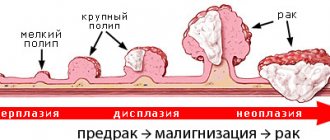
Classification by stages
In order to create a unified approach to assessing the severity of adenocarcinoma, an international classification has been adopted. It divides all intestinal adenocarcinomas into 5 stages. For each the following are defined:
- acceptable size of tumor growth;
- the presence of near and distant metastases.
In stage 0, the tumor is minimal, does not grow anywhere and has no metastases. In stage I-II – sizes are acceptable from 2 to 5 cm or more, but there are no metastases. The third stage is divided into:
- IIIa – germination into neighboring organs and the presence of metastases in the lymph nodes are allowed;
- IIIc – combines large size and the presence of metastasis only in neighboring organs.
Stage IV – is diagnosed for distant metastases, even if the size of the tumor itself is relatively small.
There is a classification of intestinal cancer, which includes such a feature as differentiation of cellular composition. It means that:
- Gx - diagnosed if the cells cannot be differentiated;
- G1 – the degree of differentiation is assessed as high, the cells are similar to normal epithelial ones;
- colon cancer grade G2 - shows an average degree of degeneration;
- G3 – tumor cells are little similar to normal;
- G4 – cell type is low differentiated and is characterized by the highest malignancy.
Outdated, but very bright classification
Possible complications: metastases to other organs, ascites and others
During the development of adenocarcinoma, the following complications may occur:
- pyloric stenosis - when the pathology is located in the area of the organ;
- metastases to the lungs, liver, esophagus and other organs - disruption of their functioning;
- growth of the formation into the wall of the stomach, damage to large vessels and perforation of the gastric wall - frequent bleeding of varying intensity;
One of the complications of advanced adenocarcinoma is perforation of the gastric wall.
- violation of venous circulation when adenocarcinoma compresses blood vessels - leads to the accumulation of fluid in the abdominal cavity (ascites).