The human musculoskeletal system is a complex system, for the full functioning of which the integrity of each element is important. Due to constant stress, the foot is most vulnerable to external physical influences, its own weight, and excess muscle tension. Injuries to this part of the body are classified as common injuries. The fifth metatarsal bone is the most commonly fractured bone, but the other four can also fracture. The success of treatment and the speed at which the patient returns to normal motor activity depend on how accurately and timely the diagnosis is made.
Signs of a foot fracture
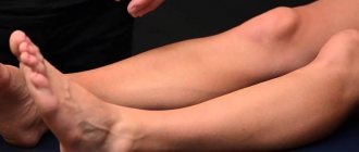
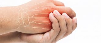
Swelling of the injured area and pain are the very first symptoms that indicate that you may have a foot fracture. The photo below shows what a sore leg looks like.
The pain can be so severe that the person cannot move. Bruising may also occur in the area of injury. A displaced fracture is characterized by a change in the shape of the foot.
Sometimes a person does not realize that he has a broken foot. Signs may not be pronounced; pain occurs only when putting weight on the affected leg. Therefore, to clarify the diagnosis, it is necessary to contact a traumatologist.
Frequently asked questions to the doctor
The feeling of pain does not go away
Hello, my name is Evgeniy. While playing football, an opponent hit his leg hard. After a while, a tumor and hematoma appeared. The pain is severe, does not increase and does not go away. 3 days have passed and the situation has not changed. I'm attaching a photo of the injured limb, is it possible that there's a fracture?
Hello, Evgeniy! I advise you to go to the emergency room to determine an accurate diagnosis; it is difficult to do this from photos and descriptions, due to the similarity of symptoms. After the x-ray, the answer to the question will be known.
First aid
If a fracture of the foot bones is suspected, the injured limb must be immobilized. You can use an improvised splint made from planks, ski poles or rods, which is attached to the leg with bandages. If you don’t have anything at hand, you can bandage the damaged limb to the healthy one using a scarf, shirt or towel.
If there is an open fracture, you should not try to set the bone yourself; first of all, you need to stop the bleeding. To do this, you need to treat the skin around the wound with iodine or hydrogen peroxide. Then you need to carefully apply a sterile dressing. After first aid has been provided, the victim must be taken to a medical facility.
What drugs are effective for leg fractures?
For leg fractures, a number of medications are prescribed:
- Analgesics. They anesthetize the patient and prevent the development of shock (Ketanov, Dexalgin, Keyver).
- Antibiotics. Prescribed for open injuries or after surgery (Ceftiriaxone, Cefosulbin, Cifran, Metronidazole, Ofloxacin).
- Calcium-containing preparations. Promotes rapid regeneration of bone tissue (Osteogenon, Calcium D3).
After surgery to reposition the bones, the wound is treated daily with antiseptics and a sterile bandage is applied.
Fracture of the talus
This bone has some special features. There is no muscle attached to the talus. In addition, it transfers body weight to the entire foot.
A fracture of the talus is possible as a result of indirect trauma, is uncommon and is considered a severe injury to the bones of the foot. Accompanied by other injuries, such as a fracture, dislocation of the ankle or other bones of the foot.
Symptoms
When an injury occurs, sharp pain is felt, swelling of the foot and ankle occurs, and hemorrhages are noticeable on the skin. If the fragments are displaced, you may notice that the foot is deformed.
To confirm the fracture, determine its location, type and degree of bone displacement, an X-ray examination is performed in two projections.
How to treat
If a displaced foot fracture is diagnosed, bone fragments are immediately compared. The fact is that the later you see a doctor, the more difficult it will be to restore their correct position, sometimes even impossible.
The plaster is applied for a month and a half. Starting from the third week, you need to release the injured limb from the splint and make active movements of the ankle joint.
Somewhat later, physical therapy, massage, and physiotherapy are prescribed. It takes two to three months to restore working capacity.
Fixation.
Fixation of the fragment is performed only in cases of fresh extensive damage. The torn osteochondral fragment is lifted, the underlying bed is subjected to microfracture or reaming, and then the fragment is fixed using submersible absorbable screws or fibrin glue. The method is technically complex and its indications are quite narrow, but when it was used according to indications, a good result was observed in 89% of cases.
Thus, the most effective and technically simple method of treating the vast majority of osteochondral injuries of the talus at the moment is removal, curettage and bone marrow stimulation. Depending on the equipment of the operating room and the skills of the operating surgeon, this intervention can be performed either openly or arthroscopically.
Scaphoid fracture
Occurs as a consequence of direct exposure. Often a fracture of this foot bone is accompanied by injuries to other bones.
Symptoms
A person cannot lean on a limb due to severe pain. Swelling and hemorrhages appear. When palpating and trying to turn the foot in and out, a person feels a sharp pain. To confirm a fracture of the navicular bone of the foot, X-ray examination is recommended.
Treatment
If no displacement is detected, the doctor applies a circular plaster cast to the damaged area. For displaced fractures of the scaphoid, bone fragments are compared; in some cases, open reduction may be necessary. The foot is fixed in plaster for a period of four to five weeks.
It is quite difficult to treat such a foot fracture in combination with a dislocation. If the dislocated fragment is not set back into place, traumatic flatfoot may develop. Displaced fragments are realigned using a special traction apparatus. Sometimes it is necessary to resort to open reduction and securing the reduced fragment with a silk suture. After such a procedure, immobilization of the injured limb should continue for up to 10-12 weeks. In the future, it is necessary to wear orthopedic shoes.
Diagnostics
The diagnosis of leg fractures is made based on the collected data from the anamnesis, examination and radiological studies.
The patient must have an x-ray of the limb. In some cases, a CT scan and MRI will be needed.
Patient management tactics are selected individually, based on X-ray data.
Stress (fatigue) fracture
Such injuries, which include a fracture of the 5th metatarsal bone of the foot, are usually found in athletes and those who lead an active lifestyle. They appear as a result of excessive and prolonged stress on the foot. Essentially, such a fracture is a crack in the bone, and it is very difficult to notice.
If a person suffers from various underlying diseases, such as osteoporosis or foot deformities, his condition may worsen significantly. A stress fracture of the metatarsal bone also occurs in those who constantly walk in uncomfortable and tight shoes.
Symptoms
The first symptom that should alert you is pain that occurs in the foot after prolonged intense exercise and disappears in a calm state. Over time, it intensifies to such an extent that any action becomes impossible. The pain persists even at rest. Swelling appears at the site of injury.
The danger is that most people with such an injury are in no hurry to see a doctor; often the person does not even suspect that he has a foot fracture. The signs in this case are not as pronounced as with other fractures; the patient walks and steps on his leg. Therefore, to avoid complications, you must consult a doctor immediately.
A fracture of the fifth metatarsal bone of the foot is an injury that occurs more often than any other.
When the foot is turned inward, an avulsion fracture may occur. In this case, the metatarsal bone is separated and displaced. Fusion is very long, so you need to contact a traumatologist as soon as possible. If not treated in a timely manner, the bone may not heal properly, in which case surgical intervention will be required.
The base of the 5th metatarsal is an area with poor blood supply. This is where the Jones fracture occurs. It occurs against the background of stress and grows together very slowly.
Features of fractures in different areas
Fractures in different parts of the lower limb have some characteristics.
Finger fracture
When a toe is broken, the patient is haunted by incessant pain. The fingers and feet swell, and a hematoma appears under the skin. The finger cannot be bent. The same applies to the foot - pain and swelling limit its mobility. The finger may deviate in an uncharacteristic direction. The finger may dangle, creating the feeling that it is supported only by the skin.
Gradually, the swelling increases, and the finger becomes visually shorter. If you feel the fracture site, you may find protruding bone. Walking with a broken toe is very painful, and the pain is felt anywhere in the foot - even if you rest on your heel.
Metatarsals
The following symptoms correspond to a fracture of the metatarsal bones:
- pain when trying to walk and when feeling the foot;
- foot deformity;
- swelling affects both sides of the foot and moves towards the ankle;
- painful sensations intensify not only when leaning on the foot, but also when turning it;
- severe deformation of the foot.
Hip fracture
Violations of the integrity of the hip bone do not occur very often - only in about 6% of cases of fractures. However, in older people such injuries are among the most common - about 40% of all injuries of this type.
In case of damage to the femoral neck, pain is felt in the groin and hip joint. Moreover, although palpation can intensify the pain, palpation does not provoke a sharp attack.
With trochanteric hip fractures, the pain is much more pronounced, increasing with changes in the position of the leg. The limb itself is turned outward. If the bone is displaced, the broken leg becomes visually shorter. The injured leg cannot be lifted from the surface, even with maximum effort.
When the integrity of an impacted bone is violated, the pain syndrome does not manifest itself so obviously. Very often, victims perceive the injury as a bruise, because, despite some pain, they can walk quite well.
Injuries in the hip area are determined only through instrumental examination using X-rays and MRI.
Treatment
If a minor fracture of the foot is detected, treatment is simply a splint. In this case, the injured limb must remain motionless for several weeks in order for the damaged bone tissue to heal completely.
When the bone is severely damaged, internal fixation is necessary. This is done using special screws.
The severity and nature of the damage determines further treatment. Any non-displaced metatarsal fracture requires immobilization. The applied plaster will reliably protect the bone from possible displacement. Due to the fact that the foot is completely motionless, the fusion of bone tissue will occur faster.
If the fragments are displaced during an injury, surgical intervention cannot be avoided. During the operation, the doctor opens the area of the fracture and compares the resulting fragments, after which he fixes them with special knitting needles or screws. A cast is then applied for up to six weeks. The patient is prohibited from stepping on the injured leg. After six weeks you can start walking. The knitting needles are removed after three months, the screws after four. The patient is recommended to wear orthopedic shoes or insoles.
For a Jones fracture, a plaster cast is applied from the toes to the middle third of the leg for up to two months. Do not step on the injured leg.
To reduce the load on the injured limb while walking, you need to use crutches. The patient must be observed by a doctor who will select the correct rehabilitation course to restore impaired functionality in the injured foot.
The recovery period for a metatarsal fracture is quite long and includes physical therapy, massage, use of arch supports, and physiotherapy.
If such an injury is not treated or treated incorrectly, complications such as arthrosis, deformity, constant pain and non-union of the fracture may occur.
Rehabilitation
The rehabilitation process after removing plaster or skeletal traction is quite long. After all, when an injury occurs, not only the bone suffers, but also muscles, tendons, nerve fibers, and blood vessels. Sometimes, the treatment process itself is much faster than restoring full functionality of the limb.
At the rehabilitation stage, the patient is recommended to:
- performing exercises (the complex is prescribed by the doctor);
- massage;
- swimming;
- measured walking, initially with support on a crutch;
- physiotherapy;
- complete nutrition enriched with calcium and vitamins.
Leg pain and partial loss of functionality can bother you throughout your life. To minimize the risk of developing this complication, you should follow all doctor’s recommendations regarding rehabilitation.
Nutrition for prevention and during treatment
Preventing the disease is much easier and cheaper; prevention does not require the expenditure of your nerves. After all, the diet - both for the prevention of damage and for a speedy recovery - does not differ.
Calcium-rich foods will help improve the condition of the musculoskeletal system, fill the bones with the necessary amount of minerals and protect against unnecessary stress.
These products include:
- milk;
- cottage cheese;
- sour cream;
- hazelnut;
- almond;
- apricots;
- currant;
- milk chocolate;
- soy;
- legumes;
- sorrel.
Important! Products containing large amounts of protein can negatively affect the body in the presence of certain diseases, so you should coordinate the diet with your doctor, after telling him about your chronic diseases.
The microelement zinc is needed to accelerate the recovery of bone tissue. The substance enters the body with vegetables (broccoli, corn, carrots), fruits (avocado), berries (raspberries, blueberries), cereals (rice, wheat), legumes, mushrooms, chocolate and nuts. From this list, you can choose a few of your favorite foods and eat them daily.
Diagnostic features
A fracture of the foot bones has a specific clinical picture. If we add the fact of injury, a preliminary diagnosis is ready. But for successful treatment and rehabilitation, this data is not enough. Information is needed about the exact location of the fracture, the absence or presence of displacement, the number of damaged bones, and the location of fragments.
X-ray of the foot allows you to see all the details of interest. Sometimes one image is not enough, the fracture line is not visualized, so an x-ray must be taken in two or three projections.
In severe cases, computed tomography (CT) or MRI is indicated for differential diagnosis and assessment of the condition of soft tissues.
Causes of fracture
Such an injury cannot be taken lightly. By the way, according to statistics, in almost all road accidents it is the talus bone that suffers first. A fracture also threatens in the following situations:
- Excessive joint flexion. This phenomenon is observed under various overloads of the body.
- When falling. Height doesn't always play a big role. When landing on your feet, the load on them increases sharply, which leads to various injuries.
- Flexion with turning outwards. Most often this happens in various accidents on the road.
Recovery
Rehabilitation after a fracture of the talus involves undergoing physiotherapeutic procedures. If the posterior process is fractured without displacement, the cast is worn for about two months. If bone displacement or fragmentation occurs, recovery takes a longer period. The main rules that must be followed when undergoing rehabilitation are as follows: it is recommended to rest the injured lower limb for the first 5-7 days. Physical activity may contribute to secondary damage to bone integrity.
When the cast is removed, you should definitely undergo a therapeutic course of physical therapy to strengthen muscle tone and restore the elasticity of the ligaments. To reduce swelling and prevent blood stagnation, a foot massage is performed with special oils. Daily diet plays an important role in recovery. It should be rich in all micro- and macroelements and nutrients necessary for the full functioning of a healthy body. Food should consist of fruits, vegetables and must contain protein.
To reduce swelling and relieve painful symptoms, the doctor may prescribe the use of non-steroidal drugs, which also have an anti-inflammatory effect. Also, if necessary, procedures such as electrophoresis, magnetic therapy or ultrasound can be prescribed.
Causes of injuries
It is important to remember that there are different types of fractures. This means that pre-medical measures taken for each type of injury will be slightly different from each other.
Let's find out why these injuries occur, how fractures are classified, and what needs to be done in each specific case.
There are 2 main factors that cause this dangerous injury:
- Impact of various physical forces on the bones: falls, blows, bruises, unsuccessful walking.
- Pathologies in the structure of the bones themselves: weakness and fragility of bone tissue caused by osteoporosis or tumors.
In addition, this injury is caused by taking antibiotics and probiotics, following a diet for weight loss, which limits the receipt of necessary vitamin complexes, excessive physical activity and stress. All this causes weakening of the connective and muscle tissue of the bone, which can lead to fractures.
By themselves, of course, the bones will not break because of this, but their condition will no longer be able to withstand various physical influences. There will be no normal load resistance. Brittle bones have the greatest risk of injury.
Clinical picture
A fracture of each of the bones of the foot has its own characteristics, but often without x-ray examination it is impossible to make an accurate diagnosis. The doctor takes into account complaints and conducts a thorough examination, which allows us to preliminarily suspect the location of the damage.
Talus
The talus is one of the largest bones in the foot. It is the connecting link between the lower leg and foot, which is why it bears the greatest load. Blood circulation is weak, only small vessels feed it. Therefore, fusion and restoration of function takes a long period.
This injury is relatively rare and is accompanied by damage to other bones, dislocations, and ligament ruptures. As a rule, it indicates severe damage to the foot.
Characteristics:
- sharp, sharp pain;
- swelling, redness of the proximal foot (closer to the lower leg);
- change in shape, deformation, hemorrhages on the skin in the presence of displacement.
Calcaneus
The calcaneus is located under the talus and has the largest volume. It is damaged when falling or jumping from a height, when the maximum load falls on the heel. Wedging of the talus occurs, which leads to splitting. There are: fracture of the foot with or without displacement, simple, comminuted, impacted.
After an injury, swelling is observed below the ankle, and a hematoma may appear. The contour of the heel changes: it becomes rounder and more swollen. When trying to touch, the patient notices acute pain, which often radiates to the calf muscle. Gait becomes a problem, the person is forced to step on the forefoot. When tapping the heel area, there is a sharp pain.
Scaphoid
A fracture most often occurs as a result of a heavy object falling on the leg and is combined with damage to other bones. The pain is mild, the person can move, but puts emphasis on the heel. There is swelling and redness of the dorsum of the foot. A protruding part of the damaged bone is visualized. The injury often goes unnoticed because signs of a foot fracture are minimal. Only with the help of x-ray examination can a diagnosis be made.
Cuboid and sphenoid bones
These are small bones on the back of the foot; fractures occur after direct injury. Falling a heavy object is a leading cause of injury. Diagnosis is difficult because the symptoms are nonspecific. Noteworthy is the swelling, redness of the dorsum of the foot, and pain when moving to the sides. Displacement occurs rarely, which simplifies treatment; it is not always possible to establish a diagnosis using X-rays; in such a situation, CT or MRI comes to the rescue.
Metatarsals
The largest percentage of fractures occurs due to damage to the metatarsal bones. This is facilitated by their structure: thin, tubular bones. They are injured as a result of a falling object, being run over by a car, a motorcycle, or falling, especially with a twisted foot. According to statistics, fractures of the first, fourth and fifth bones are more common.
The symptoms are similar: swelling of the dorsum of the foot, which can spread to the sole. The patient has difficulty walking and deliberately puts pressure on the heel to relieve pain. Support on the forefoot is sharply painful; palpation allows you to determine the deformity and the place of maximum pain.
Fractures can be single or multiple. If one bone is damaged, displacement is rarely observed, because the remaining bones do not allow the fragments to move. Natural immobilization of the area occurs, however, if the traumatic factor was of greater intensity and force of application, then displacement cannot be avoided.
Phalanges of fingers
A fracture of these small bones occurs due to a blow, an object falling on the foot, or as a result of being twisted. The patient complains of pain in the fingers, which intensifies when trying to bend or abduct it. The gait may not be disturbed, this often happens with fractures of the fifth toe. However, damage to the first digit results in significant impairment of function; discomfort and swelling in a separate phalanx are observed.
Late seeking medical help leads to malunion, and subsequently to the development of post-traumatic arthrosis and chronic pain.
Forecast
Trauma to the foot has a good prognosis. Usually, after 4–5 weeks, the ability to work is completely restored, the person returns to his normal life, and there is no trace of pain in the leg.
The speed of regeneration depends on the conditions in which the patient is: his environment, daily routine, nutrition and rest, compliance with doctor’s instructions. If there are no concomitant diseases that complicate the prognosis, then there is nothing to worry about - rest and minimal stress on the affected area will do the trick.
But to prevent recurrent fractures, it is worth taking care of the comfort of your feet for at least a period of 10 months. To do this, you need to wear comfortable shoes and buy orthopedic insoles if desired.
Remember that preventing a disease is much easier than curing it. Watch your menu and be careful when crossing the road or playing sports.
Human foot bones
The foot integrates the following sections:
- tarsus (the back part connected to the lower leg), the tarsus consists of 5 bones;
- metatarsus (the middle part that forms the elastic arch), includes 5 bones;
- phalanges of the fingers, include 14 bones.
Thus, the foot is made up of 26 bones, and each bone has its own name.
Most people also have 2 small sesamoid bones. In rare cases, the foot includes 1-2 additional, anatomically not provided bones, which often cause foot health problems to their owners.
Tarsals
The talus is the highest bone of the foot and its upper side forms the ankle joint:
- The bone has no tendons or muscles attached.
- It has 5 articular surfaces on which a layer of hyaline cartilage is located.
- The heel also has many articular surfaces (6 pieces), multiple ligaments are tied to it, the weakening of which is often associated with the formation of flat feet.
- The Achilles tendon is attached to the convex rear part.
Talus of the foot
The navicular bone forms the inner part of the foot; by palpating the joint, the doctor determines the degree of flatfoot:
- Participates in the formation of the anatomical vault.
- Connected by a joint to the talus.
- Three wedge-shaped bones are attached to it in front.
- The cuneiform bones have articular surfaces at their proximal ends for connection with the first three metatarsals.
The cuboid bone is included in the upper tarsal part of the inner side.
Navicular bone of the foot
Metatarsal or metatarsal bones
Despite the fact that these five tubular bones differ in diameter and length (the thickest and shortest is the first bone, the most elongated is the second), their structure is identical.
They include:
- head;
- body;
- base.
The bodies of these bones have the shape of a pyramid with three ribs, and the heads have rounded anterior ends. The articular surfaces on the heads of the metatarsal bones are connected with the lower phalanges of the fingers, and on the bases of the bones - with the anterior tarsal bones.
Metatarsal bones of the foot
Phalanges of fingers
By analogy with the hand, the big toes have only proximal (lower) and distal (upper) phalanges, and the remaining fingers have three phalanges (intermediate, proximal and distal), connected by movable joints. These are generally small and thin tubular bones.
Sometimes the two phalanges of the little toes grow together (which is not a pathology).
The phalanges of the feet are noticeably shorter and thicker than those of the hands. This is due to the fact that the foot is not required to have the flexibility and development of fine motor skills like the fingers, but it does require strength and the ability to withstand long-term loads.
Phalanges of fingers
Like the metatarsal bones, the bones of the phalanges of the toes are protected by a fairly meager amount of soft tissue, so they are easily palpated, especially in lean, wiry people.
Sesamoid bones of the foot
Two such bones are located in the thickness of the tendons of the big toes in the area of \u200b\u200bthe junction of the metatarsal bones with the proximal phalanges of the big toes. They affect the severity of the metatarsal arch.
When X-raying the foot, they appear on the image as grains of a foreign substance in the thickness of the ligaments. Sometimes these bones have a bifurcated shape (this can be either a given from birth or a consequence of injury).
Sesamoid bones
Accessory or supernumerary bones
The most common bone is the external tibia (12% of the population, almost twice as common in women), which is connected to the scaphoid cartilage or ligaments. Its dimensions are variable; in people with large bones, it protrudes strongly downward, which entails constant rubbing of this area with shoes. Sometimes it is found in professional athletes.
Those who have an external tibia are recommended to wear arch supports or special insoles (orthopedic shoes for large bones). Treatment of the consequences caused by the bone is determined by the particular case of the clinical picture.
7% of the population has a triangular bone. On x-ray it can be confused with a fracture. An uneven border line and clearly focused pain indicate a fracture, a smooth, even border line indicates the presence of a triangular bone.
Diagram of foot bones with captions