Definition of disease. Causes of the disease
A Bartholin gland cyst is a benign round cavity (a saccular formation with thin walls and the presence of secretions of varying consistency inside) formation in the lower third of the vestibule of the vagina as a result of a violation of the outflow of secretions from the cavity and its accumulation in it.
The cyst cavity stretched by secretions can have different sizes, reaching 7-9 cm in diameter. Bartholin gland cysts are observed mainly before the age of 30 years (hormonally active reproductive age) and account for 2% of all diseases of the female genital organs with actively functioning and hormonally dependent Bartholin glands.
Before we move on to the mechanism of development of this pathology, we will consider the normal anatomy and physiology of the Bartholin glands.
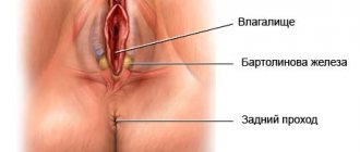
Bartholin's glands or large vestibular glands are located on both sides of the vestibule of the vagina, in its lower third in the thickness of the labia majora. They are a paired organ. They got their name in honor of the Danish anatomist Caspar Bartolin Jr. who discovered them. Their size is about 1.5-2 cm, the excretory duct of the Bartholin gland, 1.5-2.5 cm long, opens on the inner surface of the labia minora at the border of its middle and posterior third. In some cases, their atypical location occurs, for example, in the thickness of the labia minora [13]
The function of the Bartholin glands is to moisturize the mucous membrane of the vulva during arousal during sexual intercourse, which prevents it from becoming dry and painful during sexual intercourse. Due to the presence of mucin in the secretion, it has a bactericidal effect.
The main causes of this disease are frequent inflammatory processes in the genital area caused by specific and nonspecific microflora, such as staphylococci, streptococci, E. coli, and pathogens of sexually transmitted infections - gonococci, chlamydia, etc. Microorganisms that cause diseases are increasingly being isolated upper respiratory tract - streptococcus pneumoniae and influenza bacilli [2]
Ruptures of the soft tissues of the perineum during childbirth and trauma, surgical manipulations on the external genitalia (episiotomy - incision of the soft tissues of the genital slit during childbirth to prevent their rupture in an unfavorable place, followed by the application of raffia sutures, surgical perineoplasty - surgical plastic surgery of the soft tissues of the perineum, surgical labiaplasty - surgical plastic surgery of the labia in the form of reducing their size).
More often, in cultures of Bartholin gland cyst secretions, no growth of diagnostically significant flora is detected. The causative agents for inflammation of the Bartholin gland cyst and transition to an abscess (above we discussed the microflora leading to the causes of inflammation leading to the appearance of a cyst) can also be representatives of the normal microflora of the female genital organs, such as staphylococci, streptococci, E. coli, and pathogens sexually transmitted diseases - gonococci, chlamydia, etc. Microorganisms that cause diseases of the upper respiratory tract - streptococci pneumonia and influenza bacilli - are increasingly being isolated [2]
Often cysts can be preceded by acute bartholinitis - an inflammatory process of the Bartholin gland without blockage of the duct (cysts in this case develop both after treatment after some time, and in the absence of it.
Preventive actions
You can avoid the occurrence of the disease or repeated relapses by following several recommendations. These include:
- careful monitoring of hygiene procedures: you need to wash yourself as often as possible and change your underwear in a timely manner;
- using condoms during sexual intercourse;
- refusal to wear tight underwear;
- the use of herbal baths;
- strengthening immune function.
After cutting off the abscess and removing the liquid from it, experts advise carrying out a cauterization procedure with silver. This method makes it possible to completely get rid of the disease.
Any woman should know where the Bartholin glands are located in order to avoid their inflammation. The disease can be completely cured and does not pose any threat to life. But it is worth remembering that when it manifests itself, a woman experiences quite uncomfortable sensations.
Read
Also:
- There is a lump in the vagina
- How does a cervical biopsy occur during erosion: preparation, course of the operation, rehabilitation period
- Compression stockings after caesarean section
- Removal of a cyst: a lump or lump inside the vagina
https://youtu.be/PN1l4cD4i0M
Symptoms
Bartholin gland cysts are common problems in women of reproductive age. Most often, women complain about aesthetic issues - asymmetry of the labia, swelling on one side of the labia majora. Cysts are usually asymptomatic and can be detected by a gynecologist during a routine examination. But in some cases (hypothermia, acute or subacute inflammatory process of the respiratory tract, acute or subacute inflammatory diseases of the genital tract, pelvic organs), they can increase and cause significant pain. Women with larger cysts may complain of discomfort when walking, sitting, and during sexual intercourse. If the Bartholin gland is functioning on the other side, the hydration of the vagina does not change during sexual intercourse. Due to blockage and the presence of a cyst, the Bartholin gland cannot fully function.[2] .[1]
How are Bartholin gland cysts treated during pregnancy?
A Bartholin gland cyst during pregnancy does not affect the pregnancy process unless it becomes complicated. The most pressing questions are as follows.
- When to delete. If the cyst is small in size and without inflammation, surgery is not necessary during pregnancy; it is only necessary if it becomes infected. If the cyst is more than 4-5 cm, it is better to remove it before planning conception.
- Will it burst during childbirth? People often wonder whether a cyst under pressure can rupture during the birth of a child. If you follow all the recommendations of the doctor and midwife, this likelihood is minimized.
- Is it possible to remove it during childbirth? Some women want to solve two problems at once, and in the case of a natural birth, remove the cyst at the same time. However, this should not be done without indications due to the increased risk of complications (bleeding, infection).
- Does it affect the baby? A cyst in the “cold” period does not affect the growth and development of the baby. In case of inflammation, conservative or surgical treatment is necessary (chosen by the doctor).
Pathogenesis
The resulting inflammation of surrounding tissues occurs in 3 stages:
Stage 1 - Alteration the release of fluid during Stage 2 - Exudation and blood cells from the vessels into the tissue, edema occurs.
Stage 3 - Proliferation (or productive stage) does not occur, since damaging factors continue to operate and therefore the resulting swelling, thickening of the walls, narrowing of the lumen of the canal, thickening of the secretion, resulting in blockage of the duct), the secretion of the Bartholin gland being produced, accumulating, thickening, leads to the formation of a cystic cavity formation, gradually increasing in size. Local defenses fail due to concomitant diseases, decreased general immunity and the aggressiveness of the flora that causes inflammation. Reaching a size of 4 or more centimeters, squeezing the surrounding tissues, causes pain in a woman and can, turning into an inflammatory process, cause an abscess of the Bartholin gland [2] [3] [4]
The causes and mechanism of development of Bartholin’s gland abscess are described below in the “Complication” section.
Complications
1) Chronic bartholinitis - chronic inflammation of the vestibular gland for more than 3 months.
In this case, a formation in the area of the labia majora, accompanied by pain on palpation of the affected gland, redness, swelling of the tissues, can be both a complication and the primary cause of a Bartholin gland cyst. Treatment is aimed at destroying the causative agent of the disease and relieving symptoms of intoxication.
2) Bartholin gland abscess [1]
Under unfavorable conditions (secondary infection (migration of bacteria from nearby areas (genital tract, cervical canal, uterine cavity, urinary system or separate foci - oropharynx, respiratory tract), weakened immunity) the cyst suppurates with the development of a Bartholin gland abscess.
Body temperature, intoxication, and a sharp deterioration in health appear. Locally, there is an increase in the size of the formation from 10 to 12 cm, a feeling of fullness and sharp throbbing pain in the perineum. Any movement can increase the pain.
On palpation - fluctuation, increased skin temperature.
An abscess of the Bartholin gland can open spontaneously, releasing pus. Since abscess formation of a Bartholin gland cyst is often associated with sexually transmitted infections, there may be clinical symptoms of colpitis, urethritis, endocervicitis, the main symptoms of which are swelling and hyperemia of the mucous membrane, itching, leucorrhoea
The method of treatment is planned or emergency hospitalization, during which an autopsy is performed followed by drainage of the abscess, anti-inflammatory (broad-spectrum antibiotics), detoxification therapy.
3) Cyst recurrence occurs quite often in situations such as self-opening, surgical opening or puncture of the cyst.
1. Rectovaginal fistula as a complication of excision of the Bartholin gland. This is a pathological channel between the rectum and vagina as a result of ongoing inflammation and melting of surrounding tissues.
A rectovaginal fistula may occur after removal of the Bartholin gland. Case [12] illustrates a rare and serious complication of a commonly performed gynecological procedure
Patients may complain of pain in the perineal area, pain during sexual intercourse, or bowel movements. To establish and confirm the diagnosis, after collecting an anamnesis, conducting a gynecological and rectovaginal examination, additional examinations, and consulting a proctologist. The tissue defect is eliminated using an autograft, a biological collagen plug, or a titanium clip. If a fistula is detected during pregnancy, natural childbirth is prohibited. With adequate treatment, the prognosis is favorable.
2. Sepsis is a systemic inflammatory reaction in response to a local inflammatory process in the area of the Bartholin gland cyst. The response to the release of toxins formed during the destruction of harmful microorganisms in the absence of appropriate therapy is accompanied by a failure syndrome in many organs and systems, which can lead to death.
Surgical methods
If drug treatment does not produce results, and also in case of frequent relapses of the disease, surgical removal of Bartholin cysts is prescribed; several tactics have been developed for this, which involve not only excision of the cyst with a scalpel, but also removal of the formation with a laser, sclerotherapy and other equally effective methods.
Indications
It is necessary to surgically remove a cyst under the following conditions:
- if the formation progresses or is initially large;
- if the tumor causes pain and significantly affects the patient’s quality of life;
- if the cyst or the gland itself rots;
- if the pathology often recurs.
Types of operations
Surgical removal of the cyst is carried out using several methods:
- marsupialization - in this case, the cyst is opened, its contents are removed, the cavity is disinfected, and the edges are sutured to the bottom of the wound, forming a new exit channel;
- laser vaporization – cyst removal is carried out using a laser, which evaporates the contents of the cystic capsule. Such methods of intervention are characterized by anemia, painlessness and a low likelihood of relapse;
- Most often, cyst desquamation is performed, during which the capsule of the formation is completely cut out along with the membrane. In this case, there is a high risk of bleeding. The wound heals in about a month;
- cyst extirpation. This is the most radical technique, in which the entire gland is removed along with the tumor. The cyst will not appear again after the operation, but difficulties may arise in the intimate area.
Postoperative period
After an intervention has been performed to remove a Bartholin cyst, certain recommendations should be followed:
- up to 4 times a day you need to treat the wound surface with an antiseptic to prevent the development of infection;
- follow all doctor’s instructions, including taking medications, applying ointments, and attending physical treatments;
- For a month you need to maintain sexual rest, avoid hot baths, saunas, swimming pools and physical activity.
Diagnostics
The diagnosis of a Bartholin gland cyst (including asymptomatic cases) is often made on the basis of an objective examination: asymmetry of the genital fissure, an increase in the volume of one or, less often, two labia majora. If the Bartholin gland cyst is not inflamed, the skin over it retains its normal color. Upon palpation, the gynecologist detects a slightly painful cystic formation of elastic consistency in the thickness of the labia majora.
Laboratory tests: not specific [3] (in blood and urine tests, if the cyst is uncomplicated by inflammation, there will be no changes
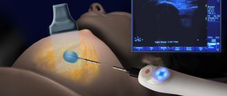
Instrumental studies: ultrasound of the external genital area reveals an anechoic or hypoechoic avascular (not reflecting or poorly reflecting ultrasound, on the screen we see a round formation with thin - light walls and completely dark or light suspended contents) formation with thin walls
List of main diagnostic measures at the outpatient stage:
1. General blood test;
2. General urine analysis;
These blood and urine tests are taken as part of the standard, to prepare for surgical treatment and to exclude concomitant pathologies from other organs with their subsequent correction)
3. Smear for microflora and degree of purity.
This is an examination method in which the test material is taken from the surface of the mucous membrane of the vagina, cervical canal and urethra. The purpose of the analysis is to assess the composition of the microflora and identify inflammatory diseases. The assessment of the standing of the natural flora has in its classification four degrees of vaginal cleanliness:
1st degree – epithelial cells and a normal number of lactobacilli in the smear, pH – acidic;
Grade 2 – a small number of leukocytes, fewer lactobacilli, gram-positive diplococci are present. pH – remains acidic;
3rd degree - increased number of epithelial cells and leukocytes, decreased lactobacilli, many coccal bacteria, pH - slightly acidic or alkaline;
4th degree – a large number of epithelium and leukocytes, pyogenic microorganisms, absence of lactobacilli, pH – alkaline.
4. Bacterioscopic examination of vaginal discharge and cyst contents - allows us to identify microbial agents that caused the Bartholin gland cyst: identification of the pathogen, sensitivity to antibiotics.
5. Testing for infections (ELISA, PCR) gonorrhea, trichomoniasis, chlamydia. [3]
The required scope of examinations before hospitalization:
1. Blood for antibodies to Treponema pallidum (this is the detection of total antibodies to the causative agent of syphilis) - a necessary test before hospitalization and surgery!!
2. Blood type and Rh factor.
3. General blood test.
4. General urine analysis.
5. Smear to determine the degree of purity.
6. Culture from the cervical canal.
7. HbsAg - surface antigen of the hepatitis B virus - to detect hepatitis B by its presence and determine its concentration, a necessary analysis before hospitalization and surgery!!
8. Anti HCV.
In women during menopause or perimenopause, an excisional biopsy is indicated to exclude adenocarcinoma, since at this age the risk of developing malignant tumors is greatest
Bartholinitis during pregnancy
During the period of gestation, the immune system of the expectant mother is greatly weakened. As a result, the body is even more susceptible to disease. Bartholinitis is no exception. As soon as a woman discovers a lump, she should immediately consult a doctor.
The main danger of inflammation of the Bartholin gland lies in the contents, which is pus. It contains pathogenic microorganisms that can go beyond the lesion and enter the vaginal mucosa and further spread throughout the pelvic organs.
If the disease manifests itself in the early stages of pregnancy, it threatens miscarriage. This phenomenon often occurs before twenty-two weeks. In addition to all this, the disease can lead to a missed pregnancy or a slowdown in fetal development. If the disease manifests itself in the later stages, then there is a risk of premature birth. In this case, an abscess may occur right during childbirth, which will lead to infection of the baby and the manifestation of diseases in the form of:
- conjunctivitis;
- lesions of the umbilical ring;
- infections of the respiratory tract and lungs;
- meningitis.
The main reasons for the development of bartholinitis include:
- candidiasis;
- inflammation of the urinary system;
- dysbacteriosis;
- presence of bacterial vaginosis.
Bartholinitis during pregnancy is characterized by immediate development. Symptoms of the disease include:
- swelling of the labia;
- pain in the intimate area;
- temperature increase.
Differential diagnosis
Includes cystic and solid lesions of the vulva, such as epidermal inclusion cyst, hidradenoma papilliferum and lipoma [3], vulvar neoplasms; - abscess of the Bartholin gland; - bartholinitis; vulvar abscess.
— Hematoma in the vulva area – there may also be complaints about a formation in the vulva area, discomfort during sexual intercourse, and pain. But when collecting anamnesis; and gynecological examination - the woman associates its appearance with mechanical trauma, childbirth; And the location is not in the area of projection of the large vestibular gland. [1]
— Paraurethral cysts – similar complaints. On examination - swelling in the paraurethral region (the area near the urethra) [1]
- Furunculosis of the labia majora. Complaints include formations in the perineal area, general malaise, hyperemia, edema, hyperthermia. Upon examination, the formation is localized in the area of the hair follicle and sebaceous gland. [1]
Stages of bartholinitis
Bartholinitis at the site of the lesion is divided into three stages as follows:
- canaliculitis;
- cyst formation of the Bartholin gland;
- abscess.
Inflammation of the Bartholin gland begins with canaliculitis. Suppuration forms on the excretory duct of the gland. There is slight redness and swelling of the skin. Externally, bartholinitis resembles a small abscess. If you try to press on it, two drops of pus come out of the pimple. After a certain time, the duct becomes clogged. As a result, pus accumulates inside the Bartholin gland. It begins to stretch and a large lump forms. Because of this, the entrance to the vagina is blocked. In this case, the patient feels pain and burning when walking.
Treatment
Small, asymptomatic cysts may not be treated except for cosmetic purposes.
Often the clinician is tempted to simply incise the cyst or abscess because this technique may be effective for other common abscesses. However, simply puncturing a cyst or abscess of the Bartholin gland can lead to a relapse, since the edges of the tissues, when punctured or cut, very quickly close again due to the rapid healing or regeneration process [6]
Only large cysts, usually 3 or more centimeters, which interfere with daily activity and sexual life, and disrupt the aesthetic appearance of a woman’s external genitalia, are subject to surgical treatment.
The main goal of surgical treatment is organ-preserving - the formation of a canal and restoration of the function of the Bartholin gland,
Catheter placement – Word
This is a modern method of surgical treatment for Bartholin’s gland cyst, especially in case of its recurrence.
Under local anesthesia, the cystic area is opened with a small incision of about 5 mm, the contents are removed and sent for bacteriological examination, the cyst cavity is washed and a Word catheter is installed in it (this is a silicone tube 55 mm long, 5 mm in diameter with a channel inside, blindly ending, with thinner walls at the end, due to which the tip can inflate into a ball, which has no analogues), inflating its rubber tip to 3 ml with a physiological solution of 0.9% sodium chloride, thereby fixing it in the cavity of the cyst. For better fixation and prevention of loss during the woman’s movements, it is recommended to apply 2-3 absorbable interrupted sutures along the contour of the catheter emerging from the cavity of the cyst. The second end of the catheter is inserted into the vagina. The catheter remains in the cyst cavity for 6 weeks. This is aimed at forming a channel for the passage of secretions, the walls of which do not grow together. The study shows that the Word catheter is an easy-to-use, low-cost outpatient procedure with acceptable short-term recurrences. Treatment costs are seven times lower than with marsupialization [7]. While the catheter is in the cyst cavity, the patient is advised to have sexual rest to avoid its loss. In a number of countries there is no such restriction, since studies have shown that the pain symptom caused by both the cyst itself and the procedure performed with the catheter in the cavity completely disappears over time (by day 6) [8]
As an alternative, a Voroda or a Jacobi ring (the catheter does not have a channel, it is harder, shaped like a ring) which is installed through 2 punctures in the mucous membrane and capsule of the cyst and the 2 ends are fastened to each other.