Causes
The main cause of the pathological condition is hormonal imbalance, a lack of hormones that are responsible for the formation of the reproductive system.
The congenital form of hypoplasia is caused by hereditary predisposition, abnormalities of fetal development and the impact of negative factors on the child during gestation. This may be due to the pregnant woman taking medications, having bad habits, or having suffered serious illnesses, including intrauterine infections.
Provoking factors for the development of the disease.
- Disruption of the endocrine system (thyroid gland, pituitary gland and adrenal glands), including tumor diseases of these organs. Diabetes mellitus, hyperprolactinemia, hypothyroidism, etc. also contribute to the development of pathology.
- Past infectious diseases (tonsillitis, severe ARVI, influenza) and chronic diseases (for example, heart defects, kidney failure).
- Chronic intoxication of the body as a result of taking alcohol, drugs or frequent contact with chemicals.
- Surgical intervention on the organs of the reproductive system.
- Excessive physical, mental and psychological stress that does not correspond to the girl’s age and development.
- Poor nutrition, lack of vitamins, anorexia and weight deficiency.
- Damage to the central nervous system, brain injury affecting the hypothalamus, toxic or infectious effects on the brain. The cause of the disease may be epilepsy.
Pregnancy
Uterine hypoplasia and pregnancy are mutually exclusive concepts. As mentioned above, a girl either will not be able to get pregnant or will not be able to bear a child. In the first degree, independent pregnancy is excluded, but in vitro fertilization is possible (while maintaining ovarian function). With a small degree, the chances of pregnancy are favorable, but careful observation by a gynecologist is necessary, because There is a risk of miscarriage and complications during childbirth.
Almost every woman dreams of becoming a mother. But not everyone can carry and give birth to a baby due to some characteristics of their body. Uterine hypoplasia is one of the conditions that interferes with the normal functioning of the reproductive system. This anomaly affects 5% of females. In this article we will talk about hypoplasia of the female organ and whether it is possible to get pregnant and carry a baby while having a small one.
Symptoms
The main sign of pathology is delayed sexual development in a girl. Menstruation begins at age 15 and later, the cycle is irregular and is accompanied by discomfort and pain. The discharge can be very abundant or scanty. There is underdevelopment of not only the uterus, but also other genital organs - the vagina and ovaries.
The clinical picture is also characterized by external signs, which is due to a delay in general physical development. Women with this pathology have short stature, a narrow chest and pelvis, and underdeveloped mammary glands. After puberty, decreased libido and anorgasmia (lack of orgasm) are observed.
Symptoms and diagnosis
She may not even suspect that a woman has a small uterus. Symptoms of the pathological condition are not always present. If the reduced size of the reproductive organ is insignificant and caused by the physiological structure of the body, which often happens in thin, miniature women, then there is no clinical picture. In other cases, baby uterus syndrome can be suspected already in adolescence, which is accompanied by:
- amenorrhea (prolonged absence of the first menstruation or long delay after menarche);
- severe pain during menstruation;
- first menstrual bleeding after 16 years;
- physical and visual immaturity (flat body, excessive thinness, underdeveloped mammary glands);
- absence or weak manifestation of secondary sexual characteristics.
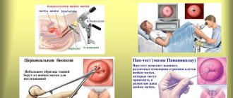
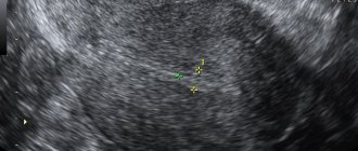
Signs of genital infantilism in diagnosis usually do not cause difficulties. During a gynecological examination, the doctor may discover that the reproductive organ is of unnatural size. Echo signs of uterine hypoplasia during ultrasound will help confirm this assumption.
Often the pathology is accompanied by disturbances in the structure of the appendages: the fallopian tubes are tortuous, the ovaries are reduced in size, and the cervix has an irregular structure. Additional research methods to learn more about the disease will be: probing the cavity of the reproductive organ, establishing bone age, x-ray of the sella turcica, MRI, hysteroscopy.
If the small size of the uterus is not detected at the stage of puberty in a girl, then later in an adult woman it may manifest itself with such signs as:
- infertility;
- habitual miscarriage;
- chronic recurrent cervicitis, endometritis;
- decreased libido;
- lack of orgasm.
Hypoplasia of the uterus 1 degree
The disease has three stages of development. Hypoplasia of the uterus of the 1st degree (germinal) is diagnosed very rarely and is a congenital pathology (the uterus was initially formed incorrectly, which does not allow it to fully develop in the future). The size of the uterus is less than 3 cm, with half of its length being the cervix.
Women with this pathology experience amenorrhea (complete absence of menstruation) and infertility. With this degree of pathology, restoration of reproductive function is impossible.
Forecast
The prognosis for life is favorable. The possibility of pregnancy and gestation is determined by the degree of underdevelopment of the fetal sac. With a mild degree of the disease, against the background of hormonal therapy, the size of the uterus quickly returns to normal, so pregnancy usually proceeds normally and ends safely in childbirth.
Hypoplasia of the uterus can occur both at the stage of intrauterine development and develop after the birth of a girl.
With stage II uterine hypoplasia, long-term treatment is required. It allows you to restore the menstrual cycle and reduce the severity of algomenorrhea. However, the prognosis for pregnancy and its successful completion is less favorable.
With grade III uterine hypoplasia, pregnancy is excluded. If the patient has preserved ovarian function, then in some cases it is possible to perform in vitro fertilization with the transfer of embryos to a surrogate mother, but a successful outcome of such a pregnancy is not guaranteed.
Hypoplasia of the uterus 2 and 3 degrees
Hypoplasia of the uterus 2 degrees is characterized by an organ size of 3–5 cm, a high location of the ovaries and elongated and tortuous fallopian tubes. A girl's menstruation begins late, her cycle is often disrupted, and her discharge is too heavy or scanty. Critical days are accompanied by a deterioration in health, dizziness, nausea and severe pain in the lower abdomen.
Pathology of the 3rd degree is the simplest and most treatable, and in some cases goes away on its own after the onset of sexual activity or pregnancy. The size of the organ reaches 7 cm, and the ratio of the uterus to the cervix is 3:1, which is the physiological norm. If therapy is started in a timely manner, the prognosis is quite favorable: the uterus develops to normal size, the menstrual cycle improves and reproductive function is restored.
Treatment methods for hypoplasia
The main method is hormonal therapy lasting from 6 months to a year. Increases the size of the uterus to normal, restores the menstrual cycle and ovulation.
Treatment of concomitant diseases is also carried out. The patency of the fallopian tubes is surgically restored, and physiotherapy is prescribed to improve blood circulation in the pelvic organs.
Gynecologists at the Medical Women's Center have many years of practical experience in treating various types of infertility. You can be confident of successful treatment.
Diagnostics
To diagnose the pathology, a consultation with a gynecologist is required. The doctor conducts a visual examination, during which warning signs include underdevelopment of the labia, mammary glands and lack of pubic hair. A gynecological examination reveals an excessively short vaginal opening and elongation of the cervix.
To confirm the diagnosis, an ultrasound of the pelvic organs is performed. This diagnosis allows us to determine the degree of development of the disease. To determine the exact size of the uterus, ultrasound hysterosalpingoscopy is performed. In some cases, cavity probing, MRI of the brain and radiography are performed.
Classic laboratory tests are required: blood test - general and biochemical; Analysis of urine; microscopic examination of a smear. To assess hormonal levels, a hormone test is performed. Prolactin must be examined, as well as TSH, FSH, LH, T4, progesterone, testosterone, estradiol, etc.
What is a baby, small uterus
You can find out about the pathology only after examination by a gynecologist. This disease is usually detected in young girls who do not have children, but have some disturbances in the functioning of the organs of the reproductive system. But you don’t need to take the diagnosis of a small uterus as a death sentence. This does not mean that it is impossible to get pregnant.
The anatomical structure of the internal genital organs is similar in all women, but it cannot be absolutely ideal. The likelihood of pregnancy is assessed based on indicators such as the severity of uterine hypoplasia, the presence of concomitant diseases, hormonal levels, and features of the menstrual cycle. In addition, the patient’s weight and her physique are taken into account.
In normal health, the female uterus is pear-shaped with dimensions of 4-5/7-8 cm, which may vary slightly depending on the day of the menstrual cycle. There are several forms of abnormalities in the structure of the uterus:
- Aplasia is an anomaly in which the size of the uterus of an adult woman does not exceed three centimeters (typical for newborn girls).
- With infantilism, the uterus does not reach even six centimeters (typical for younger girls).
- Hypoplasia is a deviation from the norm by one centimeter (the size of the reproductive organ, like in teenagers).
According to statistics, 5 percent of women have an abnormally small uterus.
A child's uterus does not interfere with intimate life, but it can become an obstacle to motherhood. The compatibility of uterine hypoplasia with pregnancy is determined by the severity of the condition. The causes are often injuries, pathologies of intrauterine development, inflammatory processes, physiological characteristics, etc. Most often, the anomaly is discovered suddenly during the first gynecological examination.
There is an opinion that many patients are diagnosed incorrectly. And often this is a “commercial” diagnosis that young girls usually hear.
If a woman has a baby's uterus, it will be difficult to get pregnant and carry a child to term.
Treatment
Treatment of uterine hypoplasia should be prescribed exclusively by a doctor after a complete examination of the woman. Hormone therapy is used, which regulates hormonal levels and normalizes the menstrual cycle. The duration of treatment for uterine hypoplasia is several months and depends on the degree of the disease. If necessary, the course is repeated after some time. Additionally, the woman is prescribed vitamin and mineral complexes.
To achieve a positive effect, physiotherapeutic procedures are used - UHF, mud treatment, laser therapy, electrical stimulation, paraffin therapy, etc. Physical therapy and special gynecological massage can be used. Women are recommended to visit specialized sanatoriums, where they have the opportunity to swim in sea water and receive salt and mud treatment.
The patient should normalize her diet. The diet should include a large amount of foods rich in vitamins and minerals. In addition, it is important to limit intense physical activity as much as possible and avoid psycho-emotional fatigue.
Causes and risk factors
Hypoplasia of the uterus can occur both at the stage of intrauterine development and develop after the birth of a girl. The causes of congenital pathology are:
- genetic diseases and chromosomal abnormalities;
- intrauterine infections (influenza, herpes, cytomegalovirus, toxoplasmosis, rubella);
- exposure to occupational hazards and radiation on the pregnant woman’s body;
- living in an environmentally unfavorable area;
- bad habits (smoking, drinking alcohol, taking drugs);
- fetoplacental insufficiency, leading to intrauterine developmental delay.
The possibility of pregnancy and gestation is determined by the degree of underdevelopment of the fetal sac.
The following factors that have an impact in childhood and during puberty can lead to an acquired form of uterine underdevelopment:
- lesions of the hypothalamus or pituitary gland of toxic, infectious, traumatic or tumor origin;
- chronic severe diseases of the lungs, liver, kidneys, heart;
- endocrine pathologies (thyroid diseases, diabetes mellitus);
- autoimmune diseases;
- dyshormonal disorders caused by ovarian tumors, severe infectious diseases (rubella, mumps);
- ovarian hypoplasia;
- weight deficiency (irrational mono-diets for weight loss, malnutrition, starvation);
- chronic stress;
- mental disorders (neuroses, depression, psychoses);
- surgical removal of the ovaries or a significant part of them;
- hereditary predisposition;
- mental overload;
- professional sports;
- smoking, alcoholism, drug and substance abuse.
Prognosis and possible complications
In the absence of proper and timely treatment, complications may develop. Most often these are obstruction of the fallopian tubes, infertility, ectopic pregnancy, weak labor and heavy bleeding in the postpartum period. There are difficulties with conceiving and bearing a child, and there is a high risk of miscarriage. In addition, pregnancy is very difficult: the expectant mother suffers from early toxicosis and gestosis, constant weakness and dizziness. Women with hypoplasia are more susceptible to gynecological diseases, including infectious ones.
Complications with hypoplasia
The presence of underdevelopment of the reproductive organ in women can provoke various kinds of complications.
The complicated course of uterine hypoplasia includes:
- infertility;
- chronic miscarriage, frequent miscarriages;
- inflammatory gynecological diseases;
- weakness of labor, uncoordinated contractions of the uterus during childbirth;
- toxicosis in early pregnancy;
- premature birth;
- ectopic pregnancy (poor tube patency);
- prolonged bleeding after childbirth.
Prevention
Following simple rules will help ensure the normal physiological development of the girl. The child must eat a balanced and healthy diet. The basis of the menu should be foods rich in vitamins and minerals. It is important to exclude excessive physical activity, strong emotional experiences and stress. You should properly organize your daily routine, set aside time for proper rest and healthy sleep.
Doctors recommend promptly treating infectious processes in the body, avoiding intoxication and monitoring physical development. To avoid the formation of congenital pathology, it is necessary to lead a correct lifestyle while carrying a child.
Description and characteristics
Hypoplasia is the underdevelopment of the uterus, which can be caused by various reasons or be congenital. Doctors also call this disease “infantilism” or “children’s uterus.”
The pathology under consideration is characterized by the fact that the uterus stops developing at some stage of formation. As a result, she does not meet accepted standards either in age or physiological terms.
A small uterus is often combined with elongated, winding tubes - this causes infertility. At the same time, the cervix is oblong and has the shape of a cone - this means that even if you become pregnant, the probability of carrying a baby is equal to zero.
Often hypoplasia of the female organ is accompanied by underdevelopment of the vagina, labia, etc.