My friend is often sick. One of her last diseases was hyperplasia of the glandular epithelium. It's good that you visited the doctor. What kind of pathology is this – we’ll talk further.
- 3 Symptoms of the disease 3.1 Before and after menstruation
- 3.2 During pregnancy
- 9.1 Secrets of traditional medicine for hyperplasia of the glandular epithelium
Causes of the disease
Pathological disruption of the endocervix is associated with various reasons. The main provocateurs of this condition are:
- hormonal imbalances,
- improper use of contraception,
- abortions,
- poor immunity,
- bad habits
- the onset of menopause,
- fibroids, ovarian cysts, endometriosis,
- improper use of hormones.
Hyperplasia is a dangerous disease, since at the very beginning of the formation of inflammation the disease may not manifest itself. With timely treatment, the result is positive.
Treatment
Treatment of endocervical hyperplasia is carried out surgically. With the help of surgery, foci of pathological epithelium are eliminated. After this event, the patient is prescribed conservative therapy.
Cervical hyperplasia is a benign pathology characterized by abnormally active tissue growth. This disease often represents the primary stage of hypertrophy. During this period, the disease can be diagnosed exclusively with the help of microscopic examination.
With prolonged refusal of treatment, the epithelial tissues of the cervical canal rapidly increase in size. Some types of pathology, in the absence of therapy, develop into cancer cells.
Types of hyperplasia
Tissues belonging to the cervical canal grow in different ways. Due to the structural features of this pathology, it is classified into separate groups.
Cervical canal
Proliferation of the endocervix is a disease that is characterized by the growth of the mucous layer of the cervical canal. It always has a benign course.
Columnar epithelium of the uterine cervix
This is a benign compaction that can transform into oncology. The following factors provoke pathogenic degeneration:
- Hormonal changes caused by puberty
- decrease or increase in hormone levels due to pregnancy.
Basal cell hyperplasia
The basal cell type of the disease is not considered an analogue of microglandular atypical hyperplasia, since it develops on the surface and not inside the cervical canal. In appearance it resembles pseudo-erosion.
Causes of cervical hyperplasia
As already noted, the true causes of the pathological proliferation of the cervical epithelium cannot always be diagnosed. Usually they talk not about the causes, but about the predisposing factors that provoke excessive growth of the epithelium of the cervical canal. These most often include:
— Hormonal dysfunction, namely a change in the normal ratio of estrogens and gestagens (progesterone). All tissues that form the cervix are highly sensitive to cyclic hormonal changes; the mucous membrane of the cervical canal reacts to them to a greater extent. The cervical epithelium changes its structural characteristics according to the phases of the cycle; depending on the level of concentration of estrogen and progesterone, the mucous membrane can increase in volume, loosen and intensively produce cervical secretions. If the hormonal balance is disturbed, the proliferation of cervical epithelial tissue may become too intense, that is, hyperplasia of the cervical epithelium is formed.
The largest number of cases of diagnosis of cervical hyperplasia occurs in two age periods: from 14 to 20 years and after 45 years. Obviously, this is due to the state of hormonal function: in adolescents and girls it is in a state of formation, and in those who have crossed the 45-year mark it naturally fades away.
Cervical hyperplasia is sometimes diagnosed in healthy patients using hormonal contraception, that is, with artificial hormonal dysfunction.
— Mechanical damage to the cervical epithelium. When the integrity of the mucous layer of the endocervix is violated, a wound appears in the cervical canal, which subsequently begins to epithelialize due to the internal resources of the mucosa. The columnar epithelium activates reserve cells that cover the damaged area. Cylindrical hyperplasia of the cervix appears if excessive growth of the epithelial layer occurs during the regeneration process. As a rule, pathological post-traumatic regeneration is provoked by repeated traumatic manipulations, for example, abortions or diagnostic curettages.
- Inflammatory processes. Infectious inflammation is a serious test for the mucous membranes of the cervical canal, which should protect the uterus from potential ascending infection.
The response of the endocervix to infection depends on the state of local immune defense. Healthy epithelium tries to eliminate unwanted microbes, so as not to allow them to penetrate deeper, through increased secretion. Cervical secretion becomes abundant and liquid in order to “wash away” the source of infection from the mucous membranes. In a negative scenario for the course of the inflammatory process, undesirable microflora penetrates into the underlying structures, damaging them too. Periods of subsidence of inflammation are accompanied by regeneration processes in the damaged mucous membranes, and then, when the infection attacks the endocervix again, the damage is formed again. Such chronic infectious inflammation with alternating periods of exacerbation and subsidence can provoke hyperplasia of the cervical epithelium.
Symptoms of the disease
The symptoms of this disease are not always noticed by women. The disease is asymptomatic in some cases. Characteristic signs of the disease are:
- Unusual cervical secretion. The mucous discharge is too excessive, the woman is forced to regularly use sanitary pads to protect her underwear.
- Intermenstrual bleeding (spotting). Not the cyclical nature of menstruation, when endocervical hyperplasia occurs simultaneously with other hyperplastic processes.
Before and after menstruation
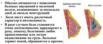
Hyperplasia is often suspected as monthly discharge changes:
- Heavy bleeding appears after a certain delay in menstruation. In such a situation, the thickness of the endometrial ball increases sharply. Menstruation has a thin consistency, with clots. Such adjustments are painful because intrauterine pressure is increased. The tissues of the uterus begin to expand, and spasms are likely due to compression of the blood vessels.
- Scanty menstruation is characteristic of uneven changes in the endometrium. The fabric becomes covered with lesions. During menstruation, these particles are not rejected. Due to the reduction of the layer, the regula are scarce.
- With hyperplasia, bleeding occurs that is not associated with menstruation. This is caused by tissue proliferation.
- Bleeding appears after sexual intercourse due to damage to the cervix. They are provoked by blood vessels that burst.
During pregnancy
Pathology during pregnancy is difficult to recognize, since cell division eliminates the symptoms of the disease. It is determined only after a certain examination. The disease can be detected when the placenta grows:
- there is a deviation in the child’s heartbeat rhythm,
- the sensation of internal movement of the developing fetus is noticeably reduced,
- with a sharp decline in heartbeat contractions, oxygen starvation of the developing fetus occurs, and its hypoxia cannot be ruled out.
When a pregnant woman is diagnosed with diabetes, the disease manifests itself as polyhydramnios. Probably vaginal burning, an increase in the amount of mucus secreted from the genitals.
Prevention
To protect against hyperplasia, preventive measures must be taken regularly. For this purpose, provoking factors should be eliminated in a timely manner. This is especially true for such pathologies as tumor lesions of the ovaries, problems with the liver, gall bladder, and inflammatory processes in the pelvic organs.
It is also important to visit a gynecologist annually for preventive examinations. In addition, it is necessary to control body weight, since obesity leads to the development of hyperplasia and other diseases. If the menstrual cycle is disrupted, then you need to examine the state of your hormonal levels.
Endocervical hyperplasia, despite its benign nature, is considered an extremely dangerous disease. The growth of the mucous membrane of the cervix occurs rapidly and intensively, as a result of which serious complications often develop.
What kind of disease is this
Significant proliferation of cells on the uterine epithelium is usually a benign process, although with untimely treatment and without medical supervision it can provoke precancerous conditions of the organ.
The types of hyperplasia are:
- Cystic. The endocervix is built from locally located glands. They have a characteristic appearance of epithelium, which has one row of its own cells.
- Atypical. Thickening of the epithelium occurs right inside the organ.
- Glandular. glandular tissue grows rapidly. Glandular hyperplasia visually resembles erosion. The doctor is obliged to accurately diagnose the problem, since “cauterization” can provoke serious complications.
- Glandular-cystic. This option combines the proliferation of a layer of glands along with the presence of small cysts.
- Micro-glandular. Accelerated growth of the mucosa occurs with uncontrolled cell division of the glandular layer.
Therapy for an atypical form of pathology should be started as quickly as possible, since it is this form that often degenerates into oncology.
https://youtu.be/t2N031WSl3E
Basal cell hyperplasia. Dysplasia. Carcinoma in situ
It is advisable to consider these states together, guided by the following considerations. First of all, they represent different steps of the ladder that leads from normal to cancerous epithelium.
But they do not necessarily have to follow each other with fatal inevitability. Only carcinoma in situ is, in all likelihood, irreversible and sooner or later begins to grow invasively; all processes preceding it may undergo reverse development.
Since malignancy is usually
- the process is staged and the epithelium acquires cancerous properties without preliminary preparation only as an exception, then in the same tumor and even in the same preparation it is often possible to catch different stages of malignancy - in one area the epithelium is only thickened, with or without symptoms of acanthosis of them, in another, hyperplasia of basal cells is visible, in the third, dysplasia, and in the fourth, carcinoma in situ.
If the latter is not detected in a given section, this does not mean that it is absent from the tumor. In cases of severe dysplasia, additional sections and searches for carcinoma in situ are required; Failure to comply with this rule cost the lives of more than one patient.
If hyperplasia, dysplasia and carcinoma in situ in their, so to speak, complete form are not so difficult to recognize, then the transitions between them, on the contrary, are very difficult to decipher, and the latter becomes a very subjective matter. The same pattern may be classified by one pathologist as dysplasia and by another as carcinoma in situ. In such a situation, making additional sections also becomes an urgent necessity. Basal cell hyperplasia.
It is carried out in acanthotic protrusions or in the bottom sections of thickened stratified squamous epithelium. Slightly elongated basal cells are arranged in several rows. Since they are stained darker than the cells of the upper layers, a kind of border appears, separating the epithelium from the underlying connective tissue.
Larynx
Acanthosis. Basal cell hyperplasia (X170).
Dysplasia.
It differs from the previous stage in that elongated dark cells populate not only the bottom, but also the middle sections of the epidermal layer; cover cells located parallel to the basement membrane are preserved. There is no atypia, polymorphism, or irregular mitoses. The lesion would be innocent if it were not for the possibility of transformation into carcinoma in situ.
Larynx
Dysplasia (X170).
Carcinoma in situ.
The most common varieties are spindle cell and bowenoid. Spindle cell carcinoma in situ, as the name implies, consists mainly of sharply elongated cells that are usually oriented perpendicular to the basement membrane, but can be parallel to it or form bizarre arcades.
Larynx
Spindle cell carcinoma in situ (X400).
It is often suggested that in carcinoma in situ, elongated cells completely replace the entire epidermoid layer, while in dysplasia - only partially.
This criterion cannot be called incorrect, but it is not universal. In many carcinomas in situ, the entire epidermoid layer is, indeed, completely rebuilt, but there are also cases when the upper parts are preserved, as in dysplasia.
When making a differential diagnosis between dysplasia and carcinoma in situ, one must take into account not only the level that elongated cells reach, but their cytological features. The spindle cells of in situ carcinomas appear more or less the same type as in dysplasia, but only at low microscope magnification. At high magnification, it turns out that they are much more polymorphic.
They differ from each other in size, in chromatin content, in shape - some of them are simply ugly. In addition, they are distributed more chaotically than in dysplasia - it has already been mentioned that they can be oriented in different directions and form arcades, which is unusual for dysplasia.
I will add that with spindle cell and other forms described below, loosening of the epithelial layer is often observed up to the isolation of individual cells, the appearance of small cavities in it, and infiltration with lymphocytes and leukocytes.
The number of mitoses varies from observation to observation, but usually there are few.
Larynx
Bowenoid carcinoma in situ (X400).
Bowenoid cancer in situ of the larynx, as well as other organs, is characterized by the presence of mononuclear hyperchromic giants, which are literally striking and make the diagnosis easy and indisputable, even if these giants are few in number.
There are combinations of bowenoid and spindle cell cancer, and the corresponding structures can be located both in different parts of the epithelial cover, and together, fitting in one field of view.
Larynx
Combination of spindle cell and bowenoid carcinoma in situ (X400).
The morphological repertoire of carcinoma in situ of the larynx is not limited to the spindle cell and bowenoid variants. There are other varieties that are rarer and less well known.
Larynx
Variant of carcinoma in situ (X400).
Thus, carcinoma in situ may consist of polymorphic, atypical, clearly cancerous cells, which, at the same time, cannot be attributed to either spindle cell or bowenoid cancer. Or such complex histological structures arise that it is difficult not only to classify, but also to describe.
Larynx
Variant of carcinoma in situ (X400).
For example, the figure shows a kind of cancer in situ, in which three floors can be distinguished. The lower one is represented by a border of elongated and hyperchromatic cancer cells, forming a kind of palisade, the middle one is also elongated cells, but located parallel to the basement membrane and very loosely, and among them there is an epidermoid alveolus with a lumen in the center; the upper one is the parakeratotic layer.
The underlying connective tissue in carcinoma in situ may remain almost intact, but more often it is somewhat edematous and infiltrated with lymphoid and histiocytic elements.
“Errors and difficulties in histological diagnosis of tumors”, D.I. Golovin
Forward →
- Beginning invasive cancer
Popular articles in the section
- Beginning invasive cancer
- Keratoacanthoma. Pseudoepitheliomatous hyperplasia
Diagnostics
The disease can be detected during a gynecological examination by a specialist. Irregular endocervical cells that extend beyond the cervical canal can be seen even with the naked eye. But for a more accurate diagnosis, other studies are recommended for the patient:
- hysteroscopy,
- strokes,
- biopsy,
- colposcopy,
- Ultrasound,
- blood analysis.
Based on the results of tests and instrumental studies, the doctor makes a diagnosis and recommends adequate treatment.
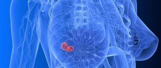
Possible complications
Mastopathy in rare cases causes serious consequences. Unpleasant symptoms often disappear after menopause.
Among the possible complications, experts highlight the risk of nodes transforming into cancerous tumors. A similar condition is observed with the development of the fibrocystic form, since it is quite difficult to diagnose it in the early stages.
When a diffuse dishormonal, focal or other form occurs, the likelihood of transformation into a malignant formation is minimal.