The role of the endometrium in female reproductive function
The structure of the endometrial layer is quite complex. The endometrium includes the following structures:
- Glandular and outer layer of epithelium;
- Main substance;
- Stroma;
- Vascular bed.
The main function of the endometrium is to ensure implantation of the embryo and its further successful development. The endometrium contributes to normal homeostasis in the uterine cavity, which ensures the normal functioning of the fetus. Proliferation of the endometrium leads to the proliferation of the vascular bed, which improves blood supply to the fetus. These vascular structures will subsequently become part of the placenta.
Throughout the month, cyclical changes occur in the uterus. They include the following stages:
- Proliferation stage;
- Presecretory stage;
- Sector stage;
- Stage of menstrual flow.
In order to correctly assess the cyclic changes of the endometrium, one should take into account the duration of the cycle and its clinical features (presence of dysfunctional uterine discharge, duration of the secretion phase, volume of menstrual bleeding during the month).
Diagnostics
Due to the similarity of the clinical picture of glandular hyperplasia with other pathologies, diagnostic measures are of great importance.
Diagnosis of proliferative type endometrial hyperplasia is carried out using the following methods:
- Studying the patient's history and complaints related to the time of onset of bleeding, its duration and frequency. Accompanying symptoms are also studied.
- Analysis of obstetric and gynecological information, which includes heredity, pregnancy, contraceptive methods used, previous diseases (not only gynecological), operations, diseases transmitted through sexual contact, etc.
- Analysis of information about the beginning of the menstrual cycle (patient’s age), its regularity, duration, pain and profuseness.
- Conducting a bimanual vaginal examination by a gynecologist.
- Gynecological smear collection and microscopy.
- Prescription of transvaginal ultrasound, which determines the thickness of the uterine mucosa and the presence of proliferative endometrial polyps.
- Determination using ultrasound of the need for an endometrial biopsy to make a diagnosis.
- Carrying out separate curettage using a hysteroscope, which scrapes or completely removes the pathological endometrium.
- Histological examination of scrapings to determine the type of hyperplasia.
Read more Tablet for thrush 1 piece name
Early stage of endometrial proliferation
Early proliferation of the endometrium occurs on days 5-7 of the menstrual cycle. This phase is histologically characterized by the fact that on the surface of the mucosa there is a cubic epithelium; the glands of the inner layer of the uterus are rectangular tubes with a narrow lumen. When performing a cross section, glands that are in the early stage of proliferation appear as oval or rounded structures. In this case, the endometrium consists of prismatic epithelium. At the early stage of proliferation, the epithelium is small in height; the cells contain oval nuclei, which are localized in the basal part of the cells. At the same time, they have an intense color. The early stage of proliferation is characterized by the fact that the stroma includes spindle-shaped cells with large nuclei. In this case, the arteries are characterized by spiral tortuosity.
Proliferation concept
Proliferation is the active process of cell division in a tissue or organ. As a result of menstruation, the mucous membranes of the uterus become very thin due to the fact that the cells that make up the functional layer are rejected. This is what determines the process of proliferation, since cell division renews the thinned functional layer.
However, proliferative endometrium does not always indicate the normal functioning of the woman’s reproductive system. Sometimes it can occur in the event of pathology development, when cells divide too actively, thickening the mucous layer of the uterus.
Endometrium late stage of the proliferation phase
Late proliferation of the endometrium is observed on days 11-14 of the cycle. The endometrium of the late proliferation phase is characterized by the fact that the glands acquire a tortuous outline. The glands have a wide lumen, and their nuclei are at different levels. In the basal sections, small-diameter vacuoles containing glycogen can be detected. The endometrium of the late stage of proliferation is distinguished by a succulent stroma with large nuclei. In this case, the kernels acquire a rounded shape and less intense color. The stroma is juicy, the nuclei are enlarged, rounded and stained less intensely. Fragments of the endometrium of the proliferation phase at a late stage have tortuous vessels.
What is proliferative endometrium?
The endometrium is the mucous layer lining the inside of the uterus.
It is he who ensures the implantation and development of the embryo, and its changes play an important role in the menstrual cycle. Such an important process as endometrial proliferation regularly occurs in the female body. Any violation of this mechanism can be the beginning of the development of pathologies of the reproductive system. The proliferative endometrium marks the first phase of the cycle - the stage that occurs immediately after menstruation and means the active division and growth of endometrial cells.
How to diagnose endometrial fragments in the proliferation phase
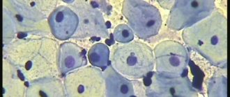
One of the most informative methods for diagnosing proliferative changes in the endometrium is hysteroscopy with biopsy. Each phase of the menstrual cycle is characterized by its own histological picture. Histologically, the early stage of endometrial proliferation is characterized by the presence of thinned pink epithelium. There are areas with foci of hemorrhage and fragments of the membrane that were not rejected.
Histologically, the middle stage of endometrial proliferation differs from the early stage in that at this stage the number of mitoses increases significantly. Single vessels with thinned walls are also observed.
Histologically, late endometrial proliferation is characterized by thickening of the functional layer of the endometrium. At this stage there is no division into division zones.
Breast
Breast changes are quite common, including among young girls and women. The organ constantly experiences the action of sex hormones, undergoes characteristic changes throughout the menstrual cycle, during pregnancy and lactation, and is therefore susceptible to various kinds of pathologies. According to statistics, up to 60% of women of reproductive age have signs of mastopathy.
Mastopathy is considered a benign process, but if it is present, the risk of malignancy increases several times. Proliferative variants are even more dangerous, they increase the likelihood of cancer by 25-30 times.
The presence or absence of proliferation is the most important sign when assessing the type of mastopathy. Non-proliferative forms are represented by foci of fibrous tissue, cystically altered ducts, the epithelium of which does not proliferate and is even atrophic. The risk of malignant transformation is relatively small.
Proliferative mastopathy is much more dangerous, because the cells of the ducts and lobules of the gland are actively dividing, creating the threat of cancerous degeneration. The more such foci of proliferation in the gland tissue and the more active the proliferation of the epithelial component, the more likely breast cancer is in the future, therefore all forms of proliferating mastopathy are considered precancerous conditions.
According to the severity of proliferation, several degrees of mastopathy are distinguished. In the first degree, proliferation is not detected, in the second, it is there, but the cells do not show signs of atypia, the third degree of mastopathy is manifested by active proliferation of epithelial cells with atypia.
Thus, proliferation of mammary gland cells is not only a criterion for the form of mastopathy, but also an indicator of the likelihood of cancer, therefore excessive division of the epithelium always attracts the attention of specialists. Diagnosis of this change is carried out by cytological examination of tissue obtained during puncture.
Diseases associated with impaired endometrial proliferation
Endometrial proliferation may be impaired in some cases. As a result, tissue proliferation is observed, which leads to the development of tumor tumors. One typical example is endometrial hyperplasia. This pathological process is a consequence of a violation of the hormonal regulation of the menstrual cycle. Endometrial hyperplasia is manifested by proliferative changes in the stroma of glandular tissue. Endometrial hyperplasia can be atypical or glandular. It all depends on how effectively early endometrial proliferation and late endometrial proliferation occur. Glandular hyperplasia is different in that here the endometrium of the late stage of proliferation stops dividing. Accordingly, this variant of hyperplasia very rarely transforms into malignant neoplasms. Atypical hyperplasia is a typical precancerous pathology. This pathology is characterized by pronounced early proliferation of the endometrium and late proliferation of the endometrium. The disease is observed mainly in older women during menopause.
Secretion phases
The secretion phase begins almost immediately after proliferation (or after 1 day) and is inextricably linked with it. It also distinguishes a number of stages - early, middle and late. They are characterized by a number of typical changes that prepare the endometrium and the body as a whole for the menstrual phase. The endometrium of the secretory type is dense, smooth, and this applies to both the basal and functional layers.
This stage lasts from approximately the fifteenth to the eighteenth day of the cycle. It is characterized by weak secretion. At this stage it is just beginning to develop.
At this stage, secretion is as active as possible, especially in the middle of the phase. A slight decline in secretory function is observed only at the very end of this stage. It lasts from the twentieth to the twenty-third day
The late stage of the secretory phase is characterized by a gradual decline in secretory function, with complete disappearance at the very end of this stage, after which the woman begins her period. This process lasts 2-3 days from the twenty-fourth to the twenty-eighth day. It is worth noting a feature that is characteristic of all stages - they last 2-3 days, while the exact duration depends on how many days are in the menstrual cycle of a particular patient.
Lifespan
In many ways, life expectancy increases due to therapy. It is therapy that improves the course of the pathology. This pathology is not a severe process.
Therapy should be early before complications occur. It is complications that can lead to serious disorders. Regeneration of the proliferation site restores the damaged structure.
Life expectancy also depends on the causes of the disease. Any cause of pathology affects a woman’s condition. Therefore, it is necessary to exclude provoking factors.
Proliferation of glandular epithelium - etiology
The etiology of this process is multiple. Sometimes they are not pathological. Proliferation may be detected after taking contraceptives.
The following reasons are also identified:
- infection;
- inflammatory reaction;
- colpitis;
- dysfunction of hormones;
- hormonal changes of a physiological nature;
- injuries;
- complicated abortions;
- difficult childbirth;
- therapeutic manipulation
Pseudo-erosion can be caused by proliferation. Cell structures are formed.
go to top
Glandular and atypical endometrial hyperplasia
This pathology occurs mainly in women of menopausal age.
The cause of the development of this disease may be hyperestrogenism or a long period of estrogen action on the endometrium, provided their amount in the blood is low. With this diagnosis, the endometrium has a thick structure and protrudes into the organ cavity in the form of polyps. The morphology of glandular cystic hyperplasia is represented by a large number of columnar (less often cubic) epithelial cells. These particles are larger in shape than normal cells; therefore, the nucleus and basophilic cytoplasm are also larger. Such elements accumulate in groups or create glandular structures. A feature of this form of proliferative type endometrial hyperplasia is that there is no further division of newly formed cells. This pathology very rarely degenerates into a malignant tumor.
This type of disease is classified as precancerous. It occurs mainly during menopause and in old age. This pathology is not observed in young women. Atypical hyperplasia is a pronounced proliferation in the endometrium with adenomatous foci consisting of branching glands. When conducting a study, it is possible to identify a large number of large columnar epithelial cells that have large nuclei with smaller nucleoli. The ratio of nucleus to cytoplasm (basophilic) remains virtually unchanged. In addition, there are large cells that have a slightly enlarged nucleus and a very extensive cytoplasm. There are also clear cells with lipids, and based on their presence, a disappointing diagnosis is made.
Atypical glandular hyperplasia develops into cancer in 2-3 patients out of a hundred. Columnar epithelial cells in this case can be located either separately or in groups. Similar elements are present during the proliferative phase of the monthly cycle without pathology, but during the disease there are no cells of decidual tissue. Sometimes atypical hyperplasia can have the opposite process. But this is only possible in the case of hormonal influence.
Prevention methods
Every woman should follow a number of preventive measures to reduce the risk of developing endometrial hyperplasia. Experts recommend following the following rules:
- Monitor your immune system. In the autumn and spring periods, you can take special multivitamin complexes.
- When planning a pregnancy, consult a doctor and undergo the necessary diagnostic methods.
- Give up bad habits, which include smoking and drinking alcohol.
- Normalize the metabolic process in the body.
- Avoid abortion if possible .
- Have regular sex life with one partner.
- properly .
- Exercise . But at the same time you need to avoid heavy physical activity.
- Treat inflammatory and infectious diseases of the pelvic organs in a timely manner. They should not be allowed to become chronic.
- Observe personal hygiene rules.
What to do if proliferation is detected?
Wherever the proliferation process is identified, the first thing a qualified specialist will do is determine the cause, and only then select the necessary therapy.
To treat proliferation in inflammatory processes, anti-inflammatory therapy is prescribed, supplemented by antiviral and antibacterial drugs that have an antiproliferative effect.
Proliferation, which indicates pathology, serves as a signal calling for immediate treatment. In this regard, patients diagnosed with proliferation are under the supervision of a doctor.
Having completed treatment of the underlying disease, an additional study (biopsy or cytological analysis) should be performed, which will assess how effectively the treatment was and determine whether there is a risk of developing a malignant tumor in the future.
Causes
Hyperplasia of the cervical canal (proliferation), according to experts, occurs as a result of hormonal imbalance. But the exact reasons have not been established. Scientists, based on the results of studies, have found that certain factors can increase the risk of developing the disease.
First of all, bad habits such as smoking and drinking alcohol have a negative impact on the state of hormonal levels.
The process of growth of the endometrium can be provoked by diseases of the urinary system and internal genital organs that have a long course and last for a long time. When inflammation is relieved, the process of epithelial cell division stops.
On this topic
- Female reproductive system
Differences between cytology and colposcopy
- Natalya Gennadievna Butsyk
- December 6, 2020
Experts have found that women's health is negatively affected by early or promiscuous sexual intercourse, sexually transmitted diseases, the HIV virus or herpes.
The condition of the mucous membrane is negatively affected by curettage or abortion, the presence of immunodeficiency syndrome, fibroids or endometriosis.
What is proliferative endometrium?
The endometrium is the outer mucous layer lining the uterine cavity. It is completely hormone-dependent, and it is it that undergoes the greatest changes during the menstrual cycle; it is its cells that are rejected and released along with the discharge during menstruation. All these processes occur in accordance with certain phases, and deviations in the passage or duration of these phases can be considered pathology. Proliferative endometrium - a conclusion that can often be seen in ultrasound descriptions is the endometrium in the proliferative phase. What this phase is, what stages it has and how it is characterized is described in this material.
Proliferation of glandular epithelium - therapy
Therapy is not used for isolated variants of proliferation. Treatment of this process is associated with the following phenomena:
- infections;
- inflammation;
- hormonal dysfunction
Antibacterial therapy is used for infection. But it is important to identify the pathogen. It is also important to carry out replacement therapy.
Forms of drugs include contraceptive pills. Methods of therapy for pseudo-erosion:
- diathermocoagulation method;
- cryodestruction method;
- laser therapy;
- radio wave destruction
When the site of proliferation is destroyed, regeneration occurs, that is, healing.
go to top
Clinical picture
Proliferation of the glandular epithelium at the initial stages of its development does not show specific signs and often occurs unnoticed by the woman. Diagnosis occurs randomly during a gynecological examination or ultrasound examination for other diseases or for preventive purposes.
At the second stage, symptoms arise that the woman does not pay attention to, attributing them to fatigue or the development of other pathologies.
Among the signs of hyperplasia are painful sensations in the lower abdomen. They may be accompanied by the appearance of bloody discharge after or during sexual intercourse. They are permanent and nagging in nature.
Proliferation is also characterized by the occurrence of spotting during menstruation. Women also complain of cycle disturbances, in which intense pain occurs in the lower abdomen.
It is important to remember that such clinical manifestations may indicate the presence of other, more serious diseases. That is why, if such symptoms appear, consult a doctor for diagnosis and treatment.
General information about the disease
Proliferation is an excessive proliferation of cells of the glandular epithelium of the uterus, which is accompanied by a change in the production of hormone levels and the occurrence of a pathological process.
At the same time, the rate of division of affected cells can increase or decrease depending on the influence of external factors and the characteristics of the woman’s body.
Proliferation is accompanied by an enlargement of the uterus. The disease is benign. But despite this, it requires treatment, since the risk of complications is quite high.
The disease is diagnosed most often in women over 35 years of age. The risk group includes patients who have previously had an abortion or undergone surgery in the internal genital area.