What and how does folliculometry measure?
This procedure consists of a series of examinations that dynamically allow us to trace regular cyclical processes in a woman’s body. Based on the results of observations, a conclusion is drawn whether ovulation occurred in the observed period (on what day of the cycle specifically); how the inner surface of the mucous membrane of the uterine body grew (it is called the endometrium).
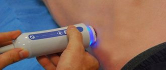
For diagnosis, equipment (scanner, sensor) is used - an ultrasound machine. Everything happens according to the usual method of ultrasound examination (US), widely used in medicine. This technique has successfully replaced the old and very approximate methods of determining the date of ovulation in gynecology.
Who can this procedure help?
Folliculometry should be done if:
- A woman has regular sexual intercourse without using any contraceptives, but pregnancy does not occur within a year.
- Deviations in the hormonal area were identified (changes in the development and functioning of the ovaries, anovulation, etc.).
- There are indications for stimulating egg maturation.
- A married couple wants to increase the likelihood of having a child of a certain gender (or twins). It has been established that with intimate intimacy during ovulation, the chances of giving birth to a boy are higher.
- You need to determine the most optimal time to conceive.
- An in vitro fertilization procedure is planned.
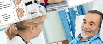
The technique is also used immediately after IVF to monitor the process of fixation of the fertilized egg inside the uterus. The interpretation of the results is carried out by an ultrasound diagnostic doctor and a gynecologist.
Goals of folliculometry
- assess the general condition of the uterus and ovaries, determine whether it corresponds to the phase of the cycle when the study is carried out;
- determine how the ovaries function and obtain confirmation of ovulation;
- determine on what day the egg leaves the follicle (ovulation), which may be required at the stage of pregnancy planning;
- identify the causes of menstrual irregularities and ovarian dysfunction, determine what causes anovulation (lack of ovulation in the menstrual cycle);
- assess cyclic changes in the endometrium and identify possible disorders;
- assess the usefulness of the luteal phase of the cycle, based on the obtained characteristics of the corpus luteum;
- monitor the process of ovulation stimulation if a doctor performs hormonal therapy.
A referral for folliculometry is prescribed by a gynecologist in order to identify or exclude a number of pathologies in the functioning of the gonads, as well as to clarify the phases of the menstrual cycle.
Research is necessary in the following cases:
- Determining the most favorable day for conception.
- Planning the sex of the child depending on the day of ovulation.
- Assessment of ovarian performance.
- Diagnosis of folliculogenesis and menstrual cycle disorders.
- Control of hormonal levels.
- Detection of pathologies of the female genital organs (cystic formations, fibroids, etc.).
- Controlling the likelihood of conceiving twins in a situation where there has already been a history of multiple pregnancies.
- Assessment of the condition of the endometrium for successful implantation of a fertilized egg.
- Control diagnostics during stimulation of ovulation and during in vitro fertilization.
- Assessing the results of taking medications during treatment.
The main indications for the study are:
- anovulatory cycle (lack of ovulation);
- hypomenorrhea (scanty discharge during menstruation);
- oligomenorrhea (the interval between menstruation exceeds 40 days);
- dysmenorrhea (menstrual cycle disorders);
- hypoplasia or dysfunction of the female gonads;
- uterine hypoplasia;
- dysfunctional uterine bleeding;
- glandular endometrial hyperplasia;
- ovarian retention cyst;
- infertility due to hormonal imbalance;
- menopause;
- hermaphroditism.
Preparatory actions
The folliculometry procedure does not require special preparation. For the entire duration of the study, the patient excludes from her diet foods that stimulate excess gas formation in the intestines (this will prevent the follicles from being seen): beans, all types of cabbage, rye bread, alcoholic and carbonated drinks. It is not recommended to drink alcohol.
On the day of the examination, you should check with your doctor about how the examination is carried out. There are two methods:
- Transabdominal (scanning directly through the abdominal wall). The patient is positioned on the couch, with the abdomen open for examination (from the chest to the groin area). The scanner moves across the body from top to bottom. Before the test, be sure to drink a liter of any still water. The internal organs are more clearly visible against the background of a full bladder.
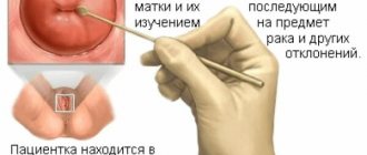
- Vaginal sensor. This is a more accurate and informative method. For its successful implementation, the bladder and intestines are emptied naturally or forcibly (medicines, enema).
How to prepare for the procedure
Preparing for folliculometry requires following certain rules for the study to be completely informative:
- Do not use:
- raw vegetables;
- dark varieties of bread;
- cabbage;
- soda.
- In case of increased flatulence:
- do not take foods with increased gas formation;
- 12-15 hours before the procedure, use medications to reduce flatulence (Espumizan, Atoxil or charcoal).
- Before examination using a transabdominal sensor, take 1.5 liters of liquid (preferably ordinary purified water) 1 hour before the examination.
- Before examination using a transvaginal probe:
- empty your bladder.
The procedure is carried out using two types of sensors, which one the specialist chooses to use.
Dates
Your doctor will tell you when to do folliculometry. This is influenced by the goals and objectives that the gynecologist sets before the procedure. A sample research protocol looks like this:
- The very first study should be done after the end of menstrual bleeding. During this period, several follicles with a diameter of at least 5-6 mm can be found in a woman’s uterus (the largest are up to 10 mm). Follicles are well visualized during transvaginal examination. The thickness of the endometrial layer in the uterus is 3 mm (it is homogeneous in structure, without foreign inclusions).
- The second procedure will take place on the 10th or 11th day of the cycle. At this time, the dominant follicle is clearly visible. It is the largest (up to 10 mm), grows and actively increases in size by 3-4 mm every day. In the inner mucous membrane of the uterus, cells of the basal layer simultaneously multiply intensively. By the end of the process, the thickness of the endometrial layer should be about 5 mm, and the size of the dominant follicle will increase to 15 mm. If the doctor does not observe such processes, then the study is completed. A woman is prescribed treatment to stimulate the ovulation process in her body.
- The third ultrasound examination is carried out on days 12-13. The dominant follicle already has all the signs of near maturation (diameter almost 25 mm). The layer of the mucous membrane of the uterine body thickens to 10-12 mm (its three-layer structure is clearly visible). If for some reason the functional layer does not thicken, the doctor concludes that the reproductive system is not functioning well. The uterus in this state is not ready to receive a fertilized egg. The chance of pregnancy is zero. The following procedure is prescribed only if the menstrual cycle is normal.
- The fourth ultrasound observes the process of ovulation. That is, there is a metamorphosis (transformation) of the dominant follicle into a “formation” with uneven contours and the release of fluid. The examination process is repeated until the gynecologist clearly sees this, or until blood discharge begins.
- The fifth examination is done if ovulation is detected on days 15-17 of the cycle. Now the star-shaped cellular formation (corpus luteum) is clearly defined, and the cellular layer on the uterine wall thickens to 13 mm.
- The last examination takes place on days 21-23. If the situation is within normal limits and fertilization has occurred, at this time the embryo should penetrate the uterine wall and begin to implant. The corpus luteum continues to grow, and the endometrial cells acquire a homogeneous structure.
Analysis of collected information
Such a long and thorough process of studying the patient’s menstrual cycle plus additional laboratory tests (determining the level of sex and many other hormones) gives the doctor the opportunity to comprehensively assess the functioning of the woman’s reproductive system and prescribe effective treatment (if necessary).
Diagnosis options are as follows:
- Ovulation is normal. This means that follicles appear normally and go through all stages until full maturation (folliculogenesis). The woman is healthy and conception can occur in the next menstrual cycle.
- Persistence means that the follicle was formed correctly and grew sufficiently, but at the right moment it did not rupture, but “delayed” until the next cycle. This process is reflected in the amount of hormones in the blood: high estrogen, low progesterone. In this case, treatment is not carried out. Usually in the next cycle the follicle goes through a chain of transformations and differentiates to the end.
- With atresia of the dominant follicle, this cellular formation suddenly stops developing and growing. The follicle sharply decreases in size and then disappears, the corpus luteum does not form. It is in this case that ovulation is stimulated.
- Luteinization means that the follicle, having skipped the ovulation stage and not releasing a mature egg, immediately turned into a corpus luteum.
- Complete absence of follicles (indicates very serious hormonal or congenital disorders in the reproductive system).
- A follicular cyst is a phenomenon where the dominant follicle has not undergone ovulation and continues to grow uncontrollably. Only ultrasound folliculometry, thanks to its monitoring, can distinguish it from healthy.
Patient reviews
They say that the examination is beneficial and sometimes helps women who have already lost hope to become pregnant.
Repeated visits to the medical center throughout the entire menstrual cycle, regardless of well-being and mood, scares off many. A large number of procedures (material factor) and their frequent occurrence (psychological factor) is the only drawback of ultrasound folliculometry. It does not require special or complex preparation; it is simple and safe on all days of your period.
For an experienced and thoughtful doctor, the results of ultrasound folliculometry will tell how the ovaries work and the uterus functions in a particular woman, whether maturation occurs at all, and in what state the inner mucous membrane of the reproductive organ (endometrium) arrives at each phase of the cycle.
Read
Also:
- Follicles in the ovaries: normal and abnormalities
- DOUBLE - How often and when can you do a pelvic ultrasound
- How is treatment and bacterial culture carried out: ureaplasmosis and mycoplasmosis in women
- How often and when can you do a pelvic ultrasound?
What abnormalities can be diagnosed during folliculometry?
In some cases, ovulation does not occur; these phenomena may be isolated or periodic. The main causes of ovulation disorders are listed below. Their diagnosis occurs after the expected date of ovulation.
Follicular atresia
It happens that the dominant follicle is visualized, clearly stands out from the others in size, often reaches the size of a mature one (over 18 mm), but suddenly stops growing and gradually regresses.
Follicle persistence
The follicle develops normally, but it does not rupture and the egg is not released (there is no release of luteinizing hormone). It retains its size throughout the entire menstrual cycle or even several.
Follicular cyst
Persistent follicle may develop into a follicular cyst. In this case, the enlarged follicle does not stop growing, but continues to grow (at the same time, follicular fluid accumulates). If the follicle exceeds 25 mm in diameter, the doctor can make such a diagnosis when performing folliculometry.
Luteinization of the follicle
In this case, the corpus luteum forms in the ovary before the release of the egg and rupture of the follicle. The condition is caused by a premature increase in luteinizing hormone during the period when the follicle does not reach adulthood, or by pathologies in the ovarian tissue.
Lack of follicular development
In some cases, follicles may not develop at all, or begin development at the beginning of the cycle, and then regress without forming a dominant one.
In the absence of ovulation, pregnancy cannot occur because the egg does not enter the uterine cavity and cannot meet the sperm. Women who are diagnosed with anovulation must undergo a comprehensive examination to identify the cause of the pathology. Restoring reproductive function is quite difficult, but with a strong desire, normalization of nutrition, lifestyle changes and compliance with medical recommendations, a woman has a chance to become a mother.