Sarcoma is an oncological disease characterized by the formation of a nonepithelial malignant tumor, formed from connective tissue cells of various types: cartilage, fat, muscle, bone, as well as from the walls of lymphatic and blood vessels. There are sarcomas of the uterus, mammary gland, vagina, bones, angiosarcoma of the liver, and so on.
The fundamental difference between such a tumor and other types of cancer is that a “regular” cancerous tumor consists of degenerated epithelial cells lining the internal surfaces of organs, while sarcoma consists of connective tissue cells.
It is characterized by a rapid course, although the changes occur the same as in other types of cancer: infiltrating tumor growth with destruction of nearby tissues, formation of metastases to organs, recurrence after surgical removal.
What is sarcoma
Many people, especially those who are faced with the growth of a tumor in any organ, want to find out what kind of disease it is, as well as whether it is sarcoma cancer or not. Despite the fact that these two neoplasms are malignant, there is a certain difference between cancer and sarcoma.
- Cancer is a tumor-like formation that affects epithelial tissue and leads to uncontrolled pathological growth of cells that have an irregular structure. In addition, cancer affects some metabolic processes, disrupting them.
- This type of neoplasm is not a type of cancer and has its own characteristics. In this case, it is not the epithelial tissue that is damaged, but the connective tissue by sarcoma. In addition, such a tumor differs from cancer in that it is not tied to any specific organ. In addition to organs, it can affect soft tissue, blood, bones and appear on any part of the body.
However, there are many similarities between the development of cancer and sarcoma, which is why people often confuse the two diagnoses. As in cancer cases, sarcoma leads to the death of healthy cells and uncontrolled division of pathological cells. Gradually, normal cells are replaced by diseased ones, resulting in the formation of a tumor. Often cells begin to divide in one place in the body, but when they enter the bloodstream and lymphatic system, they can metastasize.
Cancer is a tumor conglomerate with a bumpy surface. In this case, the tumor tends to grow rapidly. This tumor is similar in cross-section to fish meat, the structure of the tumor is elastic and soft, but in some ways it is similar to a cancerous tumor. It can also grow very quickly, destroy neighboring tissues, recur after treatment, and the pathological process spreads throughout the body. Unlike cancerous tumors, this tumor has an imperceptible transition into healthy tissue without clear boundaries.
The success of cancer treatment is higher, since it is easier to diagnose, while sarcoma is more often detected in the last two stages, which doubles the mortality rate from this disease compared to all malignant neoplasms.
General information
Sarcoma is a group of malignant tumors that consist of immature connective tissue. Characteristic of such tumors is a pinkish-white color on the section. A number of characteristics related to cancerous tumors are also inherent in sarcoma. This is infiltrating growth, in which adjacent tissues are destroyed; manifestation of relapses after tumor removal, rapid appearance of metastases that spread to the lungs (if there is sarcoma of the extremities, neck, head, torso) or to the liver (if there is sarcoma of the abdominal cavity).
Most malignant tumors that arise on soft tissues are classified as sarcomas. Such formations are characterized by an asymptomatic course, in which the general clinical picture is very similar to benign tumors and non-tumor diseases.
Malignant soft tissue tumors are a relatively rare disease. This disease occurs more frequently in men than in women. However, most cases of the disease occur in people aged thirty to sixty years.
Causes
Experimental studies have shown that the cause of sarcoma can be:
- exposure to ultraviolet radiation;
- radiation exposure;
- damage to the body by certain viruses;
- chemical substances.
We recommend reading: What is the difference between a lipoma and an atheroma?
Due to these factors, the cells of the body undergo genetic mutation. Other benign neoplasms, as well as precancerous conditions, can lead to sarcoma disease. The risk group includes:
- smokers;
- working in the production of chemicals;
- people who have had cases of cancer in their family;
- patients with impaired immune function.
Regardless of the reason for the disease, it is necessary to examine the person when the first symptoms appear and treat the foci of destruction.
Treatment
The fight against spinal sarcoma is a complex process, depending on the type of malignancy, stage of the disease and location of the tumor. Modern principles of therapy make it possible to improve the prognosis of oncological pathology and maintain the patient’s quality of life. We invite you to consider them in more detail.
Surgery. Not all types of spinal sarcomas, due to their malignancy or inaccessibility, can be eliminated with chemotherapy and radiation. Often an operation is required, during which the doctor removes the tumor, the affected tissue around it and the lymph nodes. The part of the vertebra that is removed during the intervention is replaced with an endoprosthesis. But even if it is impossible to get rid of the entire malignant neoplasm, partial resection is performed.
Chemotherapy. Typically, this method is used to combat Ewing's sarcoma, rhabdomyosarcoma and osteosarcoma. Other types of tumors are insensitive to the effects of medications. Often in this context, chemotherapy and radiation therapy are combined in order to obtain maximum effect. The result of exposure is considered positive if after the course no more than 5% of living atypical cells remain in the tumor. The drugs mainly used are Vincristine and Ifosfamide.
Radiation therapy. Its task is to destroy spinal sarcoma through radioactive radiation, namely IMRT beams with simulated intensity. The technique is considered modern and progressive; it can be used in patients of any age category. Using radiation therapy, a specialist has a therapeutic effect on the focus of the sarcoma, and in the presence of metastases in the lung tissue, on the respiratory system.
Targeted therapy. A modern method of combating malignant processes, which is targeted in nature. It is able to eliminate not only the tumor that has arisen, but also its provoking factors, without causing harm to nearby healthy tissues. Targeted therapy is considered the most effective method.
Brachytherapy. A method used in the fight against deep spinal sarcomas. It is based on the introduction of a capsule into the lesion, generating rays and gradually destroying the malignant neoplasm.
Classification
There are only two varieties of this pathology, depending on location:
- Tumor of the bones of the limbs or other bones;
- Neoplasm in soft tissues.
The first type of sarcoma most often occurs in the knee joint or shoulder. There are the following types of bone sarcoma:
- osteological;
- osteoplastic;
- mixed.
Malignant tumors occurring in soft tissues most often affect males under the age of fifteen and after forty years. This species is characterized by the rapid spread of metastases to neighboring tissues. Doctors often diagnose the presence of distant metastases. Very often, metastasis does not manifest itself in any way for two years. When a patient with symptoms of sarcoma consults a doctor several years later, the disease has already reached the last stage of development.
The tumor can be localized in:
- bones;
- retroperitoneal space;
- head;
- neck;
- uterus;
- muscles and tendons;
- soft tissues of the limbs along with the torso;
- mammary gland.
The tumor is named based on its location. For example, when tumors are localized in the head, the disease is called sarcoma of the head, etc.
Tumors in hard tissues are of the following types:
- Ewing's sarcoma;
- osteosarcoma;
- reticuloarcoma;
- parosteal sarcoma;
- chondrosarcoma.
Damage to bone tissue by chondrosarcoma and other types of neoplasms occurs mainly in the elderly or children. The classification of soft tissue neoplasms is more extensive, these are neurogenic sarcoma, liposarcoma, lymphosarcoma, synovial sarcoma, as well as Kaposi's sarcoma, etc.
Ewing's sarcoma
This type is a tumor that affects the entire bone skeleton. Due to rapid metastasis, almost all large human tubular bones are affected. Most often, Ewing's sarcoma occurs during a period of active growth and hormonal changes, that is, between the ages of twelve and seventeen years, but both young children and the elderly can get sick. Among the male half of the population, this neoplasm occurs somewhat more often than in women.
Most often, Ewing's sarcoma is localized on the bone, but it can grow into the bone and into the surrounding soft tissue. In exceptional cases, the tumor appears on the muscles without affecting the bone. This type is called extraosseous or extraosseous Ewing's sarcoma. The rate of spread of metastases is such that in every fourth patient they are detected already during the initial diagnosis.
Symptoms of Ewing's sarcoma depend on the location, so a tumor located on the thigh may not make itself felt for a long time, while a tumor in the spine can lead to paralysis. The main signs may be pain and redness at the site, mild headaches, dysfunction of parts of the body in which pathological processes occur.
We recommend reading Follicular tumor of the thyroid gland - causes, symptoms, treatment
Kaposi's sarcoma
Kaposi's angiosarcoma is a multifocal malignant tumor that completely affects the body. Most often it occurs on the skin and mucous membranes; sometimes the oral cavity and tongue are affected by this type of tumor. Damage to the lungs, gastrointestinal tract, and other internal organs often occurs.
The tumor has a purple color with various shades, the neoplasm is flat in appearance or rises slightly above the skin in the form of painless spots or nodes up to 1 cm in size. Having appeared on the surface of the skin, Kaposi's sarcoma may have a tendency to grow fascia into other layers, for example, into muscles or internal organs. The course of the disease is slow, and the symptoms are mild and similar to benign neoplasms.
Prevalence of sarcoma
Sarcomas are quite rare - they make up about 1% of all human malignant tumors. In Russia, the incidence rate among men is 2.13 and 1.83 cases per 100 thousand population among men and women, respectively.
This type of neoplasm is found in both children and adults. There are forms that occur mainly in children (Ewing's sarcoma) or mainly in adults (leiomyosarcoma, chondrosarcoma, gastrointestinal stromal tumors).
Every second case of bone sarcoma and every fifth case of soft tissue sarcoma is detected in patients under the age of 35 years. A third of cases are detected in patients under 30 years of age, but most are found between 33 and 60 years of age.
Stages
The neoplasm has several stages of development, which depend on the size of the tumor, its spread beyond the site of primary localization, germination into nearby tissues, metastasis to the lymphatic system and distant organs. There are four stages of the disease:
- A stage 1 tumor is small and does not extend beyond the site of the initial lesion. The function of the organ or segment is not impaired, nearby anatomical formations are not compressed, and there are no metastases. If the disease is detected at this stage, in most cases it is possible to completely cure the patient.
- At stage 2, there are still no metastases, but the tumor is already growing into the surrounding layers, increasing in size and disrupting the function of the organ in which it is located.
- Grade 3 sarcoma is characterized by metastasis to nearby organs or regional lymph nodes.
- The fourth stage is the most unfavorable. Stage 4 sarcoma is very large in size, which causes sharp compression of surrounding tissues and vessels by the tumor, invasion into neighboring organs, external and internal bleeding and other severe symptoms. The neoplasm metastasizes to the lymph nodes of any area of the body, as well as to distant organs - liver, lungs, bones, brain, its membranes, etc. Large tumor cells form a huge formation on the surface of the skin, and a bunch of metastases inside the body.
The higher the stage of the tumor at the time of its discovery, the more difficult the treatment and the worse the further prognosis.
Treatment of sarcoma
For soft tissue sarcomas, complex treatment is used. With this approach, a wide excision of the tumor is performed, after which the patient is prescribed a course of radiation therapy and chemotherapy . Surgery is the main element in the radical treatment of sarcoma. Depending on how much the sarcoma has spread and where exactly the formation is localized, the scope of the operation is determined.
As a standard surgical intervention, the tumor is removed within the anatomical zone. In the intermuscular spaces, areas of adjacent muscles are removed along with the tumors. If the tumor grows into the bone, nerve trunks, or great vessels, then the patient undergoes amputation of the limb.
An additional treatment method that is practiced after surgery is radiation therapy. If the tumor is small, radiation is given after surgery. This method significantly reduces the possibility of relapse.
Recently, patients with malignant tumors are often prescribed chemotherapy. When using adjuvant chemotherapy, the risk of relapses or metastases after surgery is markedly reduced.
As a result, provided that an integrated approach is used (use of radiation therapy, adjuvant chemotherapy), the effectiveness of treatment increases significantly.
Diagnostics
At the first signs of sarcoma, it is necessary to undergo an examination, which includes several diagnostic methods. Since symptoms do not appear immediately, you should not hesitate when detecting them. In order to determine the type of neoplasm, its size, stage, structure, depth of germination, etc. held:
- visual inspection;
- CT scan;
- radiography;
- ultrasound examination (ultrasound);
- radionculide diagnostics;
- biopsy.
After confirming the diagnosis, the doctor prescribes therapy.
Diagnostic methods
To confirm the presence of sarcoma and determine its type, a comprehensive examination is required, consisting of a visual examination, non-invasive, minimally invasive and laboratory research methods, different in each case.
Diagnostics necessary for damage to the mammary gland:
- mammography;
- MRI and ultrasound of the gland;
- scintigraphy.
For uterine sarcoma:
- Ultrasound;
- laparoscopy;
- hysteroscopy;
- angiography;
- hysterocervicography;
- lymphography.
For osteosarcoma:
- radiography;
- CT and MRI;
- scintigraphy
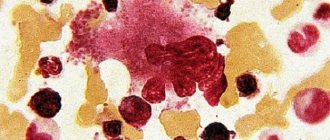
To definitively confirm the diagnosis and identify the tumor, a biopsy (aspiration, trephine biopsy, or other) is performed, with a biopsy sample taken for morphological examination.
Only on the basis of the results of cytology and histology of the biomaterial is it determined which process is developing in the body - malignant or benign. The type of cancer is determined by characteristic changes in cells. If it is a sarcoma, then its type.
Symptoms
The symptoms of sarcoma directly depend on where it is located and its size. Usually, the first manifestations are visual, that is, on the surface of the skin or under it, a person sees a gradually enlarging tumor. As the tumor grows, it involves neighboring tissues in the pathological process, and the signs and symptoms of the disease become more pronounced. Due to compression of healthy tissues and tumor growth through them, a person feels pain that cannot be relieved with analgesics. Each type may have its own symptoms of sarcoma:
- Ewing's sarcoma - night pain in the lower extremities;
- intestinal leiomyosarcoma – intestinal obstruction;
- uterine sarcoma – bleeding between cycles;
- extraperitoneal sarcoma – lymph stagnation and thickening of the legs;
- sarcoma of the face and neck – facial asymmetry, head deformation, impaired chewing function;
- Lung sarcoma – breathing disorder.
We recommend reading: What is lung adenocarcinoma and how long can a person live?
For other types of tumors, the clinical picture may vary.
Symptoms of Kaposi's sarcoma
Soft tissue sarcoma can appear in almost every part of the body. If the classic form of Kaposi's sarcoma occurs, then the foci of the disease first appear on the extremities, namely on the feet and legs. The defining features of this disease include the symmetry and multifocal nature of the lesion. Sarcoma looks like a rounded nodule, the color of which is white or gray-yellow. The surface can be smooth or with tubercles. Spots and plaques similar to nodules appear on the skin. Lesions can have different colors - from reddish-bluish to brown. The outlines of the lesions also vary. As a rule, they have clear boundaries, a dense and elastic consistency, and do not cause pain on palpation. Sometimes ulceration of the tumors occurs, and swelling of the extremities affected by the disease appears. In some rare cases, damage occurs exclusively to the genital organs, eyes, ears, mucous membranes and other organs. Other subjective symptoms include aches, pain, burning, and limited joint mobility. Due to the fact that Kaposi's sarcoma is a systemic disease, lymph nodes, bones and other organs may be involved in the disease process.
If the classic form of Kaposi's sarcoma mainly manifests itself in older men, and during the development of this disease the feet and legs are affected, then with epidemic Kaposi's sarcoma there is no talk at all about a specific location of the disease. Thus, the first foci of the disease appear on any area of the skin. Initially, a single node or spot of crimson or purple color is visible on the code. There is no pain in this case. Often several foci of the disease appear at once. In later stages, the disease can develop in different ways. So, in some cases, the shape and color of the nodes do not change for several years, but it happens that modification occurs even in a few weeks. If the tumor grows too quickly, the person often feels pain, and the skin around the tumor turns yellow-green due to constant hemorrhages. of necrosis appear in the center of the formation . The tumor may also bleed a little. The resulting nodes and plaques can merge, resulting in very severe swelling . If Kaposi's sarcoma occurs on the oral mucosa, then lesions appear on the hard palate. violet erythema appears In addition, lesions of Kaposi's sarcoma sometimes occur on the genitals.
In the process of diagnosing this disease, specialists are guided by the presence of a number of signs described above.
Both systemic and local methods are used to treat Kaposi's sarcoma. Local therapy uses irradiation, cryotherapy, injections of chemotherapeutic agents directly into the tumor, etc. Irradiation is carried out mainly in the presence of large and painful lesions.
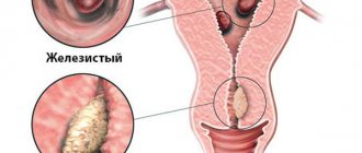
Uterine sarcoma is a malignant formation that occurs in the uterus. Today the disease is relatively rare. Uterine sarcoma most often occurs in women aged 43-53 years. With uterine sarcoma, the characteristic features of the disease are rapid enlargement of the uterus, the manifestation of disturbances in the monthly cycle, the presence of painful sensations in the pelvis, and watery discharge, sometimes having an unpleasant odor. If uterine sarcoma occurs in myomatous nodes, then the clinical picture of the disease may be similar
As the disease develops, large areas of necrosis and infection of the tumor may appear, resulting in anemia , fever , followed by cachexia . As a rule, patients turn to a specialist several months after the symptoms of the disease appear.
It is customary to define several stages of uterine sarcoma, which are differentiated according to the degree of tumor spread.
Diagnosis is carried out based on medical history, ultrasound results, as well as the use of hysteroscopy and separate diagnostic curettage.
The main treatment for uterine sarcoma is surgery. For this purpose, extended extirpation of the uterus and appendages is performed. After surgery, patients are usually prescribed a certain number of radiation therapy sessions, during which the pelvic organs are irradiated.
With uterine sarcoma, relapses of the disease occur very often, and even at the first stage of the disease, survival is very low due to the rapid manifestation of metastases.
If sarcoma develops in a myomatous node, the prognosis will be more favorable.
How to treat sarcoma
In most cases, sarcoma is treated surgically. Surgeries for sarcomas are most effective when the tumor is well located. Then you can remove it and not disrupt the functionality of the organ. However, relapse often occurs after surgery. Other treatments include radiation therapy and chemotherapy. Sometimes a person is treated using several methods at once.
Radiation therapy
Treatment of sarcoma by external irradiation is mainly carried out after surgery to avoid relapse. Although this type of treatment is effective, it is possible for a tumor to develop in another part of the body, so doctors carefully check the patient for a predisposition to the disease before starting radiation.
Chemotherapy
Treatment of sarcoma with chemicals is carried out to regress the primary tumor and to destroy metastases. Chemotherapy can be carried out before surgery, immediately during it, or after it. Chemicals can be taken orally, injected intravenously, or directly into the affected area.
Folk remedies
For sarcoma, treatment with traditional methods does not produce any results and can only be used as an auxiliary therapy to relieve symptoms. Before using any folk remedy, you should consult your doctor. By self-medicating, you can make the condition worse. Malignant neoplasms cannot be treated with herbs, decoctions, or compresses.
Features of sarcoma
Soft tissue sarcoma can appear anywhere. Most often this type of sarcoma affects the thigh. Less commonly, sarcoma occurs on the head.
A tumor of this type looks like a rounded white nodule or a yellow-gray nodule. It can have either a smooth or bumpy surface. Depending on the histological structure of the tumor, its consistency is determined. Thus, the tumor can be of a soft type ( liposarcoma and angiosarcoma ), a dense type ( fibrosarcoma ), or a jelly-like type ( myxoma ).
As a rule, a malignant tumor appears in the deep layers of the muscles. As the tumor grows, it moves to the surface of the body. The formation can grow faster under the influence of physical procedures and injuries.
Basically, sarcoma is a single tumor, but with certain types of this disease multiple lesions can appear. Moreover, they can be located even at a fairly large distance from each other.
Metastases from sarcoma spread through blood vessels . Metastases are especially common in the patient’s lungs; damage to the liver, bones, and lymph nodes occurs less frequently.
Sarcoma with metastases
Metastases are tumor cells that spread to healthy tissue through the lymph or bloodstream. In places where microcirculation is the best, metastases attach and actively grow. Metastases often affect the liver, lungs, lymph nodes, brain and flat bones. Each type of neoplasm has a specific place in the body where the tumor metastasizes most often.
There are tumors that metastasize, having already reached one centimeter in size. Sarcomas do not have a membrane that could limit their growth and reproduction. The spread of metastases to internal organs is more dangerous than metastases to the lymph nodes. Treatment of a multiply metastasizing tumor is ineffective and therefore is not carried out.
Causes of sarcomas and risk factors
Today, there are scientific works proving the role of ionizing radiation, ultraviolet radiation, chemicals, and also some viruses in the development of sarcomas. All these factors cause genetic mutations, which ultimately lead to uncontrolled growth and reproduction of cells.
The rate of bone growth and hormonal levels play an important role in the occurrence of Ewing's sarcoma. Risk factors also include smoking, work in the chemical industry, hereditary conditions, and malfunctions of the immune system.
In addition, among the reasons contributing to the formation of sarcomas are the following: the presence of benign neoplasms (they can become malignant), mechanical injuries, hereditary predisposition, etc.
Ewing's sarcoma in the knee joint
Ewing's sarcoma is considered a malignant neoplasm. Its formation is often recorded in bone tissue. In more rare cases, it is found inside the skin, subcutaneous tissue, muscles and compact parallel bundles of collagen fibers secreted within tissues by part of the nervous system. The pathology was named after James Ewing. He studied malignant neoplasms. A New Yorker first described the pathology in question at the beginning of the 20th century.
There are different types of Ewing's sarcoma. It includes the following pathologies:
- Malignant tumor of the bone skeleton;
- highly malignant sarcoma.
The pathology in question is considered to be very rapid. Lack of timely treatment can be fatal.
Sarcoma in children in the knees
This pathology is rarely recorded in children. It affects children and teenagers from 10 to 20 years old. It is also possible for neoplasms to form in infants and young children. Ewing's sarcoma can also occur in older people.
Symptoms of disorders
The differences in the signs of the disease depend on its location. But the main symptom is spasms that cannot be relieved with antispasmodics or analgesics. With the development of disorders, it manifests itself as spots on the skin, expansion of the venous network, and an increase in local temperature. Fatigue and inactivity appear. Often accompanied by deformation and swelling of body parts. Sarcoma differs from cancer in its delicate pink surface color. The tumor looks less lumpy. The difference is that this type of oncology spreads faster to neighboring healthy organs.
The following symptoms of sarcoma are characteristic of the pathology of the lymph nodes:
- rashes and swelling on the skin;
- enlarged lymph nodes;
- sudden weight loss;
- dyspnea;
- bluish color of lips;
- voice changes;
- pallor;
- high sweating;
- heat.
Signs of soft tissue sarcoma:
- the appearance of swelling;
- spasms when touching a swollen area;
- temperature;
- itching
Sarcoma difference
A feature of osteogenic sarcoma is the formation of cancer cells from bone tissue. It is in them that mutant cells are produced. The difference between tumors lies in the dominant tissue components:
- Fibroblastic;
- chondroblastic.
Experts divide the pathology in question into the following forms:
- Osteoplastic;
- osteolytic;
- crossed.
Tumor growth rate as a sign of malignancy
The progression of growth is purely individual for each individual type of sarcoma. Constrained by certain conditions, a tumor can remain at one stage of development for a long time, without delivering
The rate of tumor growth varies from person to person.
this patient hassle in everyday life. But some sarcoidal forms are characterized by intensive tumor growth, which is accompanied by intense pain, loss of functionality of an organ or limb, and general intoxication of the body. Unfortunately, the growth rate of sarcoma can only change upward.
Prognosis and prevention
Even if the doctor told the patient his life expectancy, do not forget that this is an approximate scenario for the development of the disease. It all depends on the general condition of the patient, his psychological mood, location and existing diseases. Taking into account the components of the anamnesis, a preliminary prognosis is made, indicating the possible development of the disease. The initial stages are easy to treat, but difficult to diagnose. The last stages are incurable, the doctor maintains the stability of the patient’s condition.
Clinical recommendations for preventive measures:
- Be examined by doctors for preventive measures.
- If symptoms arise, seek advice from oncologists. Especially if similar cases have already been recorded in the family.
- Do not self-medicate.
- Change your lifestyle and give up bad habits.
- Engage in physical exercise, establish proper nutrition and be attentive to the body.
Prevention reduces the risk of cancer. And reducing the amount of ultraviolet rays received by human skin reduces the likelihood of healthy cells developing into cancer.
Diagnosis of soft tissue sarcoma
As part of the diagnosis of soft tissue sarcoma, a physical examination is performed, medical imaging methods are used, followed by a biopsy and morphological examination of the obtained material.
A CT or MRI of the affected area is required. Firstly, the study helps to detect a tumor, and secondly, to monitor the radicality of the cure.
To make a diagnosis, a morphological examination of a tumor fragment is required. To obtain it, a trephine biopsy is performed using a special thick needle under ultrasound control, or an incisional biopsy, which involves surgical excision of a fragment of the tumor. In some cases, to clarify the type of sarcoma, a more detailed study is necessary, then immunohistochemical and molecular genetic analyzes are performed.
In order to determine the extent of tumor spread, a CT scan of the chest, abdominal cavity, and ultrasound examination of the lymph nodes is performed.
Book a consultation 24 hours a day
+7 (495) 151-14-53
Complications caused by the disease
Complications that malignancy entails are the main cause of death among people affected by cancer. Consequences include:
- internal bleeding;
- fistulas in hollow organs;
- penetration of affected cells into other organs;
- inflammatory process in the peritoneum;
- swelling of the brain or lungs;
- blood poisoning;
- overlap of bronchi, ureters;
- intestinal obstruction;
- compression of nerve endings and blood vessels;
- disruption of the heart muscle and breathing;
- complete poisoning of the body, exhaustion;
- coma with central nervous system depression.
Repeated relapses and metastases in distant organs increase the destructive effect on the body. The patient weakens, the immune system stops working at full strength. Such processes lead to the death of the patient.
Causes of carcinoma
The reasons for the occurrence of lesions are vague and represent a list of points that are easy to find in any large city. The patient’s lifestyle before the onset of cancer has a significant impact:
- bad habits and sedentary lifestyle;
- unbalanced diet, excess body weight;
- radiation or excess of received ultraviolet rays;
- living in a polluted area or working with hazardous substances.
Hereditary location plays a special role. If your relatives have had malignant tumors, especially of the mammary glands, then you should be very careful about your health and respond to any manifestations of any disease.
Treatment and therapy
Treatment of sarcoma depends critically on the extent of the disease after diagnosis. For small, localized tumors, surgery is the first choice. Its main goal is to completely eliminate malignant tissue. Also, part of the healthy tissue that is adjacent to the tumor is removed, since migrated cancer cells may be hiding there, which contribute to the formation of metastases.
Before large tumors are surgically removed, a course of chemotherapy is given to reduce the size of the sarcoma.
If the metastases have spread to large areas, the patient is also prescribed chemotherapy, which can be administered using tablets or by infusion and injection. If such therapy is ineffective, then only radiation will help destroy tumor tissue. Because each patient responds differently to chemotherapy agents and drugs, there is a need to develop an individualized treatment plan.
Morphological study
Morphological diagnosis is a very complex process due to the large differences in tumors, even within the same subtype. There are more than 100 types that require individual treatment.
The characteristics of soft tissue sarcomas are based on the definition from a benign tumor without metastases with slow growth to malignant with a high growth of metastases.
The tendency to complications in each type of tumor depends on the level of malignancy. Two methods have been developed for determining the characteristics of malignancy: NCI, FNCLCC.
Spindle cell sarcoma
The NCI method evaluates histological groups, the number of visible cells, pleomorphism, the number of affected organs and the presence of obvious sites of inflammation.
- G1 – low degree of malignancy (well-differentiated tumor): favorable prognosis, spread to other organs does not occur, no response to chemotherapy.
- G2 – intermediate degree of malignancy (moderately differentiated tumor).
- G3 – high degree of malignancy (poorly differentiated tumor): poor prognosis, high potential for metastasis, in most cases sensitive to chemotherapy drugs.
According to the FNCLCC system, it is determined by a three-stage method. The sum of points showing the specificity of tumor cells is considered. The degrees of both systems are the same.
Determining the level of malignancy is an absolutely subjective procedure, therefore sarcoma is classified into 2 types: high and low grade.
About the disease
Skin sarcomas are a group of malignant tumor processes that form in skin tissue from abnormal connective tissue elements.
Such formations are quite rare. They prefer to be localized on the thighs, legs, chest and abdominal area, and forearms.
Skin cancer is not considered an age-related pathology and can affect a patient of any age group, regardless of professional field, although experts note that in 30-60-year-old men, this disease is found a little more often than in other patients.
Cutaneous sarcoma accounts for about 0.3% of the total number of malignant skin tumors.
Kinds
| Types of sarcomas | Description |
| Stromal | The tumor grows inside the uterus on the soft tissue of the endometrium. Very often occurs due to radiation exposure during the treatment of other oncology. It can appear as a result of damage to the uterus as a result of abortion, polyposis, curettage, endometriosis and damage due to abnormal childbirth. It is characterized by pain and bloody discharge from the vagina outside of menstrual days. |
| Spindle cell sarcoma | Epithelioid spindle sarcoma is sometimes confused with fibroma. The nodes are dense, the structure of the sarcoma is fibrous and appears on the skin, serous tissues, mucous membranes and grows into the fascia. If detected early, the outcome is favorable. |
| Malignant | At stages 1 and 2 it does not manifest itself in any way. Develops on connective soft tissue. It grows very quickly and metastasizes: to the liver, lungs and sometimes to the brain. The sarcoma cells themselves spread through the lymphogenous route. |
| Pleomorphic | The size of such a sarcoma is at least 10 cm and often recurs. It is observed in the limbs, less often on the body. It is a dense, nodular formation resembling a lobular structure. Contain areas of necrosis and hemorrhage. A person can die in 1 year, survival rate is no more than 10%. |
| Polymorphocellular | Localized on the periphery of the soft tissues of the skin. The sarcoma metastasizes, and the spleen grows along with the growth of the tumor. Removed promptly. |
| Undifferentiated | The tumor conglomerate does not differentiate, does not belong to any type, has completely different cellular structures, and therefore always looks different. Treatment is similar to rhabdomyosarcoma. |
| Histiocytic | The structure contains polymorphic cells. The prognosis is most often unfavorable. Histiocytic sarcoma responds very poorly to therapeutic treatment and is actively growing. It is found on the skin, gastrointestinal tract and soft tissue structures. Only a doctor can tell a patient whether it is cancer or not. |
| Round cell | It grows aggressively and quickly penetrates into nearby tissues. Looks like round cell structures. Soft tissue epithelial sarcoma affects the skin of soft connective tissues. |
| Fibromyxoid | Quite favorable outcome of the disease and low malignancy. Slow growth, metastases are detected extremely rarely. Affects shoulders, hips, body. It can occur in people of all ages. |
| Lymphoid | It proceeds unnoticed and affects the immune and lymphatic systems. You can see enlarged lymph nodes, eczema on the skin, pallor of the mucous membranes and the entire integument. The malignant tumor grows, damaging nearby blood and lymphatic vessels, which leads to poor circulation and necrosis. |
| Epitheloid | It is observed to a greater extent in young people, affecting the limbs. It is considered one of the types of synovial sarcoma. |
| Myeloid | Consists of myeloblasts of the leukemic type, is a local tumor. Location: cranial, tubular, spongy bones, lymph nodes, bone skeleton, intraorganic structures, etc. |
| Clear cell | Localized in the head, cervical and torso areas. Soft structures are being damaged. Characterized by slow tumor growth, there are relapses and metastases. It can be up to 1 cm in size. It is not contagious. |
| Neurogenic | Sometimes called connective tissue carcinoma. It develops slowly, is localized on the legs, and has clearly defined tumor boundaries. The prognosis is favorable, treatment is by surgery. Survival rate is more than 80%. |
How is sarcoma different from cancer?
Cancer grows from epithelial cells, and sarcoma from connective cells, this is what differs from cancer. Typically, epithelial tissues are located at the surface of the organ - either on the inside or on the outside. Connective tissue usually connects different layers of an organ.
Symptoms of the disease
General and local symptoms of the tumor are identified. General signs of the disease indicate the patient’s condition as a whole, local signs indicate a symptom of a damaged organ.
Local signs:
- the skin and mucous membranes are modified, ulcers appear, color, structure externally become different from healthy membranes;
- bleeding of the affected organ;
- breathing problems and difficulty swallowing food;
- cough mixed with phlegm and blood;
- pain in the affected area;
- the genitals hurt during sex, intermenstrual bleeding, blood loss after intercourse;
- urinary incontinence, frequent urge to go to the toilet, bowel movements with altered color and structure;
- disturbances in voice timbre, as well as partial loss of vision, smell, and hearing;
- nausea, vomiting, headaches and dizziness.
Common symptoms of illness experienced by patients:
- loss of appetite followed by aversion to food;
- weight loss;
- weakness, fatigue, drowsiness;
- increased temperature, increased sweating at night;
- anemia and jaundice.
Classification of epithelioid sarcoma
There are 2 main types of epithelioid sarcoma of soft tissues:
- Classic, which includes distally located tumors. It accounts for about 90% of cases. Classic epithelioid sarcoma is represented by several variants, the name of which reflects their histological structure: granuloma-like (similar to granuloma), angiomatoid (similar to angioma), fibroma-like (similar to fibroma), histiocyte-like (similar to histiocytoma).
- Proximal. These are neoplasms located on the forearm, pelvis, perineum, genital area and mediastinum. Histologically, they differ from the classical forms in having larger cells, the presence of a rhabdoid component and cytological atypia. Proximal forms of epithelioid sarcoma are located deeper, they are more aggressive and difficult to treat, for this reason they are more likely to relapse and metastases form faster.
Read here: Osteogenic sarcoma
The stages of epithelioid sarcoma can be viewed in the classification of soft tissue sarcomas.
Life prognosis for skin sarcoma
When skin sarcoma develops, the prognosis depends on the stage at which the disease was diagnosed and on the morphological structure of the tumor.
Life prognosis for sarcoma by stage:
- I – 95%.
- II – 50-80%.
- III – 25%
- IV - no more than 5%.
Thus, fibrosarcomas diagnosed at stages 1-2 have a fairly favorable prognosis and respond well to treatment. Leiomyosarcoma and Kaposi's sarcoma of the skin are the most aggressive, with early spread of metastases, so the prognosis is much worse.
Course and treatment of the disease in children, pregnant and lactating women, the elderly
Children. Spinal sarcomas progress aggressively in childhood and adolescence, which is due to the rapid growth of connective tissue structures. Osteosarcoma and Ewing's sarcoma are usually diagnosed in young patients. The principles of diagnosis and treatment in children do not have specific differences from adult patients, but even successful recovery in the future often results in such undesirable effects for them as impaired bone growth, cardiomyopathy and infertility due to the aggressiveness of the tumor and therapeutic techniques.
Pregnant and lactating women. In women pregnant and during breastfeeding, spinal sarcomas are practically not found. The fact is that this malignant disease more often affects the male half of the population, mainly children and the elderly. Despite this, this oncopathology cannot be completely excluded among expectant and nursing mothers. With its development, timely diagnosis is of the greatest importance, which determines the prognosis for pregnancy and cure for the tumor process. These issues are resolved individually with an oncologist.
Elderly. When sarcoma develops in people over 50 years of age, as a rule, malignancy of a benign neoplasm or the development of the disease against the background of an inflammatory or destructive-dystrophic process is assumed. The choice of treatment principles depends not only on the type of tumor and the stage of the oncological process, but also on the patient’s health status. The prognosis for recovery is unfavorable.
Uterine cancer
Uterine carcinoma (endometrial cancer) is a tumor formation that develops from uterine cells and has the ability to metastasize. Like breast cancer, uterine cancer is the most common tumor disease, affecting hundreds of thousands of women every year. Moreover, older women are at risk of the disease.
The causes of carcinoma formation are as follows:
- Being overweight (may be caused by diabetes).
- The woman's body has never undergone labor.
- The onset of late menopause (after 50 years).
- Polycystic ovary syndrome.
- Hereditary predisposition.
- Previous history of breast carcinoma.
Symptoms of uterine carcinoma are the following:
- Prolonged bleeding during menopause. During the onset of menopause, it is also worth monitoring the appearance of bloody discharge - normally there should not be any.
- Aching pain in the lower abdomen and lower back.
- Pain and bleeding during and after sex.
- Weight loss.
Benign neoplasms
Separately, Fr. This is the name of a benign neoplasm of glandular tissue. When there is a hormonal imbalance, the adenoma develops into a malignant tumor.
Most often, the tumor is localized in the prostate area, but it also forms in other organs. Prostate adenoma – there is no metastatic phase, but it is characterized by a transition to adenocarcinoma.
Adenoma of the stomach and intestines is an acquired disease in people who previously suffered from gastritis, had an unhealthy diet, or have a genetic predisposition. An adenoma of the digestive tract is called a polyp. A single tumor is surgically removed; for multiple polyps, electrocoagulation is used.
Fibrous adenoma of the breast is a mobile formation. Women often discover changes in the breast area on their own. In the stage of rapid development, the tumor is removed. Until this moment, she is simply being watched.
Endometrial adenoma in the uterus is the growth of the endometrium to the formation of a cyst or polyp. Accompanied by bloody discharge between menstruation and cramping pain. This type of adenoma is visible on ultrasound and is removed by curettage of the uterus. Endometrioid adenocarcinoma of the uterus is the scourge of women who have no sex life, have not had childbirth or pregnancy, and also experience hormonal imbalance. As the tumor grows, the woman looks like a pregnant woman, her stomach grows, and lower back pain appears. Foul-smelling vaginal discharge indicates that the tumor is disintegrating.
Examining the adenoma of the lower extremities, it is noticeable that its degeneration resembles rheumatism. For this reason, the patient often ignores the first signs, missing favorable treatment of the disease. As the pain increases, the legs become inactive and swelling occurs. With the last stage of the disease, fractures that become chronic are more common. The lesion affects the feet, legs, heels, hip joints, and cancer of the upper extremities is less common.
Spinal adenoma is rare. Most often, the patient is faced with a cancerous tumor, which manifests itself as pain in the back. A biopsy will determine the nature of the tumor and help determine the correct treatment for the patient.
Types of soft tissue sarcomas
Soft tissue sarcoma is a collective term that unites a large number of malignant tumors, among which the most common are the following:
- Leiomyosarcoma is a tumor of smooth muscle cells.
- Rhabdomyosarcoma is a tumor of striated muscle.
- Liposarcoma is a malignant tumor of adipose tissue.
- Angiosarcoma is a malignant neoplasm that grows from the tissue of blood vessels.
- Fibrosarcoma is a tumor of fibrous tissue.
Also, soft tissue sarcomas are usually divided according to the degree of malignancy. Low-grade neoplasms rarely metastasize, but are prone to persistent recurrence. Highly malignant tumors tend to metastasize hematogenously (through the bloodstream), lymph nodes are affected less frequently.
Description and statistics, ICD-10 code
Polymorphic cell sarcoma of soft tissues is an extremely rare autonomous type of primary sarcoma of the skin. The tumor begins to develop primarily along the periphery of the dermis, without penetrating deeply into its structures, most often in the area of natural openings in humans. The neoplasm is surrounded by a characteristic erythematous rim.
As the sarcoma progresses, it ulcerates, foci of hemorrhage and necrosis form on its surface, and foul-smelling discharge occurs. Metastases spread to the lymph nodes, and at the same time there is a marked enlargement of the spleen and intense pain associated with mechanical impact on the lesion.
Histologically, polymorphocellular sarcoma has an alveolar structure.
The picture of its connective tissue section shows spindle-shaped and round cells of the embryonic type, similar to myelocytes and megakaryocytes. In cytology, this phenomenon is called polymorphism. The blood vessels in the area where the tumor is located are noticeably worn out and lack natural elasticity. Unlike other soft tissue sarcomas, such as clear cell or pleomorphic, polymorphic can only be eliminated by surgical treatment.
Pathology is diagnosed equally often among women and men, children and the elderly.
ICD-10 code: C49 Malignant neoplasm of connective and soft tissues.
Metastases
The spread of atypical sarcoma cells, which is located in the spinal column, usually begins with the third stage of the malignant process. 70% of secondary lesions go to the chest organs - lungs and mammary glands, the remaining 30% are localized in the kidneys, liver, bladder and brain.
Metastases are detected on X-ray images, secondary neoplasms have blurred contours. MRI and CT visualize their structure and nature. For small metastases, chemotherapy or radiotherapy methods are used; in advanced cases, surgical intervention is used. The prognosis for survival with the appearance of metastatic changes is significantly worse.