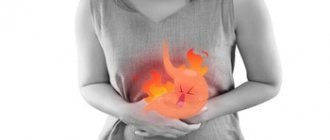
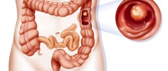
An intestinal cyst is a multiple or single benign neoplasm, which is a hollow capsule with dense walls containing exudate inside.
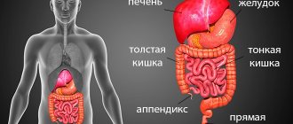
Each organ of the human body performs an important role assigned to it. The intestine is where the final absorption of substances needed by the body from food occurs, and it is also responsible for removing food debris and toxins. During its work, the intestines experience heavy loads, which can cause disruptions in its functioning and damage. At the same time, the risk of developing various ailments increases, and one of the diseases may be an intestinal cyst, which has acute symptoms, and without timely treatment can lead to an oncological process.
Three types of cystic formations can form in the intestines:
- Unilocular cyst. This type is most common and accounts for 80% of all diagnosed cases. The cavity of the node in this case has one chamber, which is formed on the mucous membrane. The germ layer is single.
- Two-chamber cyst. Diagnosed less frequently, it develops between the endoderm and ectoderm. There is an internal germ layer.
- Three-chamber cyst. This formation is diagnosed in very rare cases; in this case there are three germ layers.
In addition to the above types, experts distinguish a complex cyst, which has a large number of chambers and nodular inclusions.
Cystic formation is dangerous for the development of abscesses, fistulas and oncological processes. Therefore, if symptoms occur, you should immediately contact a specialist.
Regarding location, cysts are classified as follows:
- Small intestinal cyst. This is a benign formation that manifests itself as bloody or mucous painful stool.
- Large intestinal cyst.
- Mesenteric cyst - such cystic structures can be observed in both the small and large intestines. Most often, its formation is caused by dystonia of lymphoid tissue. The cyst in this case is thin and contains serous fluid.
- Enterocystoma (or intestinal duplication). Most often, this pathology is observed in the small intestine; it appears as a result of stratification of the intestinal tube and the proliferation of the epithelial layer.
- Cecal cyst.
- Polycystic bowel disease.
Causes
No one is immune from the formation of a cyst in the intestines, but some groups of people are more prone to this disease than others.
Provoking factors may be:
- alcohol abuse;
- smoking;
- avitaminosis;
- lack of fruits and vegetables in the diet (along with excess animal fats);
- eating large amounts of red meat;
- hereditary predisposition to colon cancer;
- sedentary lifestyle;
- excessive consumption of white bread;
- frequent and prolonged constipation;
- excess weight.
If there is at least one risk factor, the likelihood of the formation of a cystic formation in this organ increases, and therefore it is recommended to undergo periodic examinations with a doctor.
The main causes of cystic neoplasms
The main factors provoking the development of cystic diseases of the intestinal tract are considered to be poor lifestyle, unbalanced diet, and heredity.
Abuse of alcoholic beverages (especially low-quality ones) is the main cause of the development of pathology of the digestive system. This also includes smoking, drug addiction, physical inactivity and the presence of infectious diseases.
This factor includes vitamin deficiency (lack of vitamins), uncontrolled consumption of fats (especially animals), white bread and red meat. In addition, constipation, which appears mainly due to monotonous dry food and fast foods, can lead to neoplasms in the digestive system.
We suggest you familiarize yourself with: Baker cyst Vishnevsky ointment
Heredity
In this case, people who have close relatives with cancer of the digestive system (first stage or higher), as well as ulcerative colitis, are at risk of cystic neoplasms.
The above reasons can have a negative effect on the body, both individually and in combination.
Symptoms of the disease
Before considering the symptoms of an intestinal cyst, it is necessary to understand that the clinical picture will vary depending on which part of the organ the benign tumor is located in.
If there is a cystic growth in the large intestine on the right, the signs will be as follows:
- pain of varying intensity on the right side;
- dark-colored stool, sometimes mixed with blood;
- anemia.
If neoplasms of the large intestine have formed on the left side of the organ, the following signs are observed:
- bright blood on the surface of the excrement;
- constipation followed by diarrhea;
- thin stool;
- bloating and colic.
If the cyst is localized in the sigmoid region or in the rectum, then the following are observed:
- pain in the abdomen and perineum;
- sensations of incomplete bowel movement;
- bloody impurities in the stool;
- colic.
If there is a cyst in the small intestine, the symptoms are as follows:
- constant or paroxysmal pain in the epigastrium or in the umbilical region, which becomes more intense when bending the body or during physical activity;
- If complications develop, intestinal obstruction may occur.
As the cystic formation grows, the patient experiences:
- dull pain in the epigastrium without clear localization;
- frequent urination and bowel movements;
- a nodular formation in the perianal area that can be palpated.
If an inflammatory process develops, the patient’s condition worsens sharply:
- there is throbbing pain in the lower abdomen, in the tailbone and in the perineum;
- severe weakness;
- heat;
- headache, nausea.
Also, the human body can develop an intestinal air cyst (in which air accumulates), thereby causing increased gas formation and constipation.
With a fistula opening into the intestinal lumen, pus, mucus and blood can be seen in the stool.
If such symptoms are present, each person should understand that there is a problem in the intestinal tract, and it must be eliminated as quickly as possible. If blood is found in the stool, this is a good reason to immediately contact a specialist. Of course, blood in the stool is a sign of a wide variety of diseases - from hemorrhoids to oncology, so it is necessary to conduct a thorough diagnosis and establish the correct diagnosis.
Symptoms
The symptoms of an intestinal cyst are determined, first of all, by which part of the digestive tract the problem occurs. The formation can be located in the esophagus, stomach, small or large intestine. There are rectal cysts; some formations are localized in the area of the membrane that supports the intestines in the abdominal cavity (intestinal mesentery cyst).
If we talk about the large intestine, when the tumor is located on the right side of the colon, patients note:
- stool mixed with dark blood,
- pain localized in the lower abdomen,
- anemia.
The cyst, which is located on the left side of the organ, is characterized by the following symptoms:
- diarrhea alternating with delayed bowel movements,
- bright blood clots on the surface of the stool,
- bloating,
- change in the shape of the stool - it becomes thin,
- colic.
Bloating
If the problem occurs in the lower part, then the sigmoid or rectum signals this as follows:
- manifestation of blood admixture, the color of which is light red,
- intestinal colic,
- pain in the perineal area,
- a feeling that emptying has not occurred completely,
- pain in the abdomen.
Important! The manifestation of even a single symptom of an intestinal cyst is a reason to immediately visit a doctor. This is especially true if the stool contains blood clots. Often patients take the described symptoms for a long time as signs of hemorrhoids, and when it comes to hospitalization, it becomes obvious that they were mistaken. It’s sad that it’s not always possible to correct an error caused by inattention.
Diagnostics
The diagnosis can be made after the results of the examination, namely:
- Digital examination - during palpation, a specialist can detect intestinal prolapse, which is associated with pressure on it from a large cystic formation. In addition, the elasticity of the intestinal walls decreases and tone decreases. Such phenomena are associated with the presence of scars in those places where there was a fistula breakthrough.
- Colonoscopy. This examination is performed under local anesthesia. During this diagnostic study, it is possible to simultaneously detect polyps in the intestine, its narrowing and other pathologies. In addition, during the procedure, a specialist takes organ tissue for histological analysis to determine the nature of the pathology.
- Sigmoidoscopy - this study allows you to assess the condition of the mucous membrane, as well as determine the exact size of the cystic node.
- Proctofistulography – determination of the length of fistula tracts.
- Ultrasound, MRI, CT are studies that provide a lot of additional information about the sphincter muscles and the condition of other organs located in the pelvis.
Blood is taken from the patient to assess the general condition of the body.
Considering the fact that the signs of cystic formation are very similar to other intestinal diseases, it is very important to conduct a differential diagnosis regarding hernia, coccyx cyst, coccyx osteomyelitis, paraproctitis, and oncology.
Treatment tactics
Cyst treatment methods are selected according to the severity of the patient’s condition and the stage of the disease. The malignant nature dictates the need for a set of measures - this is surgery, then several courses of chemotherapy and radiation exposure.
If the benign nature of the pathology is confirmed, the cystic cavity is removed along with the affected part of the intestine. One operation is sufficient if the disease is detected at an early stage.
In severe and advanced cases, implants are used to restore gastrointestinal tract patency.
Mesenteric cysts are removed surgically without violating the integrity of the intestinal wall or local damage to the circulatory system. Emergency surgery is required if there is a threat of torsion or rupture of the cavity with the development of peritonitis.
If the pathology is benign, the prognosis is usually favorable. Late detected cysts require more radical interventions and a long rehabilitation period.
Treatment methods for intestinal cysts
Treatment of intestinal cysts is carried out using traditional medicine, conservative or surgical methods. Antibacterial and anti-inflammatory drugs are used as medications to achieve remission of the disease. As for traditional methods, such treatment is symptomatic in nature, which means they can only be used to eliminate negative symptoms.
Remission is a temporary measure that only delays the time for surgical intervention. It is possible to get rid of cystic formation only in a radical way.
The earlier the operation is performed, the more favorable the prognosis will be, since the likelihood of relapse is reduced, and it is also possible to avoid serious consequences.
Surgeries to remove intestinal cysts are performed in the proctology department. In the acute stage of the disease, the existing abscess is simply opened and drained so that the process does not spread further. Removal of a cystic node is carried out only in the remission stage to avoid complications.
As for the surgical technique, it consists of excision of the tumor and subsequent suturing of the excision site. Which method of surgical intervention will be performed depends on the location of the cyst, its nature and the extent of the lesion. In some cases, a benign tumor can be removed not through the anus, but through the vaginal wall.
After the operation, the patient is provided with a drainage outlet, which is necessary to prevent suppuration and the development of an infectious process. During the postoperative period, the patient is prescribed antibiotics and daily dressings.
If the cyst becomes malignant, it is treated in the following ways:
- part of the intestine is removed along with the tumor;
- local treatment is used - radiation therapy;
- use systemic treatment - chemotherapy.
After removal of the cyst, the patient is recommended to:
- sit and lie on your back as little as possible;
- do not overcool or overheat;
- after the surgical sutures are removed, regularly and thoroughly wash the perianal area with warm water;
- do not lift heavy things or engage in heavy physical labor;
- Do not epilate the perianal area for six months.
If the cyst and possible fistula tracts are removed surgically in a timely and competent manner, the prognosis for the patient is favorable, and relapses in this case are extremely rare.
Treatment of pathology
If it is suspected that the formation of a cyst is due to hormonal imbalance, then the level of hormones is determined. Next, drug therapy is prescribed to restore balance. Thus, education may disappear.
However, in all other cases, deletion is required. Especially if the cyst threatens intestinal obstruction and causes serious pathologies of the digestive system. Or it has festered, presses on other organs, and is predisposed to rapid growth.
The operation is carried out in different ways:
- A laparoscopic or endoscopic procedure, when the contents of the cyst are pumped out using a puncture and installation of drainage. This method is not possible if cancer is suspected. This elimination of pathology can be used to firmly establish the benign quality of the tissue.
- More often, the cyst is excised along with a piece of intestine. The ends are sewn together. If the area removed is too large, a stoma may need to be placed. And at the next stage, form the rectum.
- Radiation and chemotherapy are prescribed to patients whose removed cyst contained cancer cells. Irradiation of the site leads to the death of the remaining metastases.
The fistula tracts of the formation are also removed.
Important information! A cyst is an extremely unpredictable formation. It grows very quickly and also soon turns into a cancerous tumor. Therefore, there is no need to delay the operation.
Complications in the postoperative period
After removal, there is a risk of the formation of fistula canals that need to be eliminated. Otherwise, they can lead to abscess and necrosis of the tissues of the anus and coccyx.
In any case, the patient is observed until complete recovery. Further, it is recommended to undergo control examinations, first once every six months, then every year. The larger the cyst and the more associated problems, the longer the recovery period and the higher the risk of complications.
Diet before and after removal
The entire period of treatment of intestinal formations is accompanied by compliance with a special nutritional system. Anything fatty, spicy, smoked, that promotes gas formation, as well as alcohol is prohibited. Any adult understands what harmful products are. A patient with this pathology will have to refuse them.
The consumption of fresh vegetables, except those that promote gas formation, and fruits, except citrus fruits, is encouraged. Lean meat, poultry, fish, stale bread, dairy products, whole milk to a minimum. Soups, cereals and other similar dishes.
After removal of the cyst, the diet for a month is of a therapeutic and dietary nature and is strictly controlled by a doctor.
Attention! Diet is the basis of rehabilitation after surgery on the gastrointestinal tract. Therefore, compliance speeds up the recovery process.
Complications
In the absence of surgical intervention at the initial stage of the pathological process, complications may develop. For example, the formation of multiple fistula tracts, the development of phlegmon or an inflammatory process with frequent relapses. In the latter case, there is a high risk of abscess formation in the perianal or sacrococcygeal region, which can lead to rectal prolapse, which develops due to insufficiency of the anal sphincter. This process significantly complicates surgical intervention and the recovery period after it.
The most unfavorable complication of a cystic formation in the intestine is considered to be a breakthrough of the fistula into neighboring organs of the abdominal cavity. In this case, the complexity of surgical intervention increases several times. In addition, in some cases, the cyst can malignize (degenerate into a cancerous tumor), which entails consequences that can be fatal.
Most often, intestinal mesenteric cysts transform into malignant formations. If left untreated, such cystic structures can lead to:
- bleeding;
- suppuration and abscesses;
- ruptures with contents entering the abdominal cavity, which entails peritonitis;
- fusion with neighboring organs.
Intestinal duplication cyst: ultrasound diagnosis and morphological comparisons
Ultrasound scanner WS80
An ideal tool for prenatal research.
Unique image quality and a full range of diagnostic programs for an expert assessment of a woman’s health.
Intestinal duplication (enterogenous) cysts are quite rare. They can be localized in the central nervous system [1], pericardium [2], mediastinum [3], testicles [4], as well as anywhere along the gastrointestinal tract, mainly in the small intestine [5, 6]. In the literature, the following terms have been proposed to denote this condition: duplication (doubling), reduplication, double intestine, intestinal cyst, giant diverticulum, enterogenous cyst [7]. Currently, two terms are most often used: enterogenous cysts to refer to formations in the central nervous system [1] and intestinal duplication cysts to refer to cysts developing along the intestinal tube [8, 9]. The latter cysts are usually observed in children [2, 3].
Intestinal duplication cysts are congenital abnormalities that can develop anywhere along the intestinal tract, from the mouth to the anus. Such cysts are localized, as a rule, in the wall itself or adjacent to the wall of any part of the gastrointestinal tract. H. Dardic et al. [10] proposed the following diagnostic criteria: the presence of a lining corresponding to the intestinal or gastric mucosa, a smooth muscle layer and a close connection with any part of the gastrointestinal tract. At the same time, there are descriptions in the literature of cysts lined with respiratory epithelium [5, 11] or stratified squamous epithelium [12]. In some observations, there was no connection between the cyst and the gastrointestinal tract [13].
Several theories have been proposed to explain the pathogenesis of duplication cysts. The theory of aberrant recanalization of cavities explains the duplication of those parts of the gastrointestinal tract that pass through a solid stage in their development (for example, the esophagus, small intestine and colon) [14]. It is believed that the formation of an isolated cavity is preceded by torsion or some vascular abnormality in the proximal portion of the diverticulum. Such cavities can completely separate from the intestinal wall, forming isolated duplication cysts [5, 9, 10]. According to the theory of intrauterine circulatory disturbance, duplication cysts develop as a result of local vascular insufficiency during anoxia or any stressful situations [15].
Intestinal duplication cysts usually appear during the first years of life as a palpable formation, as well as signs of intestinal obstruction. In adults, in addition to these symptoms, arrosive bleeding may occur due to ulceration of cysts [16]. In addition, duplication cysts may become malignant [5, 13, 17].
Radiation research methods, as a rule, confirm the cystic nature of the formation. During ultrasound examination (ultrasound), duplication cysts have the appearance of hypoechoic masses with pronounced walls and good transmission due to the presence of transparent liquid contents, or the appearance of anechoic formations with hemorrhage or thickening of the contents in the lumen of the cyst.
Treatment consists of complete surgical removal of such cysts [3, 5].
As an example, we give our own observations.
Observation 1
Patient A., 33 years old, entered the institute with complaints of constant nagging pain in the epigastric region and hunger pains. According to the anamnesis, she first began to notice hunger pains about 4 years ago, and therefore underwent examination at the clinic at her place of residence. Esophagogastroduodenoscopy diagnosed reflux esophgitis. The patient was treated with a course of antisecretory therapy with a temporary positive effect. However, subsequently the pain resumed and gradually intensified, and therefore a re-examination was carried out, but no pathological changes were identified. Three years ago, an ultrasound of the abdominal cavity was performed, which revealed a cystic formation in the area of the stomach and pancreas. During the next esophagogastroduodenoscopy, a cystic formation protruding into the lumen of the stomach was revealed. The patient was admitted to the institute for further examination and determination of treatment tactics.
Upon admission, the patient’s general condition was satisfactory, the skin and visible mucous membranes were naturally colored and clean. Breathing is carried out in all parts, no wheezing is heard, the respiratory rate is 16 per minute. The boundaries of the heart are within the age norm, the heart sounds are rhythmic and sonorous. Blood pressure 120/80 mm Hg. Art. Pulse 76 beats per minute, rhythmic, satisfactory filling and tension. There is no swelling. The tongue is clean and moist. The abdomen is symmetrical and participates in the act of breathing by all sections. The epigastric region is sensitive to palpation, there are no symptoms of peritoneal irritation. Peristalsis is dried. Physiological functions are normal.
Data from laboratory research methods are within normal limits.
Ultrasound of the abdominal organs in the epigastric region on the left, in the projection of the stomach (in its lumen), reveals a round formation, 53.1×49.6×45.6 mm in size, with clear, even contours, anechoic in structure, homogeneous, with the presence of echo-dense walls up to 4.1 mm thick. When the stomach is filled with liquid, it seems that the formation comes from its posterior wall, since it is fixed to it and does not move when the patient changes her body position and coughs (Fig. 1). In the color Doppler mapping mode, no data on the presence of blood flow in the capsule of the formation were obtained. Conclusion: cyst (cystic formation) emanating from the wall or located in the stomach cavity.
Rice. 1.
Ultrasound picture of a duplication cyst in the lumen of the stomach, B-mode.
The patient was given a preliminary diagnosis: cystic formation of the stomach. In order to clarify the diagnosis, it was decided to perform an endoscopic diagnostic puncture of the formation. The formation is soft on instrumental palpation, it seems that it has liquid contents. A puncture of the formation was performed with an attempt to aspirate the contents, but its thick nature did not allow this manipulation to be performed through the catheter. Using a papillotome, the cyst was fenestrated with aspiration of a large amount of viscous, thick secretion that was “café au lait” colored and odorless. During aspiration, the cyst completely subsided.
A cytological examination of the contents of the cyst revealed tissue detritus, a macrophage reaction, and squamous epithelial cells, mainly in a state of destruction.
Biochemical study of the contents of the cyst: diastase - 5 units/l, CEA - 1887.6 ng/ml, CA 19-9 - 29707, 92 units/l.
Thus, the patient had a clinical and instrumental picture of a duplication cyst of the stomach. Due to the extremely high level of tumor markers in the contents of the cyst, it is necessary to think about its malignancy.
The patient was fully informed about the nature of the disease, the risks and possible complications of surgery, as well as in case of refusal. The patient refrained from the proposed surgical treatment and endoscopic biopsy of the cyst wall. In satisfactory condition, she was discharged under the supervision of a surgeon and oncologist at her place of residence.
Observation 2
Patient Z., 47 years old, was admitted with complaints of aching pain in the upper abdomen. He considers himself sick since the age of 17, when he began to experience periodic pain in the upper abdomen, especially after errors in diet. She received outpatient treatment for chronic gastritis. Three years ago, an attack of pain in the epigastric region developed and the patient was hospitalized in a city hospital, where ultrasound diagnosed a cystic formation of the head of the pancreas up to 2 cm in diameter. A year ago, the patient developed another attack of pain in the epigastric region, accompanied by a single attack of vomiting and loose stools. For this reason, she was treated at the Institute of Gastroenterology, where ultrasound revealed an increase in the formation in the head of the pancreas to 3.3 cm. The patient was admitted to the Institute of Surgery named after. A.V. Vishnevsky for examination and treatment.
Upon admission, the patient’s condition was satisfactory, the skin and visible mucous membranes were of normal color and clean. Peripheral lymph nodes are not enlarged. In the lungs, vesicular breathing is carried out in all sections. Heart sounds are rhythmic, no murmurs are heard. Pulse satisfactorily filled, 68 beats per minute. Blood pressure 110/70 mm Hg. Art. Varicose veins of both lower extremities are detected. The abdomen is not swollen, participates symmetrically in the act of breathing, is soft on palpation, moderately painful in the epigastric region. There are no symptoms of peritoneal irritation. Peristalsis is heard. Physiological functions are normal.
With ultrasound and duplex scanning of the vessels of the abdominal cavity, the liver is not enlarged in size, the anteroposterior size of the right lobe is 136 mm, the left lobe is 66 mm, the contours are clear, the structure of the parenchyma is homogeneous, diffusely compacted. The gallbladder is not enlarged, is visualized with an inflection, the walls are thickened, the contents are homogeneous. Intra- and extrahepatic bile ducts are not dilated. Hepaticocholedochus is determined to be 3.6 mm in diameter, its lumen is free. The pancreas is not enlarged in size, the head is 30 mm, the body is 11 mm, and the tail is 18 mm. The contours of the gland in the body-tail projection are smooth, clear, the structure of the parenchyma is homogeneous, diffusely compacted. The main pancreatic duct is not visualized. In the projection of the hepatoduodenal ligament, an oval-shaped cystic formation is determined, with clear, even contours, measuring 47.0×33.4 mm, anechoic in structure, heterogeneous with the presence of echo-dense, unevenly expressed septa in an echo-dense capsule up to 2.8 mm thick (Fig. 2 , A). Duplex scanning revealed no evidence of blood flow in the capsule and septa of the formation. The course of the common hepatic artery can be traced along the posterior contour of the formation (Fig. 2, b) without hemodynamically significant changes in blood flow velocity.
Rice. 2.
Ultrasound picture of a cystic formation (detached duplication cyst of the duodenum) in the projection of the hepatoduodenal ligament.
A)
Cyst (indicated by arrow) in B-mode (VP - portal vein).
b)
Cyst (C) in the reflected Doppler signal energy mode (A - artery involved in the blood supply to the cystic formation).
Conclusion:
avascular cystic tumor in the projection of the head of the pancreas.
The patient underwent surgery. In the area of the lesser omentum and along the anterior surface of the hepatoduodenal ligament, a tumor-like formation of dense elastic consistency was detected, intimately fused to the anterior surface of the head of the pancreas and the upper surface of the duodenal bulb. The mass was separated from the duodenum by an acute method without technical difficulties, and a planar resection of the head of the pancreas was performed along the lower contour of the mass. Education removed. A cholecystectomy was performed.
An opened cystic formation, 3.5x3x2.5 cm in size, pinkish-red in color, elastic consistency, with remains of mucus-like transparent contents, was sent for morphological examination. The wall is up to 1 cm thick, in one of the areas it protrudes into the lumen in the form of a node measuring 1.5x1.3 cm, whitish-pinkish in color (Fig. 3).
Rice. 3.
Duplication intestinal cyst, surgical material.
Sectional view.
During histological examination, the cyst wall was represented by fibrous connective tissue and a thick layer of smooth muscle fibers. The lining was determined in the form of a high prismatic epithelium, in places with a large number of goblet cells (Fig. 4, a). In an immunohistochemical study, the presence of smooth muscle fibers was confirmed by the expression of desmin (Fig. 4, b) and a-smooth muscle actin in them. In the epithelial lining of the cyst, expression of mucin type 2 (MUC2) (Fig. 4, c), cytokeratin 20 and CDX2 (Fig. 4, d) was observed in large areas, which indicated the intestinal type of epithelium.
Rice. 4.
Duplication intestinal cyst, histological examination.
A)
Cyst wall, hematoxylin and eosin staining, UV. 100.
b)
Expression of desmin by smooth muscle cells in the cyst wall, uv. 200.
V)
Expression of MUC2 in the cytoplasm of epithelial cells lining the cyst, uv. 200.
G)
Expression of CDX2 in the nuclei of epithelial cells lining the cyst, uv. 400.
Conclusion:
intestinal duplication (enterogenous) cyst.
The postoperative period proceeded smoothly. The wound healed by primary intention. The patient was discharged with recommendations to be monitored by a surgeon at her place of residence.
Observation 3
Patient S., 34 years old. During a routine ultrasound 4 years ago, he was diagnosed with a mass in the abdominal cavity. The patient's health remained satisfactory. During the next CT examination, an exorgan tumor was discovered emanating from the wall of the stomach. The patient was hospitalized at the Institute of Surgery named after. A.V. Vishnevsky for further examination and treatment.
When he received no complaints, his condition was satisfactory. The skin and visible mucous membranes are of normal color, peripheral lymph nodes are not enlarged. Breathing is vesicular, no wheezing. Heart sounds are rhythmic, sonorous, heart boundaries are within the age norm. Pulse 76 beats per minute. Blood pressure 130/80 mm Hg. Art. There is no swelling. The tongue is clean and moist. The abdomen is not swollen, participates in breathing in all parts, and is soft and painless on palpation. The edge of the liver is along the right costal arch. Physiological functions are normal.
Ultrasound with duplex scanning of the vessels of the abdominal cavity shows that the liver is not enlarged in size, the anteroposterior size of the right lobe is 165 mm, the left lobe is 88 mm, its contours are smooth and clear, the structure of the parenchyma is diffusely heterogeneous. The gallbladder is not enlarged, the walls are not thickened, the contents are homogeneous. The intra- and extrahepatic ducts are not dilated, the hepaticocholedochus is determined to be 4.2 mm in diameter, its lumen is free. The pancreas is not enlarged in size: head - 25 mm, body - 12 mm, tail - 19 mm, its contours are clear, its structure is homogeneous, the Main pancreatic duct is not dilated. The spleen is not enlarged (S = 36 cm2), its contours are smooth and clear, the structure of the parenchyma is heterogeneous. In the projection of the upper pole of the spleen, a thin-walled liquid formation is determined, in a dense capsule, traced throughout with homogeneous hypoechoic content (viscous liquid) with the presence of a fine isoechoic suspension (Fig. 5, a), measuring 52x58 mm. Polypositional examination gives the impression of its weak displacement relative to the spleen. With color Doppler mapping, vessels along the periphery of the formation and in its capsule cannot be registered. The splenic vessels pass anteriorly and below the formation (Fig. 5, b).
Rice. 5.
Ultrasound picture of a cystic formation of the spleen.
A)
B-mode, cystic formation in the projection of the upper pole of the spleen (o - duplication cyst, splen - spleen).
b)
Color flow mode, relationship between the cystic formation (o) and the great vessels of the spleen (A+VL).
Conclusion:
an avascular fluid formation located in the projection of the upper pole of the spleen. It is necessary to differentiate between a cystic tumor (lymphangioma) and a splenic cyst.
The patient underwent surgery. When examining the abdominal cavity, the peritoneum is smooth, the liver and gall bladder are of normal color and size, and the intestinal loops are without any features. The stomach is of normal shape, the wall is not deformed. The spleen is of normal size, there are no cystic formations. After dissection of the gastric colon ligament, the pancreas is lobulated, without pathological changes. Above the gland, under the posterior layer of the peritoneum, there is a cystic formation with a thin wall, not connected to the posterior wall of the stomach. When the cyst was opened, a thick mucous fluid was released. Upon completion of mobilization, it was revealed that the left contour of the formation was intimately adjacent to the lesser curvature of the stomach in the area of the cardia. The formation was removed.
An opened cystic formation of grayish-brown color, 5x3 cm in size, up to 7 mm thick, was sent for morphological examination. On histological examination, the cyst wall is represented by fibrous connective and adipose tissue with the presence of a well-defined layer of smooth muscle fibers (Fig. 6, a). The lining is represented by single-layer cylindrical and cuboidal epithelium, in some places with the formation of papillae; in some areas in the cyst wall mucous glands are detected without signs of atypia (Fig. 6, b).
Rice. 6.
Duplication intestinal cyst, morphological study.
A)
The wall of the cyst with a well-defined muscle layer, stained with hematoxylin and eosin, UV. 50.
b)
Mucous glands in the cyst wall, stained with hematoxylin and eosin, UV. 200.
Conclusion:
duplication (enterogenous) cyst.
The postoperative period (on the 6th day) was complicated by the development of gastrointestinal bleeding, the source of which was an acute ulcer of the lesser curvature of the stomach. Antiulcer and hemostatic therapy was carried out.
In satisfactory condition, the patient was discharged under the supervision of a gastroenterologist at his place of residence. The above observations are of undoubted interest due to the insufficient knowledge of this pathology. The diagnosis of duplication cyst is made extremely rarely due to insufficient awareness of doctors about this disease. We presented classic observations of a duplication cyst, when there is a connection with the wall of the gastrointestinal tract, and also provided an observation when there was no connection between the formation and the intestinal wall. The difficulties of preoperative ultrasound diagnosis are shown, especially in the absence of communication with the intestinal wall. In such cases, verification of the diagnosis is possible only after a morphological examination of the surgical material.
Literature
- Tanei T., Fukui K., Kato T. et al. Colloid (enterogenous) cyst in the frontal lobe // Neurol. Med. Chir. 2006. V. 46. R. 401-404.
- Collison SP, Tomar M., Shrivastava S., Iyer KS A rare intrapericardial enterogenous cyst presenting in infancy // Ann. Thor. Surg. 2006. V. 81. e11-e12.
- Zhang KR, Jia HM, Pan EY, Wang LY Diagnosis and treatment of mediastinal enterogenous cysts in children // Chin. Med. Sci. J. 2006. V. 21. R. 201-203.
- Mondaini N., Giubilei G., Agostini S. et al. Enterogenous cyst of the testis // Asian J. Androl. 2006. V. 8. R. 243-245.
- Hill PA, Dowling C. Adenocarcinoma arising in a retroperitoneal enterogenous cyst // Histopathology. 2004. V. 44. R. 511-514.
- Tamvakopoulos GS, Sams V., Preston P., Stebbings WS Iron-deficiency anemia caused by an enterolithfilled jejunal duplication cyst // Ann. R. Coll. Surg. Engl. 2004. V. 86. R. 49-51.
- Smith JR Accessory enteric formations: classification and nomenclature // Arch. Dis. Childhood. 1960. V. 36. R. 87-89.
- Kim SK, Lim HK, Lee SJ, Park CK Completely isolated enteric duplication cyst: case report // Abdom. Imaging. 2003. V. 28. R. 12-14.
- Steiner Z., Mogilner J. A rare case of completely isolated duplication cyst of the alimentary tract // J. Pediatr. Surg. 1999. V. 34. R. 1284-1286.
- Dardik H., Klibanoff E. Retroperitoneal enterogenous cyst. Ann. Surg. 1965. V. 162. P. 1084-1086.
- Schiller AL, Schants A. A cecal enterogenous cyst lined by cilliated epithelium // Am. J. Clin. Pathol. 1970. V. 53. R. 418-422.
- Duwe BV, Sterman DH, Musani AI Tumors of the mediastinum // CHEST. 2005. V. 128. R. 2893-2909.
- Lordan JT, Jones RL, Karanjia ND et al. A rare case of a retroperitoneal enterogenous cyst with in-situ adenocarcinoma // World J. Surg. Oncol. 2007. V. 5. R. 113.
- Bremer JL Diverticula and duplications of the intestinal trace // Arch. Pathol. 1944. V. 38. R. 132-140.
- Favara BE, Franciosi RA, Akers DR Enteric duplications: thirtyseven cases - a vascular theory of pathogenesis // Am. J. Dis. Child. 1971. V. 122. R. 501-506.
- Macpherson RI Gastrointestinal duplications: clinical, pathologic, etiologic, and radiologic considerations // Radiographics. 1993. V. 13. R. 1063-1080.
- Marrogi AJ, Chehval M., Martin SA: Adenocarcinoma arising in retroperitoneal enterogenous cyst presenting as a renal cyst: report of an unusual case // Eur. J. Surg. Oncol. 1991. V. 17. R. 300-307.
Ultrasound scanner WS80
An ideal tool for prenatal research.
Unique image quality and a full range of diagnostic programs for an expert assessment of a woman’s health.
Prevention
Since there are no specific measures to prevent intestinal cysts, doctors recommend adhering to the following rules:
- provide quality and daily care for the anorectal area;
- get rid of excess weight;
- eat properly and balanced;
- to live an active lifestyle;
- promptly treat constipation and diarrhea;
- get rid of bad habits;
- undergo regular preventive examinations.
Only careful attention to your own health and consultation with a doctor in case of the slightest disturbance can prevent the development of dangerous ailments.
Such a pathology requires adherence to a certain diet and nutrition regimen. Prohibited for use:
- sugar;
- cakes and pastries;
- potato;
- animal fats;
- canned food;
- smoked meats;
- fat sour cream, hard cheeses;
- mushrooms, cabbage, onions, beans, peas, radishes;
- grapes and pears;
- seasonings;
- roast;
- coffee, strong tea.
- pasta;
- lean meat;
- rice;
- baked fish;
- dairy products;
- beets, parsley, carrots;
- bananas, apples, citrus fruits;
- juices from permitted fruits;
- Mint tea.
Cystic formations in the intestines are a serious pathology that requires timely diagnosis and proper treatment. Putting off going to the doctor and self-medicating is not only impractical, but also dangerous. Since the clinical picture of cystic formations is similar to a large number of other possible intestinal ailments, uncontrolled use of medications and traditional medicine can provoke a deterioration of the condition, which will lead not only to various complications, but also to difficulties in further treatment.
Serious stress causes damage to the digestive tract and the development of certain diseases. These include cysts of the large intestine and rectum, the symptoms and treatment of which have been well studied for a long time. The disease is characterized by an acute course, and the lack of quality therapy often leads to the development of oncology.
Pain in the intestines
Traditional treatment of colon diseases
Due to the great importance of nutrition in the treatment of the disease, the use of traditional recipes helps improve the condition of patients at the initial stage of the disease. The main tasks of eliminating diarrhea, constipation, relieving inflammation, and improving motor skills are solved by using traditional medicinal recipes.
In the treatment of colitis associated with inflammation of the mucous membrane, oat infusion obtained by boiling oatmeal in water, as well as drinking raspberry infusion before meals, helps well.
To remove worms and infections, enemas from garlic infusion are used, with the use of onions after the procedure. Ingestion of brewed wormwood helps prevent pinworms.
An effective remedy for getting rid of dysbacteriosis is to use propolis tincture and cinquefoil herb. Oak bark tea helps stop diarrhea.
Disorders associated with constipation are treated by consuming a mixture of prunes, figs and dried apricots. Melissa and ginger root, brewed in boiling water, help get rid of flatulence. For intestinal spasms, a mixture of valerian root and fennel fruit works well.
Correcting your diet is one of the main steps in treating colon diseases. For a long period of time, it is necessary to reduce the amount of meat and exclude white bread from the diet. An excellent effect comes from daily consumption of fresh fruits and vegetables, herbs, seafood, fish, and dairy products.
Reasons for appearance
A cyst is a cavity that develops in the intestinal space. In most cases, this is a malformation of the organ, but medicine knows cases when a pathological process in the intestines became a consequence of exposure to harmful factors.
For a cyst to appear in the intestine, certain conditions are required under which pathological processes begin to develop. Every patient who considers himself at risk must understand the danger of getting a cystic tumor.
Reasons for cyst formation:
- regular alcohol consumption,
- lack of physical activity,
- malnutrition,
- overweight,
- long-term smoking
- excessive consumption of red meat,
- genetic factors
- chronic constipation,
- avitaminosis,
- a large amount of consumed fats of animal origin,
- ulcerative colitis.
The dangers of smoking
All of these factors can provoke the development of a benign neoplasm - a cyst in the intestine. However, it is worth understanding that the cause may lie in one pathological process or in several at the same time.
Transformation of a cyst into cancer
Almost every person who has been diagnosed with a growth in the digestive system is worried about the possible development of a malignant tumor. However, it is worth considering that this possible outcome is only a few percent.
The process of transformation of a cystic capsule into a cancerous tumor is observed in people over 50 years of age, and only if the original disease was advanced and no treatment was applied to it.
Cancer also forms from a three-chambered or complex type of neoplasm.
Diagnostics
When the first signs of a cyst appear, you need to get a referral from your doctor for diagnostic tests. Their results will help clearly determine the scale of the problem and its localization. First of all, stool is submitted for analysis for occult blood. This method is successfully used in studying the internal state of the intestine and searching for pathological formations in it.
To make the patient’s recovery after cyst removal as quick and easy as possible, it is also recommended to perform the following diagnostic procedures:
- Ultrasound,
- laboratory testing aimed at identifying cancer,
- radiography using a contrast agent,
- general blood analysis,
- blood test to determine the efficiency of the kidneys and liver (biochemical),
- CT.
Attention! Accurate information about the condition of the cyst and the entire digestive system can be obtained using colonoscopy. This is a guarantee of the accuracy of diagnosis and the quality of therapy.
Diagnostics of education
In addition to collecting anamnesis and external examination, additional studies will be needed to determine whether it is a cyst, tumor, or intestinal polyp. This is done using the following methods:
- Digital examination of the lower part of the rectum and anal canal. In this way, the proctologist can note the presence of a formation.
- Stool analysis. Detection of blood.
- Colonoscopy. Examination of the intestines from the inside using a special device with a camera at the end. Helps you see any formation with your own eyes.
- Sigmoidoscopy. The difference from the previous method is that it allows you to look only at a small depth - up to 25 cm. Inspection using an optical device.
- Ultrasound allows you to visualize the contours of the cyst and assess its size.
- CT scan of the abdominal region. An accurate method for diagnosing formations with assessment of the nature of the contents of the cyst capsule.
- Radiography. A dye solution is injected into the intestinal cavity and an image is taken. Tumors, polyps and other formations are clearly distinguished.
- Blood tests. General, biochemical, for the content of tumor markers.
Treatment tactics
Treatment of intestinal cysts in men and women is divided into three areas, which are used individually or in combination with each other:
- Surgical intervention.
- Radio wave exposure.
- Chemotherapy.
The severity of the disease is a factor that becomes determining when prescribing treatment. The doctor pays attention to the patient’s age, general health, and the results of diagnostic procedures.Surgical treatment of cysts is an almost indispensable condition for quality treatment. The section of intestine on which the tumor has grown is removed. For some patients, it becomes possible to preserve the entire length of the intestine; certain groups of patients have to come to terms with the fact that part of the intestine will be cut out.
Surgical intervention
Patients who have undergone surgery to remove a cyst return to a full life very quickly. If the start of treatment was delayed, then radical surgical treatment alone is not enough. In such situations, doctors may resort to implanting an artificial anus.
Radiation therapy is a way to treat the cyst locally. The method works well in combination with chemotherapy when it comes to cancer. The duration of radiation exposure varies, depending on which treatment plan is chosen for a particular patient. The duration of therapy is from seven to 50 days.
Chemotherapy as a way to eliminate an intestinal cyst is chosen if the malignant nature of the formation is determined. This treatment is systemic and affects not only the cyst, but the entire body as a whole, protecting it from new metastases. This cyst therapy is carried out periodically, with breaks.
Open approaches in the treatment of presacral cysts and tumors
Perineal approach in the surgical treatment of presacral cysts.
The patient's position on the operating table depends on the location of the tumor. The most commonly used position is the patient on his back with his legs apart (localization of the teratoma in front and to the side of the rectum) and on his side with his legs adducted (presacral localization). If the tumor-like formation is located in the midline, then it is advisable to use a skin incision along the intergluteal fold. In cases where the formation is displaced to the side, a skin incision is made along the edge of the sacrum, starting at the level of 2-3 vertebrae and further along the intergluteal fold, not reaching 2 cm to the anus. If the lower pole of the cyst is located under the skin of the perineum or in the gluteal region, the skin incision must be extended down to the palpable or visible area of the tumor. However, in all cases it is necessary to maintain the integrity of the anal sphincter.
After removing the coccyx, the fibers of the left or right gluteus maximus muscle are partially cut off at the site of its attachment to the lateral surface of the sacrum. Next, the cyst is isolated, being careful not to damage the rectum.
Technique for removing teratomas located near the lateral wall of the rectum.
Perineal access.
When localized below the levators, the skin and tissue are dissected, and the cyst is isolated using a sharp or blunt method. When the formation is localized above the levators, the levators are first retracted to the sides, and then the cyst is isolated. In both cases, to prevent injury to the rectum, it is recommended that all manipulations be performed under the control of the surgeon’s second finger inserted into the rectum.
Transanal access.
After divulsion of the sphincter, the rectum is dilated with a rectal speculum. The intestinal wall above the cyst is dissected with a longitudinal incision, the teratoma is isolated and removed. The operation ends with suturing the rectal wall and inserting a gas tube into the rectum.
Technique for removing teratomas located in the rectovaginal septum.
The perineal approach is characterized by the least invasiveness for this localization of the formation. Using the method of hydraulic preparation with a solution of novocaine, the rectovaginal septum is split, the teratoma is isolated and removed, the wound is sutured leaving a rubber graduate.
Transvaginal access. A longitudinal incision is made in the vaginal wall above the cyst, the teratoma is pulled outward using a clamp. Isolation of the posterior wall is carried out under the control of a second finger inserted into the rectum. The vaginal defect is sutured with an interrupted suture using an atraumatic thread.
The greatest difficulties arise in the surgical treatment of large presacral teratomas, which is due to limited access to this area and the proximity of the sacral venous plexuses and the rectal wall. In cases where the upper pole of the cyst reaches the pelvic peritoneum, a combined abdominal-perineal approach , which is highly traumatic, invasive and has a long rehabilitation period. In this case, as a rule, the coccyx is removed to provide full access to the tumor.
Laparotomy allows you to open the pelvic peritoneum from the abdomen, separate the cyst wall from the rectum, thereby avoiding serious complications.
Thus, my experience indicates that surgical treatment of large presacral cysts is often accompanied by serious technical difficulties due to limited access and high morbidity. A worthy alternative to traditional laparotomy and perineal approaches is laparoscopic, which has significant advantages in the treatment of uncomplicated teratomas due to adequate visualization and minimal invasiveness of the intervention.
You can read additional information on the diagnosis and surgical treatment of pararectal teratoid neoplasms (presacral cysts) in my articles and monographs:
- Puchkov K.V., Filimonov V.B., Rodichenko D.S., Martynov M.M., Osipov V.V., Karpov O.E. Laparoscopic removal of a presacral cyst // Russian Journal. gastroenterology, hepatology, coloproctology. - 1998. - T. 8, No. 5 (add. 5). — P. 300.
- Puchkov K.V., Filimonov V.B., Rodichenko D.S. Laparoscopic excision of the presacral cyst // Endoscopic surgery. -1998, T. 4, No. 4.-S. 32-34.
- Puchkov K., Filimonov V., Titov G., Chubezov D. Laparoscopic technology in coloproctology // Proktologia. – 2001. – Suppl. No. 1. – P. 97.
- Puchkov K.V., Khubezov D.A., Khubezov A.T., Titov G.M. Surgical treatment of teratoid formations in the peri-rectal region // Problems of coloproctology. Vol. 18. - M., 2002.- P.195-198.
- Puchkov K.V., Khubezov D.A.. Minimally invasive surgery of the colon: a guide for doctors. - M.: Medicine, 2005. - 280 p.
- Puchkov K.V., Ivanov V.V. and others. Technology of dosed ligating electrothermal effects at the stages of laparoscopic operations: monograph. - M.: ID MEDPRACTIKA, 2005. - 176 p.
Recommendations from nutritionists
Detection of an intestinal cyst is also a signal that special attention should be paid to the diet and, if necessary, rearranged. A well-planned diet is gentle on irritated and exhausted intestines, preventing recurrence of cyst formation.
Products that are prohibited include:
fatty baked goods,
There are also products whose use is not prohibited, but should be limited:
- bread and crackers,
- weak tea,
- dairy products,
- lean meat,
- natural fruit juices.
Important! Any symptom of an intestinal cyst that occurs repeatedly is a reason to consult a doctor. Cysts and tumors that are detected at an early stage of development are much more treatable.
An intestinal cyst is a specific neoplasm that can be localized in different parts. This is a capsule that is attached to the mucous membranes and contains liquid exudate. The appearance of a benign tumor is caused by an incorrect lifestyle, consumption of large amounts of flour and hereditary predisposition.
Indications for surgery for presacral cysts and tumors
The indication for surgical intervention is the very presence of a teratoid neoplasm (presacral cyst), even in the absence of clinical symptoms, since purulent complications often occur or malignant degeneration of teratomas occurs.
Diagnosis of presacral cysts and tumors
As a rule, a presacral cyst is detected on MRI or ultrasound of the pelvic organs measuring 2 cm or more. If the cyst is located low, it can be palpated by the doctor during vaginal or rectal examination.
Types of approaches in the surgical treatment of presacral cysts and tumors:
- laparoscopic access;
- perineal access;
- transanal access
- transvaginal access.
To correctly select the method of operation, you must send me a complete description of the ultrasound of the pelvic organs to my personal email address, indicate your age and main complaints. Then I will be able to give a more accurate answer to your situation.
Of all the possible approaches, the most modern, less traumatic, cosmetically justified and effective is the laparoscopic approach.
Types of cysts
Classifications of a cyst are related to its location, histological structure, as well as the number of chambers. A compaction can form in the following parts of the internal organ:
- large or small intestine;
- intestinal mesenteric cyst (includes serous contents);
- enterocystoma (represents a dissection of the intestinal tube, in which there is more epithelial tissue);
- cyst in the cecum.
Neoplasms most often contain one chamber, but there are cases in which the tumor has two or even three chambers. Thickenings are also distinguished by their histological structure. They can be:
- retention (occur due to blockage of the excretory duct);
- duplication (the area has been doubled);
- parasitic (associated with infection by tapeworms);
- traumatic (if there was a strong blow to the abdominal cavity);
- dysontogenetic (the embryonic canals did not resolve);
- tumoral (associated with cancer processes).
Types and causes of pathology development
Cyst of the rectum is a congenital pathology. It is formed as a result of serious disruptions at the initial stage of embryo development in the germinal membranes. Scientists suggest that this formation is a consequence of the abnormal development of twins, when one embryo in the early stages of development is absorbed by the other, but at the same time remains in the body of a stronger fetus in the form of separate tissues or rudiments of various organs.
Cyst of the rectum is a congenital pathology.
There are three types of perirectal cysts:
- single-chamber - found in 80% of cases, they are single-chamber cavities filled with ectoderm (the outer germ layer of the embryo, which during further development performs an integumentary and sensitive function);
- two-chambered - are very rare, include ectoderm and mesoderm (middle germ layer, located between the ectoderm and endoderm, with further development it performs a motor and support function, responsible for connections between parts of the embryo) or ectoderm and endoderm (inner germ layer, with further development responsible for the function of nutrition and respiration in the embryo);
- three-chambered - found in isolated cases and contain all three types of germ layers.
In addition to single-chamber formations, complex cysts with multi-chamber cavities or ribbon-like nodes are sometimes found. They can be located at the side walls of the intestine, in the rectovaginal septum, in the subcutaneous tissue of the perineum and buttocks.
Cysts may not manifest themselves for a long period of time. They do not cause any discomfort or provoke the formation of any discharge. Difficulties begin when the cyst grows or suppurates, which can cause serious complications in the form of an abscess inside the pelvis with subsequent formation of a fistula.
Causes
An intestinal cyst occurs in the presence of the following risk factors:
- excessive consumption of alcohol and tobacco products;
- vitamin deficiency;
- insufficient consumption of vegetables and fruits, which causes a lack of fiber;
- the diet contains an excessive amount of confectionery products and bread;
- lack of physical activity;
- the predominance of red and white meat, eggs in food (excess protein in the absence of fiber provokes constipation);
- hereditary factor;
- excess weight;
- constipation (most often associated with poor diet);
- concomitant intestinal diseases (in addition to constipation, a cyst can be caused by ulcerative colitis).
Symptoms
A benign tumor does not manifest itself in the early stages of formation. The patient may notice the first signs several months and sometimes years after the cyst appears. Only with a large diameter of the compaction do the first symptoms appear.
As the tumor grows, the first signs appear. Symptoms of a cyst in the intestine include:
- nagging abdominal pain that does not cause serious discomfort. May resemble colic;
- excessive gas formation, incontinence of gases, and sometimes feces;
- after defecation, there remains a feeling that the intestines are not completely emptied;
If spontaneous opening of the capsule occurs or it becomes inflamed or suppurates, the following signs appear:
- severe, paroxysmal pain in the abdominal cavity;
- nausea or vomiting;
- lethargy, drowsiness, apathy;
Symptoms of pathology
Most often, a person who has such cystic tumors of the rectum may not know for a long time that he has them. But this happens until, due to their growth, they reach a large size or become inflamed.
In the first case, the following clinical picture will be observed:
- There will be discomfort in the rectal area, and in some cases moderate pain;
- Intestinal upset will occur, constipation and diarrhea will appear;
- There will be incontinence of gases, and in severe cases, also feces.
If we talk about suppuration or the appearance of inflammation in a pathological formation, then this will also have its own characteristic symptoms. Often it has the same character for any purulent and inflammatory processes. So, pain will appear in the rectal area, which will be of a pulsating type.
A swelling will be visible on the outside, touching which will cause severe pain. In addition, the skin in such cases is hyperemic, which is why local hyperthermia will be felt during palpation. Also, pus may begin to leak from the cyst into the anal canal.
Patients with this anomaly have symptoms of general intoxication, which include:
- The appearance of body aches and a constant feeling of general weakness;
- Significant decrease in the level of performance;
- An ongoing increase in body temperature.
When, after treatment of a rectal tumor, complete recovery does not occur, this means that there is still a source of infection in the body, which is already chronic.
Types of fistulas in the anus. Increase.
In this case, there are fistula formations that have a tendency to recur, as well as abscess formation. Outside of periods of exacerbation, a person will be bothered by the following symptoms:
- Small discharge from their fistulas;
- Dull pain of moderate strength appears;
- Feeling of constant discomfort.
It is because of this that it is necessary to carry out radical treatment of such pathologies using surgical intervention. In this case, the anomaly is completely removed, and not just cleaned, which is often carried out during an exacerbation, to eliminate symptomatic manifestations in the patient. It is worth noting that such intestinal cysts are benign and almost never mutate into malignant ones.
Complications
The most dangerous complication of a rectal cyst is its degeneration into a malignant tumor. However, there are other problems that thickening of the intestines can lead to.
Only at the initial stage there is a small probability that the capsule will resolve spontaneously. However, most often there is an increase in the size of the cavity, which leads to serious consequences. A large tumor may rupture, causing purulent contents to enter the abdominal cavity. This leads to sepsis and severe intoxication of the body.
Intestinal compaction can fester and become inflamed, causing a person to experience unpleasant symptoms. It also provokes internal bleeding and the development of multiple fistula tracts. Potential complications include bowel prolapse.
Possible complications and prevention
The larger the tumor grows, the higher the risk of unwanted and dangerous complications. Cysts in the rectum can lead to the development of:
- chronic paraproctitis (fistulas);
- frequent relapses of inflammation in the peri-rectal tissue;
- dysfunction of the anal sphincter and prolapse of the rectal mucosa;
- colorectal cancer.
Mesenteric intestinal cysts in 3% of cases become malignant (become malignant), for example, lymphangiosarcoma, adenocarcinoma. If left untreated, complications may include:
- intussusception (obstruction) of the intestine;
- bleeding;
- suppuration and abscess formation;
- rupture of a cyst (for example, after injury) with spillage of contents into the abdominal cavity and the development of peritonitis;
- fusion with neighboring organs.
The appearance of congenital pathological formations cannot be prevented. In other cases, the prevention of cysts in the intestines consists of eliminating all provoking factors - organizing proper nutrition, an active lifestyle without bad habits, sanitizing foci of infection, and timely contacting a doctor for any suspicious symptoms.
Diagnosis of the disease
To make an accurate diagnosis and confirm the presence of a tumor in the small or large intestine, a number of studies are prescribed:
- the doctor performs palpation. If the capsule is large and presses on the mucous membranes, intestinal prolapse often occurs;
- A colonoscopy is performed, most often with local anesthesia. During the manipulation, the intestinal walls are examined and the presence of concomitant diseases is clarified;
- blood and urine tests are prescribed, as well as histological examination to detect cancer cells;
- sigmoidoscopy gives an idea of the condition of the mucous membranes. It is possible to clarify the size of the cyst if it is located in the large intestine;
- Ultrasound or MRI is used to examine the abdominal cavity in detail;
- proctofistulography is used if it is necessary to clarify the size of the fistula passages.
Types of disease and its classification
In medical practice, cystic neoplasms are divided into three types.
Single chamber
The most common type of pathological disease. It is characterized by a hollow single-chamber node that forms on the inside of the intestine. Differentiation (germ layer) is single.
Three-chamber
Very rare. Has three germ layers. This type of growth is dangerous for humans as it has a very low cure rate. It can only be eliminated by surgical intervention.
Difficult
In this case, four or more chambers with nodular inclusions are formed inside the intestine. A complex type of cyst is found in only 0.5% of 100% of diagnosed patients.
On this topic
- Natalya Gennadievna Butsyk
- May 27, 2020
The classification of intestinal pathology primarily depends on the place of its formation, which are the following parts of the gastrointestinal tract:
- Small intestine. It is a benign neoplasm, manifested by mucous and bloody discharge from the anus.
- Mesentery. Characterized by formations in the small or large intestine. In this case, the walls of the cystic capsule are thin, inside of which there is a serous fluid. The reason for the appearance of this type of growth is lymphoid tissue dystonia.
- Duplication of the intestinal tract (enterocystoma). A factor in this class of disease is the division of the digestive tube and the proliferation of its epithelial lining.
Regardless of the type and classification of the disease, it can lead to the development of fistulas, cancer and the development of other dangerous abscesses.
It is important to know that treating colon polyps with folk remedies can be an excellent alternative to surgery, which, moreover, is by no means a guarantee that the problem will not arise again.
- Pour 2 teaspoons of celandine into a glass of boiling water and leave for about two hours. Warm liquid is used for therapeutic enemas. The duration of treatment is from 10 to 14 days, after which you need to take a break for a week. As a rule, at this stage it is possible to get rid of the disease, but in some cases you will have to repeat the course of treatment of polyps in the colon with folk remedies again.
- Combine equal amounts of calendula, celandine and yarrow (about 1 tablespoon each). Pour 300 ml of boiling water over the herbal mixture and leave for several hours. The enema can be alternated with the previous option - the duration of treatment is also at least 10 days.
- For a comprehensive effect, it is worth using not only enemas, but also healing decoctions. Viburnum fruits – 1-2 tablespoons, pour a glass of boiling water. Once the liquid has cooled, add a small amount of honey to it. Drink instead of tea - 1 glass 3-5 times throughout the day. This is an effective folk remedy for the treatment of polyps in the colon, which prevents their growth in the future, which is especially important. The duration of the course of taking the infusion is at least 3 weeks.
To protect yourself from the active growth of polyps and eliminate inflammation of the colon by treating with folk remedies from the first symptoms, it is worth combining several recipes at once. For example, a decoction of celandine can be used not only for cleansing enemas, but also taken orally, which will significantly speed up the achievement of the desired effect
Treatment
To get rid of a cyst in the intestine permanently, the patient will need to undergo surgery. Medications are also used, but they provide temporary relief and lead to frequent relapses.
Traditional medicine recipes only slightly alleviate the symptoms, but also do not eliminate the tumor in the intestine.
Timely surgery to remove the cyst will avoid serious complications and also reduce the likelihood of relapses. The surgical intervention is performed in a proctology department.
The specifics of the procedure depend on the patient's condition. If the cyst has festered, they can simply open the capsule and pump out the contents. The cavity will not be removed until the inflammatory process is eliminated. Only after the condition has stabilized is surgery indicated.
The manipulation is carried out under anesthesia. During the procedure, an incision is made in the walls of the intestinal seal, through which the contents are pumped out, and then the tissue is sutured. The tumor is removed through the anus, sometimes through the vaginal wall. The exact method depends on the location of the cyst.
For a certain period, drainage is used to eliminate the inflammatory process on the intestinal walls. Antibacterial drugs and other medications are prescribed to speed up recovery.
After removal, the patient must limit movement. In the first time after surgery, a supine position, limiting exposure to cold or heat, physical activity and heavy lifting are indicated.
In the presence of thickening in the intestines, drugs are used in cases where surgery cannot be performed. For example, during infection and inflammation. To eliminate unpleasant symptoms, antibacterial therapy, anti-inflammatory drugs, and vitamins are prescribed.
A diet for cysts helps avoid relapses and stabilizes the condition of the victim. During the recovery period, you need to limit (or better yet completely eliminate) the following foods:
- confectionery, flour products, bread;
- potatoes (in any form);
- canned or sausage products;
- fatty dairy products (cheese, sour cream);
- heavy foods, as well as foods that cause flatulence and increased gas formation (mushrooms, cabbage, beans, grapes, etc.);
- seasonings and fried foods;
- too strong tea or coffee.
During rehabilitation, you can eat pasta (but in small quantities), steamed meat or fish, rice, and dairy products. Vegetables and fruits containing a large amount of fiber (beets, carrots, apples, bananas) are also shown. Drinking mint tea and a limited amount of juice is allowed.
Features of treatment
Treatment is surgical only. Considering that in most cases the pathology is diagnosed during the period of active growth or during the development of an abscess, the patient is prescribed a radical operation, in which the formation is excised and the rectum is fixed. It is carried out routinely after a thorough examination and preparation of the patient. Contraindications to this method is the presence of the patient in the final stage of progression of somatic diseases.
The method of surgical removal of a cyst depends on its location, the complexity of its shape, the presence or absence of fistula tracts, adhesions, and scars. A rectal cyst can be removed through the vaginal or anterior abdominal wall in one operation. After removal, the wound is sutured in layers. The patient must have a drainage system installed. Operated patients undergo daily dressings, are prescribed strong painkillers and a course of antibiotics.
The course and duration of the operation depends on the size, location of the cyst, as well as the presence or absence of a purulent process in it. After excision of cysts of the rectum that are not complicated by an abscess, complete recovery occurs. In complex pathologies, sometimes it is not possible to completely remove cysts due to the complexity of the fistula tracts.
For rectal cysts, there are no preventive measures to prevent their occurrence, since pathological formations exist in the human body already at the time of birth.
We recommend reading
A rectal cyst is a pathological cavity developing in the retrorectal space. Often it is a congenital malformation, but sometimes it can also be the result of fiber dysplasia in the rectal area. The only effective treatment option for such a formation is surgical removal.