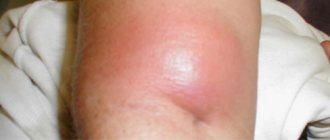
Symptoms
Symptoms and treatment of pulpitis depend on its form. It is worth noting that the signs of pulpitis of different forms are similar, although they are expressed differently.
Acute form
Acute pulpitis manifests itself in pronounced pain, which is paroxysmal in nature and especially bothers patients at night. They appear without irritants, but can sometimes be triggered by cold or hot food and water. It is worth noting that after the irritant is eliminated, the pain remains for some time. Often it covers the entire jaw. As the disease develops, the pain symptoms become pulsating, shooting, and the intervals between them become shorter and then disappear altogether.
Chronic form
Chronic pulpitis is also characterized by pain, but it is not so pronounced and occurs periodically.
In rare cases, no pain symptoms appear at all. During an exacerbation of the disease, the symptoms of chronic pulpitis are the same as in the acute form: paroxysmal pain, which is especially noticeable at night. The form of pulpitis depends on:
- the degree of pathogenicity of microorganisms that caused the disease;
- the body's immune response to the action of pathogenic microorganisms.
With age, the risk of developing chronic pulpitis increases.
Symptoms of pulpitis
How to determine inflammation with pulpitis? At an early stage, the symptoms and signs of tooth pulpitis are reduced only to a painful increase in sensitivity to irritants: hot, sour foods and drinks, cold. Subsequently, the patient begins to experience attacks of acute pain, often appearing suddenly. This means that the disease is progressing and you need to urgently consult a dentist. Often, painful sensations affect the entire side of the jaw and it is difficult to understand which tooth hurts more. In this case, you need to focus on graying enamel, bleeding gums, and the presence of a carious cavity with growing tissue.
Causes of pulpitis
Pulpitis in children and adults occurs for a number of reasons. Among them, the following stand out:
- Advanced caries. If caries treatment is not started on time, it will begin to affect the deep layers of dental tissue until it reaches the pulp. Once inside, microorganisms will cause inflammation.
- Improper treatment of caries. In this case, pulpitis occurs almost immediately after caries treatment has been carried out, and there may be several reasons for this: The dentist did not remove all carious tissues, and they continue to destroy the tooth;
- During the treatment, the pulp received a thermal burn due to insufficient cooling;
- Before starting the filling, the dentist dried the bottom of the tooth cavity with an air jet;
Indications
Elimination of pathology using a biological method is recommended in the following cases:
- Fibrous pulpitis without pain.
- Deep caries.
- Acute pulpitis with discharge of serous pus.
- Hyperemia of the pulp due to the spread of caries.
- Opening the pulp during the treatment of a carious cavity.
Surgical intervention is permissible only if there are no results with conservative treatment. Tooth extraction is carried out when complications arise after surgery and in the absence of positive dynamics.
Contraindications
Contraindications to conservative treatment:
- Exacerbation of the disease.
- Location of carious tissues in the cervical area.
- Purulent-necrotic form of pathology.
- Intolerance to medications and anesthesia.
Symptoms of rare types of pulpitis
Traumatic pulpitis. It differs from the classical forms of pulpitis in the cause of its appearance: the neurovascular bundle is damaged due to tooth trauma. As a result of a traumatic factor, inflammation of the tooth begins, sometimes accompanied by an infection from the oral cavity.
Despite the possibility of infection, in their pure form such pulpitis is non-infectious, but causes quite typical clinical symptoms: severe pain from irritants (cold, hot), as well as spontaneous and paroxysmal pain. Traumatic pulpitis occurs often in childhood and adolescence, as well as in antisocial categories of the population.
Retrograde pulpitis is a rare form of pulpitis, which sometimes causes bewilderment at a dentist’s appointment, when there is no carious cavity, but the symptoms of pulpitis (attacks of pain, night and prolonged pain) are present. In such cases, the infection enters the tooth through a hole at the top of the root.
Common causes of retrograde pulpitis are:
- Sinusitis
- Periodontitis
- Osteomyelitis
- Actinomycosis
and etc.
Concrete pulpitis is also a rare form of pulpitis, in which there is no infectious factor in the development of spontaneous or long-term aching pain in the tooth. Inflammation of the pulp develops as a result of prolonged compression by stones - wall deposits that are present in some people in the dental canals.
It is also useful to read: How to remove a nerve from a tooth and what problems may arise during this process
Most often, in old age, the lumen of the canals is narrowed by denticles or petrification - salt deposits. Clinical signs of concremental pulpitis are often blurred: there may be prolonged aching spontaneous pain, or only from irritants (hot).
Note: How to find a diseased tooth with retrograde or concremental pulpitis...
Unfortunately, in most clinics there is no EDI that allows you to check which teeth are healthy and respond to a current of 2-6 μA, and which diseased tooth responds to 20 μA or higher. X-ray diagnostics can sometimes only help with concremental pulpitis, where areas with signs of channel blocking by accumulations of salts are identified. Almost always, the bone tissue around the root is not affected by pulpitis.
Large dental centers and research institutes have equipment for EDI, and in most clinics they can search for the causative tooth based on the patient’s complaints, pulpitis clinics, but in fact - by the “poke” method, when the patient can partially help the doctor by pointing approximately to the diseased tooth. Sometimes 1-2 healthy and innocent teeth are treated, but it is worth emphasizing the point that for any doctor, an encounter with retrograde or concremental pulpitis is a very rare phenomenon that may not occur at all during his entire practice.
Diagnosis of pulpitis
Correct diagnosis of pulpitis is a primary task for any dentist, since this disease can be confused with caries, trigeminal neuralgia and exacerbation of chronic periodontitis. Each of these diseases is characterized by pain symptoms, so in order for the diagnosis to be correct, it is necessary:
- identify the causes of pain;
- determine the frequency of pain;
- determine the intensity of attacks;
- conduct a visual inspection;
- carry out x-ray diagnostics.
What to do in case of tooth pulpitis?
First of all, at the first painful sensation, you need to go to the dental clinic to undergo a differential diagnosis. An experienced doctor will determine whether pulpitis is reversible—whether the pulp can be cured. The dentist’s task is to preserve the appearance and functionality of the entire dentition. The tissue “eaten” by caries is removed, the cavities are filled, and the tooth is restored. If irreversible processes have occurred in the pulp, it is better to remove it, clean the canals and seal it. In difficult cases (for example, with the accumulation of pus), with inflammation of wisdom teeth, access to which is difficult, removal is carried out. A tooth correctly removed due to pulpitis, or rather, the hole remaining from it, heals within 3 - 4 days.
How is pulpitis treated?
Treatment of pulpitis is carried out under local anesthesia, which helps prevent pain. The dentist removes the affected tissue and neurovascular bundle, thoroughly rinses and disinfects the canals with antiseptic agents, calibrates them, mechanically treats them, and dries them. After this, the canals are sealed using filling pastes or special pins. The process requires the use of materials that perfectly match the shape and dimensions of the canal. This will eliminate the presence of voids in which infection can develop.
Biological method - based on the creation of therapeutic pads with a high calcium content. Healthy areas of the tooth with deep carious lesions are strengthened using linings and special pastes. After this, a temporary filling is installed. The result is monitored after 2-3 months using x-rays. The lack of positive dynamics is a serious reason for surgical intervention.
Note! The biological method is not recommended for people over 30 years of age. It is great for children and teenagers due to low trauma.
Surgery - performed for local or complete removal of the pulp. In this case, the pulp in the roots is preserved and removed from the crown. This allows you to maintain tooth health and adequate blood supply. This operation is relevant in the treatment of temporary teeth that have not yet fully formed.
Treatment for exacerbation - the only correct solution is to remove the pulp, expand and clean the canals, and install a filling. Specialists use the devital or vital method, depending on the medical history. When choosing the vital method, the nerve is removed, antiseptic treatment and canal filling are performed.
The devital method allows you to achieve excellent results. The operation is performed under local anesthesia. The carious area is opened, cleaned, and an arsenic-free paste is placed inside, which is effective for a maximum of 7 days (then it is removed). Pastes with a high arsenic content are left for no more than 2 days. After devitalization, the opening and removal of dead tissue from the root canals is performed. For children and adolescents, fillings are used to restore the natural shape of the tooth. In case of severe damage, adults are given a metal or ceramic crown depending on the location of the tooth.
Causes of pulpitis
Pulpitis most often begins when exposed to natural factors, which include infection and traumatic injury. A rarer reason is an iatrogenic effect, that is, caused by the intervention of a dentist. The vast majority of cases of pulpitis result from the penetration of infectious microflora into the pulp.
Deep caries, periodontal pathology, abrasion of enamel with exposure of dentinal tubules - these are the main ways through which the infection enters the pulp chamber. Theoretically, it is also possible for microorganisms to penetrate hematogenously, through the bloodstream.
Factors of traumatic origin are cracks and fractures of teeth, chipped enamel, and damage to crowns. In all these cases, dentin and pulp are exposed, an open gate appears for any microflora, so acute pulpitis always manifests itself brightly and violently. The formation of a blood clot in a tooth cavity during trauma is very undesirable; it becomes an ideal breeding ground for the rapid proliferation of microorganisms. In most cases, necrosis and complete death of the pulp occurs within the first day.
A parallel mechanism for the development of traumatic pulpitis is a disruption of the normal blood supply to tooth tissue. This is especially true for fractures, dislocations, and contusions of teeth. Trauma and death of the capillary network inside the tooth causes pulp ischemia followed by necrosis. But if the tooth is in the growth stage, then the resulting pulpitis can heal itself, as revascularization occurs (restoration of capillaries and full blood flow inside the tooth).
Pulpitis of iatrogenic nature deserves special attention. Many dental procedures, although they do not contribute to the penetration of infection into the pulp, can cause irritation. Such pulpitis is neither infectious nor traumatic. It can develop after treating a carious cavity (its preparation and drying), when exposed to vibration, heat or cold, when filling a tooth or taking an impression for prosthetics. These causes of iatrogenic pulpitis are called physical.
There is a group of factors of a chemical nature, that is, the use of various reagents during dental procedures. These are means for cleaning, disinfecting and drying the treated carious cavity, for the antiseptic treatment of opened canals. A variety of varnishes, gaskets, filling and adhesive materials can also cause iatrogenic pulpitis.
Complications of pulpitis
Failure to see a dentist in a timely manner will lead to inflammation spreading to bone tissue and to the development of periodontitis. This will make the treatment more complicated, and the most likely outcome will be tooth extraction. This complication can also occur with improper treatment. If the doctor introduces an infection into the canals or the tightness of the fillings is insufficient, complications cannot be avoided. Experts recommend using treatment technologies with insulating curtains. Cleaning and processing of canals is carried out much better when using a visiograph, operating microscope, endomotor or binocular.
So, what is pulpitis?
Pulpitis is an inflammation of the dental pulp, although it is popularly called inflammation of the nerve of the tooth. Yes, the nerve is part of the pulp, but in addition to it, the pulp also includes venules or thin arteries, thin veins, the outer layer of the pulp is a layer of odontoblasts - specific cells that participate in the formation of dentin. That is, the pulp is a complex part of the tooth in its structure.
But for ease of understanding, we will also use this term - inflammation of the nerves of the tooth = inflammation of the pulp.
What happens to the pulp when teeth wear away?
Even the grinding of teeth affects the condition of the pulp.
What happens to the pulp when teeth wear down? From daily mechanical stress, first the enamel is worn down, then the dentin. The top layer of the tooth becomes thinner and on the pulp side, odontoblasts begin to form replacement dentin. This causes the pulp to become smaller and walled off.
Recommendations after treatment
To consolidate the result and avoid complications, follow 5 rules:
- Do not eat hard, sweet or sour foods for 2 days. Reduce the chewing load and give preference to soft foods.
- Do not consume tea, coffee, carrots, beets and other coloring foods for 2-3 days. This will lead to staining of the filling.
- Visit a specialist exactly at the time agreed upon during the consultation. You will often need to visit your doctor 2-3 months after surgery to monitor your progress.
- Visit your doctor after 7 days if the pain and burning sensation does not go away. Do not use painkillers - consult a specialist immediately.
- Prevent the development of caries and other diseases. Visit the dentist 2 times a year to eliminate pathologies at an early stage.
The occurrence of pain when eating or brushing teeth within 5-7 days after therapeutic or surgical treatment is normal. If the pain does not go away, visit a specialist.
You can undergo a course of treatment for pulpitis of any form and complexity in the dental department of the CELT clinic!
Make an appointment through the application or by calling +7 +7 We work every day:
- Monday—Friday: 8.00—20.00
- Saturday: 9.00–18.00
- Sunday: 9.00–17.00
The nearest metro and MCC stations to the clinic:
- Highway of Enthusiasts or Perovo
- Partisan
- Enthusiast Highway
Driving directions
Causes of tooth pulpitis
Pain during dental pulpitis is a consequence of inflammation in the pulp. In world practice, the number of patients with this problem reaches 20%. Inflammation is caused by an infection that penetrates the pulp through the outer bone tissue. What is the difference between pulpitis and caries? Caries, unlike pulpitis, is accompanied by the destruction of hard, rather than soft, dental tissues.
The most common causes of pulpitis are:
- caries;
- periodontal trauma during operations;
- sudden changes in temperature when consuming food and drinks;
- exposure to chemicals.
These factors cause a violation of the integrity of the bone tissue; microcracks form in it, through which bacteria easily enter the pulp. As a rule, the pathology affects the chewing teeth. Pulpitis of the anterior teeth is a rarer phenomenon, but the disease progresses faster because the layer of hard tissue is thin.
Treatment with folk remedies
The specifics of the development of pulpitis do not allow it to be cured completely using traditional methods, since the inflammation is localized in a limited space of the tooth. But it is possible to stop the development of the inflammatory process and reduce pain with the help of home remedies. Many recipes involve long-term preparation of medicinal infusions, over several days. But since inflammation usually develops rapidly, you should consider those recipes that allow you to quickly treat pulpitis at home.
- 2 tbsp. crushed willow bark is poured into 200 ml of boiling water and left under the lid for an hour. Rinse the aching tooth with the strained infusion.
- 1 tbsp. crushed laurel leaves pour 1 tbsp. boiling water and leave for an hour. After which the liquid is filtered and used for rinsing.
- Calamus and St. John's wort need to be mashed in a mortar, transferred to a glass jar and poured with alcohol so that the liquid covers the plants. After two hours, the infusion needs to be decanted. Directions for use: instill 3 drops. into the carious cavity every 3 hours.
- Brew 1 tbsp. boiling water 2 tbsp. sage The infusion, brewed for 20 minutes and subsequently strained, is used for rinsing. The procedure is performed every 2-3 hours.
- 20 g of fresh crushed ginger root are brewed with 150 ml of boiling water and left to infuse for 20 minutes. The infusion is used for baths. Single serving – 50 ml. The procedure is performed every 2 hours.
- Spruce or pine resin is used as a compress on the gum next to the sore tooth. Use the product 3 times a day until the resin is completely dissolved.
- Half a teaspoon of soda is mixed with 15-20 drops. peroxide and add 5 drops. freshly squeezed lemon juice. The paste is used to wipe a sore tooth.
- After rinsing, a garlic filling is placed in the cavity, which is prepared from garlic crushed to a homogeneous mass and beeswax, taken in the same quantity. It is not recommended to keep the filling for longer than a day.
- Do the same with propolis. As the “filling” dissolves, the procedure is repeated.
- The healer's method of treating pulpitis involves applying a garlic compress to the pulse on the arm. If the tooth hurts on the right side, then apply the compress to the left hand, if on the left side - to the right.
What does it look like on an x-ray?
Inflammation during pulpitis is practically not characterized by radiological signs, since it does not provoke changes in the hard tissues of the diseased tooth. An indirect symptom may include a deep carious cavity, which is connected to the tooth cavity. The diagnosis is made by a specialist based on the results of electrical excitability and probing.
Denticles, which are located near the walls of the dental cavity and root canal, are identified on an X-ray image in the form of dense tissues, single or multiple, which have a rounded shape. As for chronic granulomatous pulpitis, it can be easily seen on an x-ray, which has a rounded contour.
Stages of pulpitis treatment
- The doctor performs anesthesia and drills out tooth tissue softened by caries, providing access to the internal structures.
- Removing the pulp - first, the doctor amputates the pulp using a bur or excavator, then performs extirpation - removes the neurovascular bundle from the root canals using thin instruments.
- Channel processing. Using small “needles” - files, reamers - the specialist passes the canals along their entire length, expanding and at the same time treating their walls with antiseptics. In this case, everything “unnecessary” is consistently washed out of the canals: bacteria, canal particles, pus, blood impurities, pulp residues, etc. Drug treatment ensures sterility - the likelihood of complications in the future depends on how carefully it is carried out. After treatment, the doctor dries the canals, places cotton pads impregnated with antiseptics there and installs a temporary filling.
- Filling. During the second visit, the dentist removes the temporary filling and medications from the canals. The treated root canals are filled to the apex - a natural narrowing or the narrowest point. For this purpose, pastes and gutta-percha pins are used that are mixed with the patient.
- Control. X-ray diagnostics are performed to assess whether the canals are filled correctly and along their entire length. Then the doctor places a filling or inlay on the crown of the tooth.
Control makes it possible to prevent some serious errors in endodontic treatment. Removal of the filling material beyond the root apex or insufficient filling are causes of possible complications in the form of the spread of the inflammatory process to the peri-root tissue, so it is important to monitor compliance with the treatment technology.
Pulp removal with preliminary devitalization is carried out according to the same principle, but in this case an additional stage is added and two visits to the doctor are required. On the first visit, the doctor applies a special paste - arsenic or arsenic-free - to the exposed pulp horn. In the first case, you need to come for an appointment within 24 hours, and if devitalization of the pulp of a multi-rooted tooth is carried out, after 48 hours. This type of therapy is more difficult to control by the doctor, because the patient may come for an appointment later than scheduled, which increases the risk of toxic effects of arsenic on the tooth root. That is why modern dentistry suggests using alternative solutions - using a paste without arsenic in the composition.
Why does the disease occur?
Pulpitis appears in a patient as a result of many provoking factors, the main one of which is considered to be caries at the third stage. The inflammatory process in the pulp begins due to the penetration of infection (streptococci, staphylococci, lactobacilli). These pathogenic microorganisms damage not only the surface of the tooth, but also affect the nerve.
The complicated form of pulpitis is necessarily accompanied by pronounced signs of pathology and has a transient course. In this case, the patient’s condition deteriorates sharply and therefore urgent medical intervention is required. If you refuse therapy, this can provoke the formation of a fistula, that is, a fistula tract.
You can identify pulpitis yourself due to the characteristic symptoms of the disease and its stage, but as for treatment, it must be prescribed by a dentist based on the results of a preliminary examination.
Treatment of pulpitis
To avoid complications of pulpitis, it is important to start treatment in a timely manner. In rare cases, this allows you to save the pulp and avoid its extraction from the canal, but partial or complete removal of the neurovascular bundle is still more often practiced, and here’s why.
Removing the pulp allows you to completely eliminate the source of inflammation, which means preventing the spread of pathogenic microorganisms into the tissue surrounding the tooth. This is the basic principle of preserving the tooth and tissues from possible complications.
Features of comparative diagnosis of acute focal pulpitis and deep caries
Similarities:
- The tooth begins to ache from exposure to any irritant, especially cold.
- The carious cavity is closed and has considerable dimensions.
- The pain is felt in one place without spreading to other areas.
Acute focal pulpitis and deep caries are visually similar, but differ in the location of pain, irritants and duration of the painful period.
Differences:
- The focal form of the disease causes pain that occurs on its own and is interspersed with rather long periods of calm.
- A small irritant is enough to renew the pain.
- Painful sensations do not go away immediately after the cessation of exposure, but are still present for some time.
- If the patient has acute focal pulpitis, during probing he feels pain in the projection of the inflamed pulp horn, while with deep caries pain is observed in the area of the dentinoenamel border and at the bottom of the cavity.
Additional methods of treating pulpitis
Today, it is possible to ensure impeccable disinfection of root canals during treatment using modern equipment - ultrasonic, laser.
In addition, copper-calcium hydroxide depophoresis is an effective method of therapy. It has a bactericidal and fungicidal effect - it destroys pathogenic bacteria and fungi.
Physiotherapy for pulpitis is more often used as a means of treating pain after filling - it can occur due to adaptation to the material. In general, physiotherapy is used relatively rarely.
Possible causes of the disease
Dentists identify 5 reasons that can cause the development of pulpitis in humans . Among them:
- untreated caries;
- ineffective treatment;
- damage when grinding a tooth;
- gum disease;
- mechanical dental injuries.
Now we’ll look at each cause of pulpitis in more detail so that you don’t have any additional questions.
Untreated caries
An advanced form of caries in 95% of cases leads to further development of pulpitis.
A carious tooth cavity is simply an ideal place for the emergence and subsequent development of pathogenic microbes. Over time, microbes penetrate even deeper into the pulp of the tooth, causing the development of pulpitis .
It should be remembered that the depth of the carious cavity is directly related to the rate of development of pulpitis.
Incorrect dental treatment
Even timely treatment of caries does not mean that inflammation under the gums will be avoided in the future. Such situations arise especially often when all procedures are performed by young and inexperienced dentists.
That is why the success of treatment depends on the professionalism of the doctor in 95% of cases.
The most common reason for the development of pulpitis is the doctor leaving carious particles of hard tissue under the filling, which cause further development of the disease and its transition to a chronic form.
Tooth damage during grinding
In 10% of the total number of cases of pulpitis, it is caused by improper grinding of the tooth for subsequent installation of a crown. The fact is that sometimes orthopedic doctors grind living teeth under anesthesia and cause a burn to the pulp. In this case, the patient does not feel any discomfort or pain.
This may be due to hasty work by the dentist or poor cooling of the drill.
Pain due to the development of pulpitis appears only a few days after the procedure.
Gum diseases
Gum disease such as periodontal disease often causes acute pulpitis. The exposure of the tooth root as a result of the destruction of bone tissue allows pathogenic microbes to penetrate into the very depths of the pulp and provoke and provoke the development of the disease.
Periodontal disease can only be detected by taking an x-ray.
Features of the course of pathology in children
Acute pulpitis of temporary teeth develops much more rapidly than permanent teeth. In addition, the pathology develops with different dynamics. Thus, damage to the nerve of primary teeth can occur already at the stage of development of the middle stage of caries, because Dentin of milk units has a very high permeability and susceptibility to any infections. The focal form of the pathology can very quickly transform into diffuse, from acute to chronic, from serous to purulent. Moreover, with a purulent lesion, the infection can very quickly spread with lymph and blood to neighboring organs.
Despite the seriousness and rapidity of the disease, children complain of pain less often. Firstly, they are less intense, and secondly, children do not always understand and can explain what is happening to them.
The following circumstances should be a reason for parents to suspect a disease: the child has begun to eat poorly, chews mainly on one side of the jaw, complains of pain from cold and hot foods, or expresses concern not with words, but with tears. Visually, you can notice that the baby’s lymph nodes have enlarged and swelling of the cheek has appeared. Also, when dental problems arise, children often begin to sleep on the side opposite to the disease, on the left or on the right - it all depends on where the diseased tooth is located.
Important! Acute pulpitis of mammary units must be diagnosed and treated as soon as possible, because otherwise, the infection may spread to the rudiments of permanent teeth, which will provoke serious malocclusions and disrupt the order of eruption (maybe some units will not erupt at all, or will have an irregular shape or dark color).
Features of comparative diagnosis of acute focal and chronic fibrous pulpitis
Similarities:
- Patients complain of pain that appears from exposure to various factors. The pain does not disappear for quite a long time. Cold causes more intense pain.
- When probing the bottom of the cavity, the patient feels pain in one point.
Differences:
- When interviewing a patient, the dentist usually finds out that previously a patient with suspected focal pulpitis had not noticed spontaneous pain, but in the chronic fibrous form there were such phenomena.
- Pain in acute focal and chronic fibrous forms of the disease manifests itself differently: in the first case it occurs involuntarily, and in the second only during exacerbation of the disease.
- Attention! In the acute focal form, the pulp chamber is usually not opened, but in the chronic fibrous form it communicates with the carious cavity.
It may not be visible at first, but after the doctor removes dead tooth tissue and cleans the cavity, the pulp chamber can be seen.
- EDI indicators for acute focal pulpitis are up to 20 μA, and for chronic fibrous pulpitis – up to 35–40 μA.
- The duration of the disease varies: the acute focal form lasts up to twelve days, and the chronic fibrous form can last several years. Typically, pulpitis, which a doctor discovers during a patient’s routine examination, is chronic.
Kinds
There are several classifications of pulpitis according to various criteria depending on the etiology. In dental practice, the species names proposed by domestic scientists in 1989 are more often used.
Classification according to ICD-10
This division of pulpitis is official and is used for diagnosis in most dental clinics in Europe. For each type there is a code designated K04-K04.3. Pulpitis itself is designated in the classifier under the code K04.0, all subsequent numbers indicate its features and localization. Below are the types of pathology according to the international standard:
- Elementary;
- Spicy;
- Purulent;
- Chronic;
- Chronic ulcerative;
- Pulp polyp;
- Other specified pulpitis;
- Unspecified;
- Necrosis (gangrene) of the pulp;
- Pulp degeneration (this also includes denticles and petrification of the pulp chamber);
- Incorrect formation of hard tissue in the pulp.
The classification allows you to correctly read and analyze medical history prepared by dentists from another country, since the disease code is unified and accurate.
Spicy
It is considered the most common complication of deep caries. It is often accompanied by severe pain when pressing on a tooth, which intensifies at night. Usually diagnosed in adults, it practically does not occur in children. It can occur in two forms:
- Focal pulpitis . The initial stage of development of the disease lasts no more than 48 hours. The source of inflammation is always located in the part of the pulp close to the caries. Accompanied by severe outbreaks of pain, which is spontaneous. Unpleasant sensations are localized near the affected tooth. Read more about the treatment of acute focal pulpitis.
- Diffuse . Occurs when the disease spreads to the coronal and root portions of the pulp chamber. The pain spreads to different parts of the jaw apparatus, attacks lengthen and intensify. Treatment of acute diffuse pulpitis can be carried out exclusively during the first 2 weeks after the onset of symptoms. After this period, it becomes chronic.
Since the disease can be assessed by pain, which is always subjective, special scales are used in dentistry. They allow you to accurately differentiate symptoms and select treatment.
Purulent
This type of pulpitis is called “pulp abscess.” This is an acute inflammatory process in which purulent exudate accumulates in the pulp chamber or nearby space.
Appears due to a prolonged course of the disease or poor quality dental treatment. The key symptoms of purulent pulpitis are severe swelling, pain and increased sensitivity to various influences.
Chronic
Chronic pulpitis can last from 2 weeks to several years. The pain is usually dull and only worsens when exposed to heat, cold or touch. In the chronic form, the gums and the pulp itself can bleed, and the hard tissues of the tooth are destroyed. Periodic exacerbations often occur, accompanied by increased symptoms. There are several types of chronic pulpitis:
- Fibrous . The first stage of the disease, in which there are no acute manifestations. Tissue destruction occurs slowly, exacerbations are rare. The carious cavity is not always connected to the pulp; bleeding occurs periodically. For details regarding fibrous pulpitis, see here;
- Gangrenous . Formed during prolonged exposure to an infectious agent on fibrous pulpitis. Against the background of bacterial activity, the camera takes on a gray tint. Frequent attacks of pain and halitosis (bad breath) appear. The destruction of hard tissue is extensive, caries merges with the pulp chamber;
- Chronic hypertrophic pulpitis . The last stage of chronic pulpitis. The carious area is completely connected to the pulp, a granuloma or polyp is formed - a neoplasm that occupies the entire space inside the tooth. When pressed, acute pain and bleeding occurs; without pressure, pain does not occur.
In 90% of cases, chronic pulpitis is an irreversible disease and leads to tooth loss. This is why it is important to contact your dentist at the first signs of illness.
With a long course of pathology, the risk of complications increases significantly. In case of complete destruction of the pulp, tissue necrosis develops, which can only be cured surgically.
Wisdom tooth
Pulpitis on eights or wisdom teeth is common in dentistry. It is formed as a result of deep caries, as well as trauma and infection of soft tissues during the eruption process. The disease can develop due to incorrect position of the tooth in space, problems with eruption.
The greatest difficulty in treating pulpitis in this case is the inaccessibility of the third molars; they are more difficult to care for and clean regularly. All this increases the risk of infection entering the pulp chamber with the subsequent development of inflammation. Read more about the treatment of wisdom tooth pulpitis here.
Features of comparative diagnosis of acute focal and acute diffuse pulpitis
Similarities:
- Pain can appear on its own, without any external influence.
- Any irritation of the pulp causes pain.
- At night, the pain becomes stronger and does not allow the patient to sleep.
- The carious cavity is large and closed.
Focal and diffuse forms of pulpitis bring severe discomfort to the human body, but still, with diffuse pulpitis, the pain is longer lasting and is not limited to the oral cavity, spreading discomfort to the ears, eyes, and frontal part.
Differences:
- Focal and diffuse pulpitis differ in the duration of painful attacks: with the focal form, attacks of pain are much shorter than periods of calm, and with diffuse pulpitis, the patient complains of long periods of pain and short periods of calm, and attacks appear spontaneously and can last for several hours.
- Acute focal and diffuse pulpitis can be distinguished by the reaction to temperature factors: in the focal form of the disease, a person experiences pain from cold food or drink, while the diffuse form is characterized by a painful reaction of the tooth to hot, especially when the disease enters the purulent stage. Cold pain, on the contrary, decreases.
- The duration of these forms of the disease is also different: acute pulpitis in the focal form lasts no more than twelve days, and the diffuse form lasts for at least fourteen.
- Focal pulpitis is characterized by a clear localization of pain, so the patient can accurately show the problem tooth, and a feature of diffuse pulpitis is the spread of pain along the branches of the trigeminal nerve, as a result of which the patient cannot understand which tooth is hurting. The results of probing for focal and diffuse forms will be different: in the first case, pain is felt at the site of the projection of the inflamed pulp horn, and in the second - along the entire surface of the bottom of the cavity.
- Focal pulpitis responds to percussion painlessly, while diffuse pulpitis causes pain.
- Electrodiagnostic indicators also differ: focal form – up to 20 µA, diffuse – up to 30–45 µA.
How to recognize pulpitis yourself
In order to prevent serious complications in time, it is useful to be able to independently recognize pulpitis at its first symptoms. Diagnosing yourself is not that difficult.
When toothaches occur, you should first of all understand their nature: with caries there is no acute spontaneous pain, it appears only from external irritants. With acute pulpitis and its exacerbation, both of these signs are present. And with acute periodontitis, most often you cannot touch the tooth at all, your health may worsen, and hot food sharply intensifies the already unbearable throbbing pain.
Chronic pulpitis is more difficult to identify, but if it is possible to examine the carious cavity, it becomes easier to recognize pulpitis. If the carious cavity is occupied by some soft tissue, then chronic hypertrophic pulpitis most likely develops. If, when food gets into a tooth, a prolonged aching pain occurs, then there is a high probability of an exposed nerve, as with fibrous or gangrenous pulpitis.
Chronic periodontitis has much in common with chronic pulpitis, but only in chronic forms of periodontitis can there be fistulas on the gums near the diseased tooth. Only with periodontitis, the gums around the tooth can “swell” and hurt, and pus often comes out from under it.
Despite the possibilities of self-diagnosis, only a dentist can make a final diagnosis after carrying out a set of diagnostic measures (examination, palpation, percussion, thermometry, EDI, radiography).
The photo below shows a visiograph for radiography:
Prevention
The main condition for the prevention of pulpitis is compliance with oral hygiene. This primarily includes brushing your teeth. This activity should be given a few minutes a day, starting from a very early age. You need to brush your teeth in the morning and evening.
Diet is of great importance. To keep your teeth healthy, you should eat foods rich in vitamins A, B, C, D and calcium:
- Vitamin A prevents bleeding gums and prevents the development of periodontitis.
- Vitamin B is essential for healthy tooth enamel and gums.
- Vitamin C reduces the sensitivity of enamel and gums.
- Vitamin D is involved in the absorption of calcium.
- Calcium is the main element of bone tissue. Additional intake of calcium supplements along with vitamin D is beneficial.
The key to successful treatment of pulpitis is timely consultation with a doctor. Moreover, it is advisable not to wait for inflammation to affect the pulp, but to treat caries in the early stages. It is much cheaper, faster and safer.
Pulpitis
- Symptoms
- Causes
- Chronic pulpitis
- Acute pulpitis
- Treatment
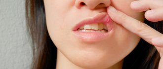
Almost every person experiences toothache. It is then that all the warnings regarding oral hygiene, caries prevention and visits to the dentist once a year come to mind. The most common cause of toothache is pulpitis.
Pulpitis is an inflammatory lesion of the soft tissues of the pulp. Pulp damage causes attacks of intense pain, which can intensify at night and torment a person for a long time without regret.
The so-called pulp is the connective tissue inside the tooth. It is in the pulp that the nerve endings and blood vessels feeding the tooth are located. The expressions “nerve hurts” and “nerve inflamed” refer specifically to the pulp.
Acute inflammation of the pulp requires consultation with a dentist who can remove it and fill the root canals through which infection could enter the tooth. A tooth treated in this way can continue to serve a person, although it will become more fragile than a healthy tooth with living soft tissue.
Symptoms of pulpitis
Despite the fact that the main companion of pulpitis is severe pain, which cannot be confused with anything, in reality this disease has similar symptoms to other pathologies. For example, external signs of pulpitis may resemble trigeminal neuralgia, advanced caries or periodontitis.
Main and general symptoms:
- 1 Acute pain syndrome when eating cold or sweet foods. The pain in this case bothers you even after eliminating the irritant.
- 2 Spontaneous attacks of intense pain that worsen at night
- 3 Acute pain can spread to the temples, radiate to the ear or to neighboring teeth
A specialist can determine the true nature of the disease based on the patient’s complaints or from a targeted photograph of the diseased tooth. For example, the localization of pain may indicate the form of the disease: with an acute focal form, it is easy to point to a diseased tooth, while diffuse pulpitis radiates to the ear or the back of the head, and then the patient feels as if several teeth hurt at once1.
Causes of pulpitis
The main cause is considered to be caries, which dissolves hard dental tissues. From the outside, a carious cavity can be practically invisible, which is why a person is in no hurry to visit the dentist until he feels acute pain.
Other reasons include:
- 1 Untreated caries. In some cases, the reasons for the development of pulpitis lie in the work of the dentist. For example, during the treatment of a carious process, a filling was installed in a patient, but the cavity was poorly treated beforehand and the filling sequence was not followed. Or, when preparing a tooth for a crown, the doctor ground it too much
- 2 Infections. Sometimes the disease can occur due to the fact that bacteria penetrate into the pulp through blood and lymphatic vessels from chronic foci of infection, for example, with sore throat or sinusitis.
- 3 Traumatic damage to teeth. A fracture of the dental crown is often accompanied by a rupture of the neurovascular bundle, which always ends in pulpitis1
Chronic pulpitis
Most often, chronic dental pulpitis is caused by the same deep caries or the penetration of pathogenic bacteria into the tooth pulp through blood vessels.
Symptoms of the chronic form:
- Periodic attacks of pain caused by cold or hot food. In this case, the pain may be delayed in nature, that is, it does not occur during eating, but after some time.
- Feeling of “heaviness” in the tooth
- Mild pain when pressing on the tooth or chewing
Despite the fact that chronic pulpitis may have mild symptoms and may not bother the patient much, it is a dangerous disease. Untreated chronic pulpitis very often causes the development of periodontitis due to the further spread of infection through the dental canals. Therefore, if pain symptoms occur, it is important to see a specialist in a timely manner.
Diagnosis of chronic pulpitis does not present any particular difficulties for a practicing dentist. To establish a final diagnosis, a specialist must review the results of an x-ray examination of the causative tooth2.
Acute pulpitis
Acute pulpitis is characterized by a bright clinical picture. The occurrence of painful attacks is associated with the formation of pus in the tooth cavity. In acute pulpitis, pain can become constant, but the most advanced stages, on the contrary, can be characterized by a decrease in pain. This is due to the destruction of nerve fibers.
Symptoms of acute pulpitis:
- The patient complains of intense spontaneous attacks of pain.
- The pain is throbbing, acute, worsens when lying down and is very annoying at night.
- Changes in temperature and contact with cold or hot can also increase pain. In this case, the painful symptom does not disappear after the removal of the stimulus.
Diagnostics includes:
- Finding out the medical history, in which the doctor clarifies the patient’s complaints
- Visual and instrumental examination of the oral cavity
- Probing of a carious cavity showing a closed pulp chamber
- Determination of tooth sensitivity to temperature stimuli in the form of cold water
- Measuring the electrical conductivity of dental tissues, which increases significantly during the inflammatory process
- X-ray, in the image a specialist examines the size and shape of the carious cavity, the presence of chronic tissue damage
Treatment of pulpitis
Treatment consists of thorough mechanical cleaning of the carious cavity, which ends with root canal filling and tooth restoration.
Treatment of pulpitis, depending on the chosen method of therapy, can be carried out in one, two or three visits. The main ways to eliminate inflammatory processes include:
- 1 Biological method. The dentist eliminates inflammation with the help of medications and physical therapy. First, the doctor cleans the carious cavity from softened dentin and pathological microorganisms. Then anti-inflammatory and hormonal agents are applied to the closed pulp cavity. The tooth remains alive
- 2 Pulp amputation. Mainly used in pediatric dentistry. This treatment allows for the preservation of the root portion of the pulp. As a result, the dental tissue is still partially nourished and remains harder than with complete removal of the soft tissue. The specialist covers the remaining pulp with a calcium-containing agent, and fixes a filling on top
- 3 Depulpation. The most common method of treatment. The pulp is removed under local anesthesia, or a special paste is first applied to necrosis the soft tissue. Then the root canal is filled with a filling mass. The service life of the sealed tooth depends on the quality and tightness of the root canal sealing3
Regardless of how pulpitis is treated, upon completion of therapy, the doctor takes an additional x-ray of the filled root canals. This allows you to promptly identify and eliminate treatment errors, avoiding the development of complications.
After completion of treatment, the patient may experience tooth pain to the touch. This symptom disappears spontaneously after 1-2 weeks.
Sometimes even cured pulpitis can cause complications. Most often, unpleasant side effects are the result of poor-quality treatment, for example, improper application of a temporary dressing or an overdose of a drug used to treat the pulp.
A sign that the treatment has caused unwanted complications is pain that persists for more than 1-2 days and is clearly manifested when biting. In this case, an urgent repeated consultation with a doctor is required, since lost time threatens the need to remove the damaged tooth.
A pulpless tooth has a shorter service life than a completely healthy one. Therefore, treatment of pulpitis itself is a last resort, which is best prevented by timely treatment of caries.
Measures to prevent pulpitis include:
- Regular visits to the dentist at least once a year
- Brushing your teeth and tongue at least 2 times a day
- Using anti-inflammatory mouthwashes and dental floss
- It is preferable to eat foods rich in fluoride, calcium, vitamin C4
Pulpitis is a disease that requires immediate treatment. Inflammation of soft tissues, in any form, is treatable, but it all depends on the stage of the disease. Therefore, timely therapy will keep the tooth as intact as possible and avoid unwanted consequences.
Sometimes inflammatory processes still become complications of pulpitis. In such cases, the doctor may prescribe medications to fight the infection or as a preventive measure after treatment. One of these drugs is Imudon®, a local immunostimulant5.
Direct indications for the use of Imudon® include oral diseases such as periodontal disease, periodontitis, stomatitis, glossitis, gingivitis, dysbiosis and infections after tooth extraction or implant installation. Imudon® activates the body's defenses (local immunity) to fight pathogens of the inflammatory process. Lysates of lactobacilli contained in the drug have an additional anti-inflammatory effect and accelerate the healing processes of oral tissue6. The drug also has a convenient form of lozenges, which ensures action at the very site of oral diseases5.
The article was prepared based on materials:
1. Danilevsky N. F. Pulpit / N. F. Danilevsky [etc.]. – M.: Publishing House of Health, 2003. – 168 p. 2. Kalinina E. S. Treatment of chronic fibrous pulpitis with preservation of pulp viability / E. S. Kalinina, P. S. Kravchuk // Bulletin of new medical technologies. – 2010. – No. 2 (17). – 201-202 p. 3. Bondarenko T. N. Comprehensive and formalized assessment of the dental status of a patient diagnosed with pulpitis in the context of the applied treatment methods / T. N. Bondarenko, S. V. Melekhov // Kuban Scientific Medical Bulletin. – 2009. – No. 7. –19-21 p. 4. Grechishnikov V.V. Prevention of periodontal complications in the treatment of acute focal pulpitis / V.V. Grechishnikov // Bulletin of the Volgograd State Medical University. – 2006. – No. 2. – 23-27 p. 5. INSTRUCTIONS FOR MEDICAL USE OF THE MEDICINE Imudon® dated 07/02/2018. 6. Kalyuzhin O.V. Topical bacterial lysates in the prevention and treatment of respiratory infections / O.V. Kalyuzhin // Practical medicine. – 2019. – No. 2 (94). – 69-74 p.
The material was developed with the support of Abbott to improve patient health awareness. The information in the material does not replace the advice of a healthcare professional. Contact your doctor.
RUIMD192193 dated 11/12/2019
To learn more
Cost of pulpitis treatment
The cost of treating pulpitis is determined by several factors, including the geography of the clinic. In a good clinic, the price already includes anesthesia, canal treatment, fillings and other materials.
The total amount is added up taking into account the number of root canals, their anatomical features, the degree of destruction of the crown and the method of its restoration, if we are talking about caries. You can find out the exact cost during a consultation with a dentist-therapist at the Dentist-K clinic.
It is important to remember that you can avoid expensive treatment by consulting a doctor in a timely manner. It is necessary to maintain hygiene and undergo professional teeth cleaning on time - this will avoid the need for pulpitis treatment.
Canal and tooth filling
With the biological method, special dental preparations are used. They are applied to a thin partition of hard tissue between the pulp and enamel. The medicine is secured with a filling. The condition of the tooth is under the supervision of a doctor. If necessary, an x-ray may be required. With this method, as with surgical treatment, one cannot avoid the procedure of tooth drilling, which is so unloved by many.
All tissue damaged by caries is removed. Further treatment methods differ. According to one of them, arsenic is applied. A few days later - a second visit to the dentist. The nerve of the tooth is removed. The canals are cleaned and sealed.
Tooth canals are cleaned and filled
When using arsenic-based pastes, there is a concern that the patient will not always be able to show up for treatment on time. In this case, the drug may have a toxic effect on the tooth root. Therefore, the patient is warned about the time when the drug needs to be removed. And if he is not able to visit the clinic, this must be done at home.
This method is effective in detecting chronic pulpitis. The decision to use arsenic is made by the doctor when, during the treatment of advanced caries, the pulp suddenly opens.
In the second case, local anesthesia is possible. Under its action, the same removal of the nerve occurs.
When the decision is made to completely extract the pulp, pain relief with anesthetics is carried out immediately. High-quality preparations allow this procedure to be carried out for a long time, even with lower molars.
Preservation of the pulp carries the risk of further development of infection. The voids in the canal allow microflora to live in comfortable conditions and subsequently affect bone tissue. Therefore, at the final stage, an x-ray is taken.
At the present stage, combined treatment is practiced, when the root pulp is removed from accessible canals, and the nerve is removed only in canals that are difficult to access for this process. With proper filling, the tooth will “live” for quite a long time.
A qualified dentist understands that it is better to save a tooth than to remove it. Therefore, he will take all necessary measures for this.
In modern conditions, when filling a tooth, materials that are guaranteed are often used.
Cleaning the canals is a rather responsible procedure; special needles are used for this. During the procedure, the patient can even feel how carefully the specialist fills the canals with filling material. With high-quality anesthesia, he will not feel pain. The doctor cleans the entire length of the tooth canals. At the same time, it treats them with antiseptic solutions.
Pulpitis on x-ray
Since the nerve is removed with needles only from the main canals, and its remains are difficult to completely eliminate, antiseptics allow you to sterilize the canal and dissolve the remains from the tooth.
Diagnostics
The first thing a dentist will do if he suspects tooth pulpitis is to ask the patient many questions: when did the first attack occur, how long did it last, whether the pain intensifies at night, and what helps relieve the pain. All these points are clarified by the doctor not out of idle curiosity, but because each form of the disease has its own symptoms.
This will be followed by a thorough examination of the oral cavity, which includes probing, palpation and percussion (tapping) of the tooth. Since it is often difficult to accurately identify the culprit of pain, temperature tests are widely used by dentists. During them, the tooth is exposed to carbon dioxide (-700C) or dichlorodifluoromethane (-400C). After sensitivity is restored, the test is repeated, and if the pain subsides from the cold, the doctor can confidently speak about the beginning of the process of pulp death.
X-ray and electroodontodiagnostics have proven themselves well in identifying pulpitis. The image allows the dentist to see exactly how close the decay is to the nerve. And EDI is considered the most accurate diagnostic method, which can accurately tell whether the soft tissues of the tooth are affected.
Interesting! According to statistics, 60% of all diagnosed pulpitis are chronic. And only 4 out of 10 people come to see a dentist in the acute phase of the disease.
Signs of illness
Typically, pulpitis manifests itself as a pain syndrome, which can have varying severity.
Depending on the manifestations of the disease, pulpitis is distinguished:
- acute - accompanied by severe paroxysmal pain (they intensify at night), occurring both spontaneously and as a result of exposure to cold or heat, the pain often radiates to the neck, ears, temples and does not go away even when taking analgesics;
- chronic – the pain is aching, almost imperceptible, and appears quite rarely.
Acute pulpitis can be serous and purulent. If a purulent process develops, the pain becomes constant and acquires a shooting and pulsating character.