Ectopic pregnancy, according to statistics, develops in 2-5% of cases and poses a serious threat to the health and life of a woman. How to determine that an embryo has begun to develop outside the uterus? What to do if the diagnosis is confirmed? How is an ectopic pregnancy treated? Is a new pregnancy possible after an ectopic one?
Normally, a fertilized egg reaches the uterine cavity and is implanted into its wall. However, in some cases, the embryo attaches and begins to develop outside the uterus: in the ovaries, fallopian tubes or abdominal cavity, that is, in one of the areas along the path of the egg after fertilization. According to statistics, ectopic pregnancy is detected in approximately 2-5% of pregnant women. In this case, the probability of repeated ectopic pregnancy increases to 15%.
Would you like to make an appointment?
Depending on the location, ovarian, tubal and abdominal (abdominal) ectopic pregnancy are distinguished, respectively. In the vast majority of cases (97%), the fertilized egg is implanted in the fallopian tubes. Treatment of ectopic pregnancy in all cases is carried out only by surgery.
Causes of ectopic pregnancy
- Developmental anomalies of the pelvic organs
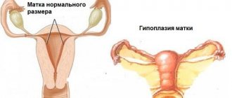
Hypoplasia (insufficient development) of the uterus and abnormal development of the fallopian tubes (too long, tortuous, thin) cause the development of ectopic pregnancy, since the oocyte is not able to reach the uterus or implant in its cavity.
- Pathological changes and dysfunction of the fallopian tubes
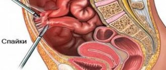
As a result of previous infections, abortions, and surgical interventions, the fallopian tubes may become deformed, and adhesions may develop. What it is? During inflammation, the outer shell of organs (peritoneum) becomes covered with fibrin, which glues adjacent tissues and thereby prevents the spread of the source of infection. Gradually, adhesions (adhesions) appear between the glued areas, which provoke deformation and displacement of organs in relation to each other. The penetration of infection into the fallopian tubes leads to the fact that peristalsis is disrupted (the inflammatory process destroys the special cilia-fimbriae that push the egg, and the muscle layer is replaced by scar tissue), resulting in their dysfunction. Deformation of the tubes leads to the fact that mechanical obstacles arise in the path of the egg, preventing it from moving further. As a result, the embryo attaches to the wall of the tube. An ectopic pregnancy develops.
- Tumors of the uterus and ovaries
An enlarged tumor provokes a displacement of the organs of the reproductive system in relation to each other, the fallopian tubes become longer and begin to contract less and less actively.
- Hormonal imbalance
A low level of the “pregnancy hormone” progesterone causes weak contractile activity of the fallopian tubes, preventing the movement of the oocyte towards the uterus.
- Other risk factors
- the use of intrauterine contraceptives (many experts argue that it is not the fact of this method of contraception that matters, but the inflammatory process that can be associated with the use of an IUD);
- smoking (according to the results of some studies, nicotine affects the function of the fallopian tubes, the ovulation process and the general state of the immune system, increasing the risk of ectopic pregnancy by 2-3 times); - endometriosis; - mature age. In some cases, it is not possible to identify the causes of ectopic pregnancy in the early stages.
Causes
Based on clinical experience, we can say with complete confidence that the following reasons can serve as an impetus for the development of tubal ectopic pregnancy:
- diseases of the reproductive system of an inflammatory nature;
- infectious diseases of the reproductive system;
- abortions;
- complicated childbirth (with the addition of an inflammatory process);
- intrauterine contraceptives;
- in vitro fertilization (IVF) procedure;
- infertility.
As a result of inflammatory or infectious damage to the fallopian tubes, swelling of their mucous membrane occurs, gluing of folds, deformation and decreased contractility.
An equally important provoking factor for the development of tubal pregnancy is infantilism. With this pathology, the fallopian tubes have an excessively elongated, tortuous shape. The lumen of such tubes is significantly narrowed, and contractility is practically absent. An atonic and narrow tube is not able to fully advance the fertilized egg to the entrance to the uterine cavity, during which implantation of the embryo occurs inside the tube.
The contingent of women who have crossed the age barrier of 36 years are at risk for developing an ectopic pregnancy.
Find out more about the causes of ectopic pregnancy.
Signs of an ectopic pregnancy
If a woman develops an ectopic pregnancy, there may be little or no signs in the early stages.
Up to 3-5 weeks from the date of the last menstruation, the patient may not notice any symptoms at all. In some cases, if a woman develops an ectopic pregnancy, early symptoms may be similar to those present during intrauterine pregnancy (delayed menstruation, breast engorgement, nausea, fatigue, increased appetite).
If the embryo is implanted into the tissue of the fallopian tube (which, as we have already written, happens in 97% of cases), the chorionic villi gradually begin to melt its walls, destroying the tissue and blood vessels. At this stage, the woman begins to experience sensations indicating an ectopic pregnancy. Depending on which part of the fallopian tube the fertilized egg is attached, signs of an ectopic pregnancy may be more or less pronounced.
- Pain in the lower abdomen, which can be acute, dull or throbbing. At first occurring sporadically, over time the pain becomes constant. If internal bleeding has already begun, the pain may radiate to other parts of the body (under the shoulder blade or into the rectum). It should be noted that pain in the lower abdomen is noted by up to 95% of patients with ectopic pregnancy;
- Unpleasant/painful sensations when urinating and defecating;
- Brown spotting from the genital tract (due to a decrease in progesterone concentration);
- Internal bleeding into the abdominal cavity (occurs in approximately 70% of cases and is accompanied by weakness, decreased blood pressure, and vomiting).
It should be noted that the clinical picture varies depending on how the ectopic tubal pregnancy is terminated.
The symptoms are most pronounced when the fallopian tube ruptures - in the case when the fertilized egg is attached to its narrow section. The patient feels acute pain, significant weakness, up to a short-term loss of consciousness. The pulse becomes weak, blood pressure decreases, and hemorrhagic shock is possible. Abdominal bloating is noted. There is a significant release of blood into the abdominal cavity.
With tubal abortion, which occurs in approximately 60-70% of cases, the clinical picture is not so obvious, and therefore 8-12 weeks may pass before surgery. Typically, the patient experiences repeated attacks of pain localized in either one or the other half of the abdomen (depending on which tube the fertilized egg was implanted in). Often this is accompanied by faintness and dizziness. In the intervals between attacks, the woman’s condition returns to normal. Internal bleeding in this case may either be completely absent or be much less profuse. The fact is that there is not a one-time rupture of the tissue of the fallopian tube in a certain area, but gradual damage to individual vessels. At the site of the rupture of the vessel, fibrin is formed, which “clogs” the damage and prevents the development of bleeding.
Ectopic pregnancy: how is it classified?
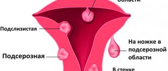
An ectopic (ectopic) pregnancy is a pathology characterized by the fact that the embryo is localized and grows outside the uterine cavity. Depending on where the implanted egg was “located,” tubal, ovarian, abdominal, and pregnancy in the rudimentary uterine horn are distinguished.
Pregnancy in the ovary can be of 2 types:
- one progresses on the ovarian capsule, that is, outside,
- the second directly in the follicle.
Abdominal pregnancy occurs:
- primary (conception and implantation of the egg to the internal organs of the abdominal cavity occurred initially)
- secondary (after the fertilized egg is “thrown out” of the fallopian tube, it attaches to the abdominal cavity).
Case study: A young nulliparous woman was brought to the gynecology department by ambulance. All the symptoms of bleeding into the abdominal cavity are present. During a puncture of the abdominal cavity, dark blood enters the syringe through the pouch of Douglas of the vagina. Diagnosis before surgery: ovarian apoplexy (no missed period and test negative). During the operation, an ovary with a rupture and blood in the abdomen are visualized. Ovarian apoplexy remained as a clinical diagnosis until the histological results became known. It turned out that there was an ovarian pregnancy.
Diagnosis of ectopic pregnancy
Diagnosis of ectopic pregnancy involves:
- taking anamnesis;
- examination by an obstetrician-gynecologist, during which a dough-like tumor-like formation is determined on one side of the uterus. In addition, the doctor determines the size of the uterus and correlates it with the duration of pregnancy;
- transvaginal ultrasound of the pelvic organs, in which the fertilized egg is not detected in the uterine cavity;
- blood test to determine the level of hCG in dynamics - what matters is the increase in the level of the hormone against the background of the absence of the fertilized egg in the uterine cavity;
- possible additional studies (clinical blood test, puncture through the posterior vaginal fornix, diagnostic laparoscopy).
Why is it developing?
Ectopic tubal (with the placement of the fertilized egg outside the uterus) pregnancy is a pathological process provoked by a number of reasons. The main one is a violation of the movement of the fertilized egg due to the abnormal structure of the tubes, inflammation, and the use of the uterine device.
More often, the main role in the appearance of pathology is played by acute salpingitis or inflammatory processes in the organ. Thus, after treatment of inflammation of the appendages, the likelihood of becoming pregnant ectopically with the localization of the fertilized egg in the fallopian tube increases several times. Poor patency of the tubes with impaired synthesis of substances responsible for the unimpeded “delivery” of the egg to the uterus disrupts the functionality of the organ, which is also a prerequisite for the development of pathology.
***
Women who use an intrauterine device after several abortions with a history of unsuccessful pregnancies are at risk. Additional risk factors are neoplasms in the uterus and appendages, organ infantilism and endometriosis.
Treatment of ectopic pregnancy
Treatment of ectopic pregnancy involves surgery. Depending on the specific situation, the following can be done:
- Tubotomy – cutting of the fallopian tube. As part of this organ-preserving operation, a tube is cut, the fertilized egg is extracted and removed.
- Tubectomy is the removal of the fallopian tube, the functions of which cannot be preserved.
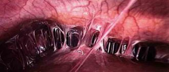
As a rule, laparoscopic intervention is practiced for ectopic pregnancy, but in case of extensive bleeding, abdominal surgery is performed.
Treatment of ectopic pregnancy in the early stages should be carried out as quickly as possible. Therefore, if a woman notices the characteristic signs described above, she should immediately consult a doctor.
Principles of treatment
Treatment is surgical only:
- Laparoscopy - through three small punctures in the abdominal wall, the fertilized egg is removed; one tube may have to be removed, but in some cases it is possible to save both tubes.
- Laparotomy - this intervention is rarely performed, only in severe cases and with severe bleeding. In this case, the fallopian tube is completely removed.
As for rare forms of ectopic pregnancy, the scope of surgical intervention is determined individually, in accordance with where exactly the embryo is attached. Both complete removal of the affected organ and organ-preserving surgery are possible.
Pregnancy after an ectopic pregnancy
Many patients ask the question of whether it is possible to get pregnant after an ectopic pregnancy.
Yes, of course, but before planning the next pregnancy, a woman must undergo rehabilitation aimed at restoring fertility. Doctors must prevent the possible development of adhesions and stabilize hormonal levels. In the near future after surgery, physiotherapeutic procedures, anti-inflammatory, restorative and hemostimulating drugs are prescribed. Patients are recommended to take COCs for a period of approximately three months to six months - the optimal drug and course duration are determined by the attending physician on an individual basis. Hormonal oral contraceptives are the most effective modern means of preventing pregnancy.
It is necessary to undergo a thorough diagnosis to find out all possible causes of ectopic pregnancy, including testing for infections and hormones, excluding tumors and cystic formations in the uterine cavity, and examining the fallopian tubes (or the remaining tube in the case of a tubectomy). Thus, planning a pregnancy after an ectopic pregnancy should only be done after some time. If it is impossible to restore reproductive function, IVF (in vitro fertilization) may be recommended.
Still have questions?
At what stage can an ectopic pregnancy be detected?
The disease is most easily diagnosed after the pregnancy is terminated (either a tubal rupture or a completed tubal abortion). This can happen at different times, but usually within 4 to 6 weeks. In case of further growth of pregnancy, it is possible to suspect its ectopic localization if the probable period is 21–28 days, the presence of hCG in the body and the absence of ultrasound signs of intrauterine pregnancy. A pregnancy that has “chosen” a place in the embryonic horn of the uterus can be interrupted later, at 10–16 weeks.
Why does the fallopian tube rupture during an ectopic pregnancy?
The fallopian tube ruptures when a woman does not go to the doctor, although she knows that she is pregnant. It is untimely registration that in 80% of cases leads to this internal catastrophe against the background of an ectopic pregnancy. A woman does not take the necessary tests, does not undergo an ultrasound scan in a timely manner, etc. If the patient comes to see a doctor early, he will be able to suspect an ectopic pregnancy, which will help avoid serious health complications.
The fallopian tube, unlike the uterus, is not capable of stretching to take on the size of a growing fetus. The tubes consist of dense muscles and connective tissue, so they are absolutely unsuitable for carrying a child. As the embryo grows, the walls of the tube stretch and it bursts.
Signs and symptoms
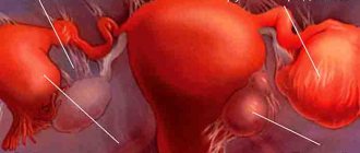
The most common form of ectopic pregnancy is tubal. 98% of all ectopic pregnancies occur in the fallopian tube. In this case, implantation of the embryo occurs not in the uterine cavity, but in the tube (see figure).
This dangerous condition occurs when the egg fertilized in the fallopian tube fails to reach the uterus in time.
The main cause of ectopic pregnancy is blockage of the fallopian tube or disruption of its contractions.
This can happen when:
- inflammatory processes in the genitals (ovaries and tubes) - for example, after an abortion;
- congenital underdevelopment of the fallopian tubes;
- hormonal disorders;
- tumors of the internal genital organs
Regardless of the form, an ectopic pregnancy is accompanied by all the signs of a normal pregnancy:
- cessation of menstruation,
- toxicosis,
- enlargement of the mammary glands.
In addition to these symptoms, ectopic pregnancy often causes severe, day-by-day pain and colic in the lower abdomen , as well as sometimes unusual discharge , which should not be confused with menstruation. An embryo that has begun to develop in the tube, unfortunately, has no chance. The fallopian tube cannot replace his uterus, and the tube itself cannot stretch as well as the uterus in accordance with the growth of the baby.
What is the danger of ectopic pregnancy?
Ectopic pregnancy is scary due to its complications:
- severe bleeding – hemorrhagic shock – death of a woman
- adhesions in the pelvis
- secondary infertility
- inflammatory process and intestinal obstruction after surgery
- recurrence of ectopic pregnancy, especially after tubotomy (in 4–13% of cases)
Case study: A woman was admitted to the emergency room with classic symptoms of an ectopic pregnancy. During the operation, the tube was removed from one side, and upon discharge the patient was given recommendations: to be examined for infections, treated if necessary, and to abstain from pregnancy for at least 6 months (the pregnancy was desired). Less than six months have passed, the same patient is admitted with a tubal pregnancy on the other side. The result of non-compliance with the recommendations is absolute infertility (both tubes were removed). The only good news is that the patient has one child.
Is it necessary to save the fallopian tubes during an ectopic pregnancy?
Since an ectopic pregnancy requires emergency surgery, doctors most often perform a salpingectomy, which involves removing the fallopian tube. It is not possible to save it, since it usually has serious deformations. Even if the appendage is retained, the next pregnancy carries a high risk of being ectopic again.
However, sometimes the surgeon decides not to remove the tube, but to make a longitudinal incision on it, remove the fertilized egg and apply stitches. It is possible to preserve the appendage when the fertilized egg has a diameter not exceeding 5 mm, the woman feels well and expresses a desire to have children in the future.
In some cases, the fertilized egg can be simply squeezed out or sucked out of the appendage. But fimbrial evacuation is possible only if the embryo is located in the ampullary part of the tube.
Another option for surgery to remove the fallopian tube while preserving the appendage is its resection. In this case, part of the pipe is cut out and the ends are sewn together.
If ectopic pregnancy is diagnosed in the early stages of its development, then it can be eliminated with medication. In this case, Methotrexate is injected into the epididymal cavity, which promotes the dissolution of the embryo. Access to the tube is gained through the posterior vaginal fornix. The entire procedure is carried out under ultrasound guidance.
In order for a woman to have the opportunity to become pregnant naturally after surgery, the following conditions must be met:
- To prevent the formation of adhesions, the patient should be raised early and given the opportunity to move. Physiological treatment is also indicated for the woman.
- Rehabilitation after surgery must be high-quality and comprehensive.
- It is important to prevent the development of infectious processes after surgery.
Early fallopian tube rupture
Sometimes a rupture of the fallopian tube occurs during an ectopic pregnancy of less than a month. However, this happens extremely rarely and only when the fertilized egg is fixed in the narrowest part of the fallopian tube (in its isthmic part). This situation threatens that the appendage may burst even before the woman finds out about her situation. In this case, it will be possible to save the patient’s life only if she seeks medical help in a timely manner. Therefore, if signs indicating a rupture of the fallopian tube appear, you need to call a medical team as quickly as possible.
If the victim gets to the hospital on her own, then it is best for her to be in a horizontal position. This will reduce pain and reduce bleeding.
