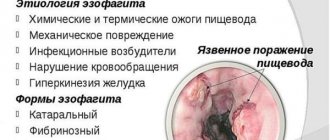
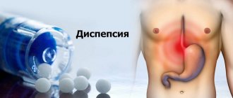
Etymology of the condition and its causes
An increase in blood flow to the vessels located near the surface of the gastrointestinal tract of the stomach provokes edema. This happens under the influence of external and internal factors. The mucous membrane is markedly swollen and painful. At the initial stage, the patient does not express complaints. It is possible to notice changes in the human stomach only on the basis of data from an endoscopic examination.
On a note! Doctors say that a hyperemic mucous surface of the stomach is a harbinger of many diseases. Some of them pose threats to human life and health. It is advisable to regularly undergo preventive gastroscopic examination.
The size of the focus of inflammation depends on the intensity of the factor that provoked a sharp increase in blood flow to the vessels. In a healthy person, the surface of the gastrointestinal tract has a pronounced pale pink color. It reflects the glare of a device called an endoscope. The doctor assesses the condition of the mucous membrane based on how severe the hyperemia is. Based on the analysis of the data obtained, the physician records the causes of excess blood flow:
- a sharp increase in pressure in large vein trunks;
- mechanical impact on the mucous surface, which led to irritation;
- excess body weight;
- insufficient blood flow rate provokes an active form of hyperemia;
- excessive blood flow leads to passive hyperemia;
- excessive activity of the heart muscle, which directs excess blood volume into the vessels;
- pathology of work in the cells of the central nervous system - dilation of blood vessels occurs and at the same time paralysis of the nerves that are responsible for the narrowing of blood vessels.
The disease can end not only with ever-increasing swelling, but also with recovery. Patients diagnosed with an active form of pathology have a greater chance of regaining lost health. Patients with a passive form of hyperemia require constant monitoring. There is a high risk of slowing down the restoration of cells on the surface of the gastrointestinal tract.
Physiology of the antrum
The specific function of the antrum is to finally grind food into a pulp state so that the maximum size of a food particle is no more than 2 mm. During mechanical grinding there is also constant mixing of food. After receiving a portion of uniform consistency, the food mass rushes through the pyloric sphincter and undergoes further processing in the duodenum.
The mechanical function of the antrum is not the only one. If the main section of the stomach produces more hydrochloric acid, then the task of the antrum is reduced to neutralizing acidity by producing alkaline mucus, concentrated in the pylorus area. This action is necessary to prepare the food mass for processing in an alkaline environment that will be created in the duodenum. The transition from acidic contents to an alkaline environment should not be too abrupt.
Another function of the antrum should be considered endocrine: individual cells produce the hormone gastrin, which has an effect on hydrochloric acid.
Insufficient gastric peristalsis contributes to food stagnation, fermentation and rotting, which causes the acidity of the environment to increase to a greater extent. The gastric mucosa is designed for a certain acidity level, corresponding to the normal production of hydrochloric acid by the parietal cells. When acidity increases, the mucous membrane is destroyed, which is accompanied by diseases of the entire digestive system of varying degrees of severity. If the effect of too acidic gastric juice is not stopped in a timely manner, the pathological condition becomes chronic.
Clinical manifestations
Patients whose gastric mucosa is severely hyperemic rarely realize this. Some symptoms may not be related to the gastrointestinal tract at all. At the initial stage, the heart rate increases. Drowsiness or problems with urination appear. Depending on where the foci of severe hyperemia are localized, the patient experiences:
- fatigue;
- obesity;
- swelling of the face.
Atrophy of the gastric antrum mucosa
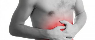
Atrophy of the antrum mucosa leads to a sharp decline in the functionality of the department, which is expressed by a number of clinical signs: diarrhea due to dysbacteriosis, intolerance to dairy products, constant flatulence and rumbling in the stomach. The patient feels an unpleasant taste in the mouth, heaviness is felt in the stomach area, but there is no sharp pain even on palpation. Typically, heaviness in the abdomen is accompanied by aching, mild pain.
In the treatment of mucosal atrophy, general means of treating stomach diseases are used, and specific drugs are also used: natural gastric juice and drugs to stimulate the secretion of hydrochloric acid.
Diagnostic stage
Patients who have hyperemic gastric mucosa undergo mandatory endoscopy. Additionally, the doctor asks for an extensive blood test. After analyzing the data obtained, the doctor may prescribe additional tests to clarify the previously made proposal:
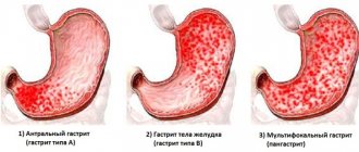
- Initial stage - the surface of the organ is covered with white foamy mucus. Partial compaction of the folds is noticeable.
- Stage II – the beginning of cell death. The surface of the gastrointestinal tract turns pale. The endoscopy results show a vascular network.
- Stage III – symptoms of the superficial form of gastritis are noticeable. The results of endoscopy show that the mucous surface and membrane are hyperemic locally or throughout. A diffuse location of lesions indicates an exacerbation of a chronic disease.
- Stage IV – signs of fibrous gastritis are noticeable. Redness is noticeable not only diffusely, but on almost the entire surface of the mucous membrane.
The results of the diagnostics allow one to draw one of the following conclusions. The first is that the patient has focal hyperemia at one specific stage. The course of treatment is aimed at eliminating the cause. The second possible conclusion is that the patient has a complicated type of pathology. This means that the increase in blood flow to the vessels is caused not only by gastrointestinal diseases, but also by a number of associated factors. For example, impaired renal function, constant stress, and depressive states provoke hyperemia.
Symptoms
If pathology begins to develop, the following symptoms appear:
- the mucous membrane becomes thinner or, on the contrary, thickened;
- redness appears;
- it swells;
- ulcers appear on the surface of the mucosa.
If the inflammatory process begins, the gastric mucosa becomes hyperemic either in one place or diffusely. Visually, upon examination, you can see that the mucous membrane is red, swollen, and blood fluid appears in the vessels.
Excessive filling of blood vessels can be a consequence of such problems:
- dysfunction of blood outflow from the walls of the stomach;
- excessive filling of the walls with blood.
By the way, active hyperemia can be considered a completely positive phenomenon, because it is a signal of recovery, and if there is a lack of blood supply and the regeneration function is inhibited, then the pathology of the stomach walls worsens. These negative phenomena are accompanied by oxygen starvation of tissues. Only with the help of an examination can a specialist determine the severity of the pathology and prescribe a competent course of treatment.
Therapeutic stage
Hyperemia of the mucous surface is a pathology that does not require immediate medical intervention. An increase in blood flow accelerates the rapid renewal of gastrointestinal tissue. If the results of the tests indicate that there is no threat to human life and health, the situation is exclusively positive. In some cases, doctors artificially provoke an increase in blood flow to speed up recovery.
On a note! If a patient has concomitant diseases, this means that he will require specific treatment. It is aimed at eliminating symptoms and subsequent prevention of gastritis, ulcers, and so on.
In situations where swelling of the mucous membrane is caused by psychological factors, further treatment is carried out by a psychotherapist. As soon as a person stops exposing himself to permanent stress, the situation returns to normal. No medications are required. The mandatory course of treatment and prevention includes diet. As you might think, there are no universal solutions. The menu is prepared by the doctor on an individual basis. General recommendations are as follows:
- the temperature of consumed food should not be less than +15 C and more than + 60 C;
- meals should be in small portions 5 to 6 times a day;
- wheat bread;
- no more than 1 egg per day;
- it is allowed to eat low-fat milk and cottage cheese (not sour);
- increase the consumption of low-fat fish varieties;
- emphasis on dietary meats: chicken, turkey;
- refusal of fast food;
- the emphasis is on boiled and stewed;
- increase the amount of vegetables and fruits consumed in your diet.
If the patient follows the doctor’s recommendations, the prognosis is favorable. After 2-3 weeks, the mucous surface returns to normal. After some time, a re-inspection is carried out. The results may mean that the pathology has been eliminated or that the patient requires additional treatment. Complications are possible in 2% of clinical cases. An increase in blood flow to the vessels provokes internal bleeding. The problem can only be solved by urgent surgical intervention.
Treatment options
In many cases, hyperemia is not treated, as it is considered a sign that the body is effectively fighting damage through self-regeneration. Hyperemia helps accelerate metabolic processes, which triggers self-healing and tissue healing. But such a diagnosis is considered the norm when it comes to arterial hyperemia. Sometimes doctors artificially induce blood flow to stimulate healing.
But more often, redness of the epithelium indicates gastritis, which is treated over a long period of time and comprehensively: diet, medication (for example, antibiotics for Helicobacter pylori infection). For gastric pathologies, the use of home remedies is indicated - herbal remedies, honey, a special diet. Diet therapy for hyperemic mucosa is based on therapeutic nutrition according to the principle of Professor Pevzner.
First aid and prevention
As soon as discomfort in the gastrointestinal tract begins to bother you, you should immediately make an appointment with a doctor. Until then, here are a few tips to follow. The first tip is to take a horizontal position. This will reduce the pain a little. The second piece of advice is to take 1 tablet of No-Spa or any other antispasmodic. If severe pain bothers you, apply a heating pad to the stomach. There is a year inside it. The cold will reduce tension.
On a note! It is strictly forbidden to apply heat to the stomach. This will only make the condition worse. There is a high risk of causing complications.
The doctor’s task is to eliminate risk factors that provoke an increase in blood flow to the vessels located near the gastric mucosa. If the patient has chronic diseases, then they should be treated. In other cases, preventive recommendations are targeted. A healthy person should avoid foods that irritate the mucous membrane:
- onion;
- radish;
- radish;
- horseradish;
- mustard;
- spices;
- spices.
Foods that cause constipation are prohibited. The more tension is required during defecation, the higher the risk of increased stress on the blood vessels. A mandatory rule is to refrain from self-administration of medications. Only a doctor can determine whether a particular medicine can be used or not. Some medications are not compatible at all, so you should not risk your health. Other recommendations:
- those with a sweet tooth should moderate their appetite;
- quitting tobacco and alcohol;
- Minimize the consumption of canned food.
Refusal of products containing a large amount of dyes.
Preventive actions
To avoid atrophy of the gastric mucosa and other complications, as well as to completely get rid of unpleasant symptoms, it is necessary to consult a doctor at the first signs of a clinical picture to make an accurate diagnosis and determine the causes of the development of the pathology.
Twice a year it is recommended to undergo diagnostics using gastroscopy to avoid recurrence of the disease. Take preventive measures prescribed by your doctor in a timely manner. Do not neglect your health, follow all the recommendations of your doctor and do not ignore unpleasant symptoms. If the disease is recognized in time, serious consequences and long-term treatment for advanced pathology can be avoided.
Dietary course according to Pevzner
The doctor may prescribe a dietary course. Manuil Pevzner’s development takes into account individual pathologies of the gastrointestinal tract. Once the doctor has determined the root cause of the disease, the doctor gives specific recommendations. The first rule is a ban on all indigestible foods. The second rule is, as mentioned above, avoiding food that negatively affects the mucous membrane. The diet includes the following elements:
- stewed poultry;
- stewed fish;
- oatmeal;
- buckwheat;
- rice;
- cream;
- condensed milk;
- fresh fruits and berries.
The Pevzner diet includes a mandatory recommendation. The listed products are consumed fresh. If this is not possible, then steaming or stewing is allowed. The temperature of food consumed should not exceed + 20 °C + 25 °C.
Stages of the malignant process
Stomach cancer can have the following stages of development:
- 1A: T1, N0, M0.
- 1B: T1, N1, M0; T2, N0, M0.
- 2: T1, N2, M0; T2, N1, M0; T3, N0, M0.
- 3A: T2, N2, M0; T3, N1, M0; T4, N0, M0.
- 3B: T3, N2, M0.
- 4: T4, N1-3, M0; T 1-3, N3, M0; any T, any N, M1.
Read here: Liver carcinoma: developmental features
T (tumor size):
- T1 – the tumor infiltrates the gastric wall to the submucosal layer;
- T2 – there is infiltration of cancer cells to the subserous layer. Possible involvement of the gastrointestinal, gastrohepatic ligament, greater or lesser omentum, but without penetration into the visceral layer;
- T3 – neoplasm that has spread to the serosa or visceral peritoneum;
- T4 – tumor growth into organs adjacent to the stomach.
N (metastases in regional lymph nodes):
- N0 – no metastases.
- N1 – metastases in 1-6 regional lymph nodes.
- N2 – regional nodes 7 to 15 are damaged.
- N3 – metastases in more than 15 lymph nodes.
M (distant metastases):
- M0 – no distant metastases.
- M1 – metastases in distant organs.
Classification
Gastritis of the antrum is divided into several types.
Catarrhal
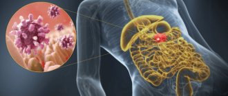
Catarrhal gastritis of the antrum is most common in medical practice and is the easiest. The inflammatory process affects only the upper layer of the epithelium. If the irritating effect ends, the person’s well-being improves. Catarrhal antral gastritis is caused by:
- Fatty and spicy foods.
- Overeating and irregular meals.
- Insufficient chewing of food.
- Poor quality products.
- Alcohol poisoning.
- Incorrect use of certain medications.
Basically, the catarrhal form of the disease occurs in an acute form. Swelling, thickening, redness and small areas of hemorrhage can be observed in the mucous membrane.
Diffuse
This type of gastritis differs in that the entire surface of the mucous membrane is affected. When food enters the stomach, it takes a long time to digest, irritating the walls of the organ. The patient appears:
- Heartburn.
- Feeling of heaviness.
- Nausea.
- Vomit.
- Diarrhea.
- Fatigue and increased irritability.
Before starting therapy for diffuse antral gastritis, fasting and taking antibacterial drugs are prescribed. Hunger and antibiotics allow the gastric epithelium to be restored. After fasting, a person is prescribed a diet. Antibacterial and anti-inflammatory therapy continues. The danger of this form of gastritis lies in its rapid progression and development of complications.
Surface
Superficial gastritis is one of the forms of chronic gastritis. It is characterized by atrophy and inflammation of the mucous membranes in the antrum of the stomach. Most often, the disease occurs due to uncontrolled use of non-steroidal anti-inflammatory drugs. These include:
- Aspirin.
- Diclofenac.
- Paracetamol.
- Nimesil.
Follicular
A special type of disease in which follicles appear on the walls of the stomach. This position promotes the accumulation of large amounts of lymph. Doctors say that this form of pathology appears against the background of chronic antral gastritis.
When the epithelium of an organ is damaged, the body’s protective function is activated, as a result of which lymph is directed to the damaged tissue. This promotes the production of antibodies that can eliminate pathogenic bacteria. However, this interferes with the appearance of gastric juice. A large amount of lymph leads to the formation of follicles, which cover the mucous membrane with a dense layer. The production of gastric juice deteriorates and after a while stops completely.
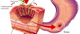
Erosive
Erosive antral gastritis is a chronic disease characterized by the appearance of surface defects on the gastric epithelium. The main difference between this form and catarrhal form is that against the background of tissue swelling, foci of erosion form on the mucous membrane. They lead to ulcers and bleeding.
Hyperplastic
A pathology in which thickening and proliferation of the inflamed mucous membrane of the organ occurs, occurring with low acidity of gastric juice. Decreased acidity is caused by excess mucus production. The disease promotes the development of tumors, and there is a high chance of the formation of a malignant neoplasm.
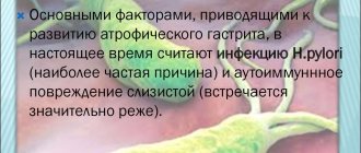
Atrophic
With atrophic gastritis, working cells die and are replaced by others. Modified glands produce mucus instead of gastric juice. The death of parietal cells without subsequent restoration begins in the antrum of the stomach. Over time, the pathology progresses, the functioning glands are replaced by a layer of epithelium. Atrophic gastritis in its chronic course is dangerous due to the appearance of malignant tumors.
What is the problem
If the results of gastroscopy indicate that the gastric mucosa is focally hyperemic, this indicates the development of the initial stage of the inflammatory process in the gastric walls. This is not a separate disease, but an additional symptom of the main pathological processes developing in the epigastric region.
It is very important to consult a doctor in time, do not ignore pain in the upper stomach, nausea, and heartburn. Focal hyperemia of the gastric mucosa accompanies most diseases localized in this area, but it is detected only during diagnosis.
Normally, the gastric mucosa is pink, smooth, and reflects the shine of endoscopic equipment. The thickness of the folds should not be less than 5 mm, more than 8 mm. When expanded with air, the folds should be completely straightened.
Diagnostic methods
A gastroenterologist will help diagnose the problem. He first examines the patient and collects anamnesis.
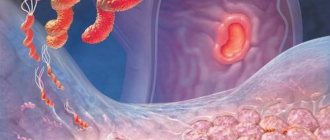
After a medical examination, a gastroscopy is performed. It is performed using a special device - an endoscope. It is equipped with viewing optics and a camera.
This diagnosis is an unpleasant and painful procedure, but it allows you to accurately determine the condition of the organ, identify the causes of hyperemia, thanks to which the doctor prescribes the appropriate treatment tactics. In addition, using this method, a biopsy is performed, i.e., tissue is taken for examination.