Keratoma is a benign neoplasm in the superficial stratum corneum of the skin. It is small in size (1–2 cm in diameter). The shade can vary from dark brown to black. Over time, the growth becomes larger and begins to peel off, turning into a plaque. Foci of the disease can appear on the face, head or body, and can be single or multiple.
These formations appear after 40 years and are equally common in both men and women. They are not contagious, and predisposition to them is transmitted at the genetic level. Sometimes they disappear or fall off on their own.
In this article we will talk about what seborrheic and senile keratomas are, what to do when they appear, what are the symptoms of the disease, whether it can be treated with folk and modern remedies and where to remove the unpleasant tumor.
Causes
The pathological process is polyetiological. The reason for the formation of neoplasms is, first of all, age-related degeneration of skin cells. The pathology is caused by two mutually opposite processes - aging and stabilization of cell activity. The lifespan of each cell depends on the balance of these processes. With age, skin cells partially lose their ability to withstand external negative factors, their RNA and DNA become vulnerable, the likelihood of tumor transformation increases, and changes in adaptation mechanisms are observed.
The main trigger for keratoma is ultraviolet radiation, the excess of which is neutralized by melanin. The pigment accumulates in the keratinocytes of the epidermis and is retained in the upper layers of the skin due to the cumulative effect. With age, melanin loses its ability to accumulate due to a slowdown in metabolic processes, while the intracellular secretion of melanin in keratinocytes increases with the simultaneous prevalence of hyperkeratosis processes. As a result, a keratoma is formed. With age, the immune system also loses some of its protective and control functions, which leads to intensive growth of epidermal cells and, as a consequence, the formation of keratinization areas.
Genetic failures provoke the formation of keratomas. In addition, the appearance of neoplasms is stimulated by neuroendocrine pathology, vitamin A deficiency, and impaired synthesis of sex hormones. Somatic diseases are important, affecting more than 70% of patients over 50 years of age. Keratoma also occurs against the background of pathogenic effects on the skin of chemicals, juices of poisonous plants, and long-term use of medications. In this case, the protective mechanisms of humoral and cellular immunity are activated, which, through macrophages and T-lymphocytes, pro-inflammatory cytokines and interleukins, activate inflammation with a predominance of proliferative processes and the development of hyperkeratosis.
Symptomatic picture of the disease
Initially, the patient notices a smooth spot on the body, the color of which varies from light yellow to dark brown. The neoplasm does not protrude above the skin and does not show a specific structure. Keratoma is characterized by slow growth, so new lesions will appear spontaneously and unnoticeably. After 50 years, the lesions tend to unite.
Over time, the spots transform into papules and nodules. They have clear boundaries and rise above the level of the dermis.
Then the papules become keratinized and become seborrheic in nature. The tumor is convex. The color becomes dark. The shape is round with a rough surface, resembling a wart. The size of the formed keratome does not exceed 3 cm.
The new growth is greasy and rough to the touch. The scales are easily separated, exposing the inside of the node. If the keratoma is accidentally caught, pain is felt and bleeding is observed. The tumor can become inflamed, which can lead to infections and fungi. Trauma occurs frequently, since the nodes are voluminous.
Classification
Benign hyperkeratotic skin neoplasms in dermatology are classified according to clinical manifestations and the degree of risk of malignancy.
There are senile, seborrheic, horny, follicular, solar keratoma and angiokeratoma.
Seborrheic keratoma
Seborrheic keratoma appears approximately as often as senile keratoma. Its characteristics are unhurried growth with the formation of multilayer crusts.
The characteristics are as follows:
- by type - spots;
- by color - yellow;
- in size - no more than 3 cm in diameter;
- by location - most often on the chest, shoulders, back, scalp.
At the same time, there is a disruption in the functioning of the sebaceous glands - because of this, the spots in the lesion are covered with loose scales, which are easily separated.
With the development of seborrheic keratoma, the spots rarely remain separate from each other - they are grouped and can sometimes merge into a single whole. Also, such spots tend to grow along the edges. Crusts grow along with them - at the same time they begin to peel off and become covered with cracks. The thickness of such crusts in advanced cases can reach 1 cm. The keratoma becomes brown and sensitive to mechanical stress - even slight damage to it causes bleeding and pain.
Seborrheic keratoma is not prone to spontaneous disappearance or malignant degeneration.
Senile (senile) keratoma
The first sign of the disease is the appearance of a light yellow or brown spot, which looks like a small hyperpigmented area of skin. As the spot develops, it acquires a darker color (burgundy, brown, gray), while increasing in size.
The growth of the neoplasm is combined with a change in its structure: it becomes loose, soft to the touch, while including keratinized cysts in the tissues of the base, which appear when the hair follicles are blocked. Infiltration, as well as accelerated growth of individual areas, leads to the formation of a lumpy surface with alternating protrusions and depressions, layers, veins, dark spots, etc. Later, the keratoma becomes rough; the layer of cells covering it flakes off, exfoliating in the form of small grayish scales.
Senile keratoma can range in size from 0.5 to 6 cm, most often 1-2 cm in diameter. Some keratomas may lighten over time, becoming pale brown or grayish. Often, age-related keratomas have a multiple distribution pattern, localized on the arms and lower extremities, face, neck, and occasionally on the body. If damaged, the tumor is prone to bleeding and inflammation, and can cause pain.
Senile keratomas, which can be treated in several ways, are often carried out in several stages, since the disease often recurs. Among the most effective methods of treating senile keratomas are:
- laser removal;
- electrocoagulation;
- radio wave method;
- cryodestruction;
- solcoderm;
- surgical excision.
If the keratoma is multiple in nature, the patient may be additionally prescribed a course of therapy with aromatic retinoids.
Other types of disease and their symptoms
In addition to age-related and seborrheic nodes, experts identify the following types of pathology:
- Follicular keratoma. This type of disease occurs near the hair follicles. The most common locations are the face, lips, and scalp. The appearance of small, colorless nodules on the body with a rough surface and a cone-shaped depression in the center is a sign of such a disease. As the papules grow, multiple clogged hair follicles (horny cysts) appear. Such a tumor is prone to recurrence even after removal.
- Solar keratoma (actinic keratosis). The main group that is susceptible to developing this form of the disease is men with fair skin. This precancerous disease is dangerous because the process of malignancy occurs hidden. At the initial stage, the patient may notice small multiple pink nodules on the body (most often on the face). As they grow, the sun seals become brown or brown in color and are covered with dense scales that come off easily.
- Angiokeratoma. This compaction does not have clear boundaries, often occurs in the scrotum area, and does not resolve on its own. Externally, it looks like a skin hemangioma. It may contain blood vessels, which is why the color of the neoplasm is blue or even black (depending on the number of capillaries). Nodes can be either single or multiple.
Large tumors covered with a thick crust can be damaged and torn off by clothing. These lumps often bleed and hurt.
Differences between keratoma and other skin pathologies and diagnosis
It is important to distinguish a senile keratoma from a pigment spot. During dermoscopic examination, horny cysts are identified on the surface of the senile neoplasm.
In old age, it is possible to develop a pigmented form of basal cell carcinoma. The disease affects the same areas of the body as with keratomas, but the edges of the papules will resemble ridges. An ulceration is noted in the middle of the neoplasm.
Seborrheic neoplasms are covered with multi-layered crusts that come off easily. The tumor is characterized by peeling and itching. The color is light yellow. Senile nodes are distinguished by the presence of black dots and veins.
Seborrheic keratoma
Keratoma is often mistaken for papilloma. The keratoma looks as if it was glued to the skin. When the second type of neoplasm appears on the body, light or pink processes with a smooth surface on the stem appear. Papillomas occur at any age. Warts are flat in shape. They arise against the background of the development of human papillomavirus. They grow from the epidermis. They have a “ragged” surface.
The malignant form of senile keratoma is extremely rare, but the risk of malignancy of the tumor cannot be excluded.
To determine the degree of cell differentiation, the neoplasm is removed. Excision of the node is directly related to histology. During the study, a section of the keratoma is examined under a microscope to determine the nature of the tumor.
Among the methods for diagnosing keratoma are:
- Ultrasonography;
- Dermatoscopy;
- Histology.
Diagnostics
A clinical diagnosis is made by a dermatologist based on medical history, symptoms of the disease and additional research data.
The priority is oncological vigilance. The appearance of a large number of keratomas on the skin, a sharp change in the color and size of neoplasms is a reason to consult a dermato-oncologist and take a biopsy.
Diagnosis uses dermatoscopy, ultrasound of keratomas, and siascanning.
Differential diagnosis
Differential (distinctive) diagnosis of keratomas is primarily carried out with such diseases and pathological conditions as:
- basal cell carcinoma (basal cell carcinoma) is a malignant skin tumor that develops from epidermal cells;
- Melanoma is a malignant tumor that occurs due to the degeneration and proliferation of pigment cells (melanocytes). It is one of the most aggressive malignant tumors of the human body;
- papilloma is a benign neoplasm of viral origin that affects the skin and mucous membranes, and has the shape of a papilla on a narrow base (pedicle);
- Bowen's disease is a neoplasm that appears inside the skin or mucous membranes and can develop into invasive cancer; hemangioma;
- nevus (or mole) is a benign pigmented formation on the skin that can be congenital or appear during life;
- wart is a pathology of a viral nature, which is characterized by the appearance of small round protruding growths on the skin; keratosis - a violation of keratinization of the skin;
- lymphangioma is a neoplasm that develops from the walls of lymphatic vessels.
Keratoma - what is it? Photo
photo of keratoma on the face
and development of education
The epidermis consists of five layers: basal, spinous, granular, shiny and the uppermost - horny. Skin diseases can affect any of them.
A keratoma is a benign skin tumor that forms from the stratum corneum, which is made up of dead cells called keratinocytes.
The latter normally gradually peel off and are washed off from the skin with water, they are replaced by new cells - these two processes occur at the same speed. If new cells of the stratum corneum are formed faster than they have time to exfoliate, then benign neoplasms are formed - keratomas. They vary in appearance and can appear in different places on the body and face.
The main cause of keratoma is the effect of sunlight on the skin. In this case, the cellular structure of the epidermis can undergo changes over many years, gradually forming a skin tumor.
An important factor is the total dose of solar radiation received by a person over a long period. Therefore, tan lovers are primarily at risk.
Other factors:
- neuroendocrine disorders;
- metabolic diseases;
- long-term use of diuretics or antibiotics;
- exposure to plant poisons and other chemical compounds on the skin;
- vitamin A deficiency;
- imbalance in the production of sex hormones;
- regular rubbing of the skin by clothing and tight underwear.
What is the danger of keratoma?
Keratomas are both dangerous and safe. This means that in general, keratomas are safe neoplasms because they are benign, but at certain moments they can become dangerous due to malignancy and their transformation into a cancerous tumor. That is, until the process of malignancy and degeneration into cancer begins in the keratome, it is safe.
Based on the fact that the keratoma itself is a safe formation, and becomes dangerous only with malignant degeneration, it is very important to monitor the condition of the tumor and record possible signs of its transformation into cancer. Currently, the signs of malignancy of keratomas are the following changes in it:
- She began to grow quickly;
- It began to bleed without injury;
- She started to itch.
This means that if these signs are detected, you should consult a doctor as soon as possible and remove the suspicious keratoma.
In addition, the danger of keratoma lies in the fact that in appearance some forms are similar to skin cancer, as a result of which even experienced doctors cannot always distinguish one formation from another. In such situations, it is recommended to remove the suspicious tumor as soon as possible and send it for histological examination. If the results of histology reveal that the formation was indeed a cancerous tumor, then for a complete recovery you should undergo a course of chemotherapy.
Finally, the indirect danger of keratomas is that with the simultaneous appearance of a large number of such tumors on the skin, there is a high probability of developing cancer in any internal organ. In such a situation, it is necessary to consult a doctor and undergo a detailed examination, which will detect a growing cancerous tumor and remove it at an early stage.
Prevention
Seborrheic keratosis of the skin can be prevented by following these recommendations:
- observe the rules of personal hygiene;
- avoid sunburn, use protection against ultraviolet rays;
- use only high-quality skin care products;
- take vitamin complexes, especially E and C;
- drink plenty of fluids;
- lead a healthy lifestyle.
Also, these preventive measures will help delay the appearance of plaques.
Seborrheic keratoma does not pose a threat to human health, but you should not self-medicate. Throughout the course of the disease, it is necessary to differentiate the neoplasm from malignant forms of the tumor.
Author: Oksana Belokur, doctor, especially for Dermatologiya.pro
How to treat keratoma?
There is no perfect cause, and therefore there is no cure as such. But after studying this problem, doctors came to the conclusion that with various complex simulations, the treatment gives results.
Methods of traditional elimination of the disease are used for 3-4 types of keratomas, the rest do not respond to this effect - they are removed.
As a conservative treatment for keratomas, the following are prescribed:
- Taking immune stimulants and antioxidants. That is, taking vitamin C in large quantities. This method does not get rid of keratosis, but it will reduce the size of the keratomas.
- Lubricating stains with hormonal ointments. This reduces the risk of contracting fungal infections due to mechanical damage.
- Fruit and vegetable diet. Proper nutrition will enrich the body with vitamins and restore the vitamin complex.
- Reducing stress. With normal health of the nervous system, the risk of keratoma developing into skin cancer is reduced.
- A way to avoid sun exposure. If you avoid ultraviolet radiation, this will not only get rid of new formations, but also improve the functioning of uninfected areas of the skin.
- Full sleep. Will provide a healthy look and rest for the whole body.
Preventive measures and forecasts
To prevent skin diseases in old age, it is recommended to avoid contact with direct sunlight. This is especially true for vacationers on the sea coast. Maximum solar activity is observed from 10 a.m. to 4 p.m. At this time, it is not advisable to leave the room. If you need to stay in the sun, it is important to use sunscreen with a high spf factor and move to shaded areas.
Preference should be given to loose, light-colored clothing made from natural fabric. A wide-brimmed hat or cap is suitable to protect your face. Glass sunglasses will keep your eye skin healthy.
Keratoma rarely affects the body of people with strong immunity. After 30 years, it is important to be physically active, perform breathing exercises, and maintain a sleep and work schedule.
Special attention is paid to the quality of food. It is recommended to include fresh vegetables or fruits in every meal. To replenish vitamin A deficiency, boiled or baked pumpkin, liver dishes, carrots, apricots and dandelion leaf salads are suitable.
If the keratoma has developed into cancer, survival rate is reduced due to the spread of atypical cells throughout the organs and systems of the human body. Fortunately, senile pathology rarely malignizes and a person lives to a ripe old age. If unknown changes appear on the body, you should consult a dermatologist to exclude malignant pathology and preserve a full life.
Surgical treatment with karat
In cases where the affected area is significant and represents a cosmetic defect that leaves no hope of cure by non-surgical methods, or treatment with traditional means has not brought the expected result, radical excision methods are recommended.
Today there are several of them:
- Radiosurgical excision. The most modern method of getting rid of skin problems. With its help, you can remove any tumors - the method has gained popularity due to the absence of any postoperative scars. The subtle and precise impact of the radioknife allows you to make any incisions and excisions. The radio wave method is especially preferable for keratomas on the face.
- Laser removal. It is a short-term effect on the center of the tumor with a beam of medical laser rays (erbium or neodymium). As a result, the body of the keratoma instantly dries out and the layers evaporate. Highly skilled execution leaves almost no subsequent scars on the skin. The duration of the operation is 10-15 minutes, healing occurs in 1-2 weeks.
- Cryodestruction. A very popular method due to its low-traumatic and painless nature. However, only single solar or seborrheic keratomas can be removed with liquid nitrogen; for other types, its use is problematic. After freezing with nitrogen, a crust forms; after a week it comes off, revealing healthy tissue. The disadvantages of the method include the inability to control the completeness of coverage of deep keratotic cells; for this reason, relapses often occur.
- Electroexcision. Excision with high-frequency current using an electric knife that burns the tumor locally. Small keratomas are removed in this way. Precision execution allows you to preserve healthy layers of skin as carefully as possible. After the procedure, a crust remains, healing in 1-1.5 weeks.
- Surgical operation. Indicated in cases of large or rapidly spreading, uncontrollable keratomas. It is also recommended in cases of degeneration into cancerous conditions - then the areas of the skin adjacent to the keratome are also excised. It is performed under anesthesia and healing occurs more slowly than with other methods.
You should choose a treatment method that is suitable specifically for a given keratoma after diagnostic procedures, assessing all the risks and consequences for the body.
Removal
For senile keratoma, the most effective method of combating neoplasms is surgery. Hospitals use different methods to remove keratoma depending on its location and size. Seborrheic keratoma can be removed using:
- surgical scalpel;
- liquid nitrogen or carbon dioxide;
- electric currents;
- radio wave radiation;
- laser light beam.
We recommend reading Basal cell carcinoma - causes, types, symptoms, treatment
Removal gives a good result and prevents relapses.
Folk remedies
As a rule, home treatment for keratomas is carried out in the period before removal and is intended to relieve the discomfort associated with the appearance of keratomas, as well as to disinfect, moisturize and relieve inflammation. A complete cure with folk remedies is impossible.
Recipes for home treatment of keratoma:
- Fresh potatoes. In addition, fresh potatoes are used. To do this, the tubers are grated. Apply the paste onto the keratomas, and place a clean cotton cloth on top. After 30–40 minutes, rinse off the product with warm water.
- Walnut balm. You can also make a balm from walnuts: mix 1 part of unripe walnut fruits with 6 parts of vegetable oil, heated to 45°C. Next, the balm is infused for 24 hours in a thermos and filtered.
- Celandine. For growing keratomas that threaten to degenerate into a malignant tumor, before surgery they are treated with celandine leaves (mix powdered dry celandine leaves with rendered pork fat). To make this ointment last longer, add 10 drops of carbolic acid to it.
- Bay leaf ointment. Therapeutic ointment from bay leaves is prepared as follows: take 6–7 bay leaves and one juniper leaf, chop and mix the ingredients. Add unsalted butter to the mixture). Oils are taken 10–12 times more than leaves. When the product is ready, add 15–20 drops of fir or lavender oil for every 100 grams. The ointment is carefully rubbed into problem areas of the skin.
- Walnut oil. When injuring a keratoma, walnut oil is used. To do this, the nut pericarps are crushed into powder and mixed with baby cream (in a ratio of 1:5). The mixture is rubbed in carefully.
- Aloe leaves. Before use, place in the freezer for 2-3 days. After thawing at room temperature, the leaves are applied to the affected area overnight. In the morning, wipe this area with salicylic alcohol. Course 3 weeks.
- Vegetable oil. To soften keratinized areas, keratomas can be gently wiped with warm vegetable oil (calcined in advance). Use sea buckthorn, fir or sunflower oil.
Bibliography
- I. A. Lamotkin. Clinical dermato-oncology: atlas/M.: BINOM. Knowledge Laboratory, 2011.
- Kennedy C, Bajdik CD, Willemze R, De Gruijl FR, Bouwes Bavinck JN. The influence of painful sunburns and lifetime sun exposure on the risk of actinic keratoses, seborrheic warts, melanocytic nevi, atypical nevi, and skin cancer. J Invest Dermatol, 2003.
- Yeatman JM, Kilkenny M, Marks R. The prevalence of seborrhoeic keratoses in an Australian population: does exposure to sunlight play a part in their frequency? Br J Dermatol, 1997
- Hafner C, Vogt T. Seborrheic keratosis J Dtsch Dermatol Ges. 2008 Aug; 6(8):664-77. doi: 10.1111/j.1610-0387.2008.06788.x.
- Vun Y., De'Ambrosis B., Spelman L., Muir JB, Yong-Gee S., Wagner G., Lun K. Seborrhoeic keratosis and malignancy: collision tumor or malignant transformation? Australas J Dermatol. May 2006; 47 (2): 106–8.
- Rajabi P., Adibi N., Nematollahi P., Heidarpour M., Eftekhari M., Siadat A.H. Bowenoid transformation in seborrheic keratosis: A retrospective analysis of 429 patients. J Res Med Sci. Mar 2012; 17 (3): 217–21.
- Thomas I., Kihiczak NI, Rothenberg J., Ahmed S., Schwartz RA Melanoma within the seborrheic keratosis. Dermatol Surg. 2004 Apr; 30 (4 Pt 1): 559–61.
- Birnie AJ, Varma S. A dermatoscopically diagnosed collision tumor: malignant melanoma arising within a seborrhoeic keratosis. Clin Exp Dermatol. July 2008; 33(4):512-3. doi: 10.1111/j.1365–2230.2008.02715.x. Epub 2008, May 6.
- Zabel RJ, Vinson RP, McCollough ML. Malignant melanoma arising in a seborrheic keratosis. J Am Acad Dermatol. 2000, May; 42(5 Pt 1): 831–3.
- Terada T., Kamo M, Baba Y., Sugiura M. Microinvasive squamous cell carcinoma arising within seborrheic keratosis. Cutis. 2012, Oct; 90 (4): 176–8.
- Mitsuhashi Y, Kawaguchi M, Hozumi Y, Kondo S. Topical vitamin D3 is effective in treating senile warts possibly by inducing apoptosis. J Dermatol 2005; 32:420–423.
- Herron MD, Bowen AR, Krueger GG. Seborrheic keratoses: a study comparing thestandard cryosurgery with topical calcipotriene, topical tazarotene, and topical im-iquimod. Int J Dermatol 2004; 43: 300–302.
- Herron MD, Bowen AR, Krueger GG. Seborrheic keratoses: a study comparing the standard cryosurgery with topical calcipotriene, topical tazarotene, and topical imiquimod. Int J Dermatol. 2004 Apr; 43(4): 300-2.
- Lebedeva UV, Davidov AB Clinical assessment of prevalence of seborrheic keratosis face skin and a neck among oncological patients. Dentistry 2009.
Ask them to me!
Free consultations
Other articles:
- Removing warts at home: a review of drugs
- Skin cancer: symptoms, stages, photos
- Removal of moles on various areas of the skin
- Vitamin D, tanning and melanoma
Useful article? Repost on your social network!
Complications of senile keratoma
Due to the slow but constant growth of the nevus in length, a noticeable cosmetic defect can form, especially if it is located in a visible place. The course of the disease can be significantly complicated by the constant appearance of new keratomas. This brings discomfort to the patient, since keratomas have a convex shape and are very easily injured.
The most severe complication of senile keratoma is considered to be its malignancy. Senile keratoma is a benign neoplasm of the skin, but it is possible to transform the keratoma into basal cell carcinoma or squamous cell carcinoma of the skin. Such a change from a benign to a malignant formation can be facilitated by regular trauma to the keratoma or excessive ultraviolet irradiation.
Treatment of keratosis using traditional medicine methods
Small neoplasms that do not change in size or appearance may shrink or disappear altogether after using the following folk remedies:
- Finely grate fresh potatoes, apply as an application to the sore spot, cover with a sterile gauze cloth and cling film for half an hour. Wash off after half an hour.
- Cut an aloe leaf and apply it to the keratome overnight as a compress. In the morning, remove the aloe and treat the skin with salicylic alcohol. Treatment course – 3 weeks.
- Castor oil is suitable for removing senile warts. They need to wipe the affected area every day, after preheating it in a water bath to a comfortable temperature.
- Celandine juice, used to remove warts, also helps in the fight against keratomas. In the cold season, you can prepare a rich decoction of the dry stems and leaves of the plant (2 tablespoons per half cup of boiling water) for applications and rubbing.
Signs of senile keratoma
The formation of senile keratoma begins with the appearance of a yellow or brownish spot on the skin. At first, the resulting spot does not differ from the color of the surrounding skin, but after some time it turns brown or red. Along with the change in color, the spot also grows, this leads to the appearance of a small papule that protrudes above the surface of the skin. The surface of the papule has small indentations and looks like a thimble. With the further development of the disease, senile keratoma acquires the characteristic appearance of a round and convex plaque. Its size in some cases can reach 6 centimeters in diameter. Sometimes the surface of the plaque becomes covered with scales. Multiple horny cysts can be localized on it, which appear due to blockage of hair follicles.
Common locations for senile keratoma are the back of the hands, face and neck area; less commonly, they can be located on the torso. The formation of keratomas is often multiple in nature, although single formations also occur.
The disease is characterized by a long course and very slow development. There were cases when it spontaneously disappeared. Some patients experience transformation of the keratoma into a cutaneous horn, which slowly grows in length.
Laser excision of senile keratoma
Removal of keratoma with a laser is carried out in two well-known ways: by laser valorization and by using a laser beam as a scalpel. The laser scalpel method allows, after excision, to conduct a histological analysis of the removed material. Laser removal is a very simple and affordable method; it is widely practiced in all clinics. The laser burns and simply evaporates the tissue. In the place where the keratoma was, a small crust will remain, which after some time will fall off on its own and healthy skin will remain in its place. Senile keratomas are removed in a few minutes.
Symptoms
Typically, a skin keratoma develops as a small, vague spot of light brown or gray color that soon begins to peel off. Gradually, the plaque increases in size and rises above the skin, resembling a nevus. The color of the spot becomes richer, and a dense crust grows on the surface. When it is torn off, the keratome becomes inflamed and bleeds.
Such symptoms in themselves are not terrible. The tumor becomes dangerous if the growth swells and hurts. This may indicate degeneration into a malignant form, although it is difficult to predict the likelihood of such a transformation.
Keratoma on the skin has several manifestations:
- pigmented spots;
- scaly plaques;
- nodules;
- skin growth.
Such keratinization of cells causes significant aesthetic discomfort to patients. Women are especially affected by keratomas.
When is medical help needed?
When the first signs of seborrheic keratosis appear, you should immediately consult a doctor to exclude the diagnosis of cancer.
If the benign nature of the keratoma is established, the doctor recommends treatment of the wart in the following cases:
- skin growths cause discomfort or itching (spread on the face, injury from clothing);
- increased risk of warts turning into skin cancer;
- presence of manifestations;
- blackening of the growth;
- active proliferation of keratomas.
If you don’t yet know which doctor treats warts, then be sure to read the material at the link.
Characteristic symptoms
As mentioned, the symptoms are largely determined by what kind of keratosis is shown in the photo. Thus, the actinic appearance is expressed by multiple formations with gray scales on open areas of the body. Seborrheic keratoma appears in the photo as single spots that crack, peel, and bleed.
Types of keratomas
This skin disease can vary both in external characteristics and in the patient’s sensations. In modern medicine, six varieties of this disease are defined.
Seborrheic keratoma
At the initial stage, the growth has a regular round shape from pale yellow to brown. Over time, the skin formation changes color, structure and grows. If the keratoma is initially smooth, then with age its surface becomes covered with keratinized and loose cells. There is also a significant increase not only in the area of the lesion, but also in its volume.
Subsequently, the surface begins to peel off. If the lesion with seborrheic keratoma is multiple, then the skin becomes uneven, with the presence of depressions and bumps. If the growths are damaged, they begin to bleed.
At the next stage, skin formations begin to change their color again. In some cases they pass without a trace. Seborrheic keratoma often occurs on the face, cervical region or hands.
Follicular keratoma
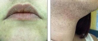
The growth is colored pink or natural flesh-colored. The shape is lumpy with an uneven surface. The size of the neoplasms is small and in rare cases exceeds a diameter of 1.5 cm. As a rule, it appears on the patient’s face and head. The cheeks and edges of the lips are especially affected. Less commonly, follicular keratoma is diagnosed on other parts of the body.
Solar keratoma
Another name is actinic. In the vast majority of cases, it manifests itself in men and is characterized by growths on the surface of which small pale gray scales are observed. Next, plaques with rich red tints appear. Actinic keratoma is localized mainly on the face and back. Often found on the hands and feet. It can either disappear on its own or reappear in the same place. The main cause of the disease is excessive exposure to the sun and other ultraviolet radiation. It can develop into a malignant tumor and for this reason can be considered a precancerous condition.
Senile
The disease is characterized by abundant formations of white or pale gray color with thin crusts. The surface of the affected skin area is sensitive to inflammatory processes. Occurs in people who have crossed the threshold of 30 years. The growths can occur all over the body, but the most vulnerable areas are the face, neck and hands.
Angiokeratoma
The only type of keratoma that can occur in newborns. It is characterized by new formations of nodules under the epidermis from red to black. Their size ranges from 1-10 mm. Agiokeratomas, in turn, are divided into papular and limited. In the vast majority of cases, the formations affect the back and abdomen. Occasionally they are found on the surface of the arms, legs, or even the human genitals.
Horny keratoma
This skin disease is known as cutaneous horn. At the initial stage, a brownish or pale yellow spot appears, which is covered with small scales. The shape of the growth can be very different, and its volume contributes to a significant elevation above healthy areas of the skin.
Horny keratoma is sometimes a secondary disease and can coexist with diseases such as tuberculosis or lupus erythematosus. Most often, the head, face and mucous membranes are affected by horny keratoma. In some cases, lesions occur on the genitals. Externally, the formations look covered with small plaques or horn-like growths.