Every year the number of women who cannot have children increases.
This is often due to pathologies of the fallopian or fallopian tubes. A little-known to non-specialists, but effective method called “hysterosalpingography” will help to identify this problem and cope with it. Let's find out the features of this method.
- What is it? Which is better: ultrasound or x-ray?
- Indications
- Contraindications
Are HSG and ultrasound the same thing?
HSG and ultrasound are two different procedures. An ultrasound examination involves an overview of the patient’s internal organs and detection of possible pathology by changes in their structure and density. The picture is displayed on the monitor. To perform an ultrasound, there is no need to perform any additional procedures. It is enough to simply lubricate the surface being viewed with a special gel.
HSG involves injecting a contrast fluid into the uterus. After its distribution, the doctor performs a series of images using an X-ray machine (but it is possible to examine the internal organs using an ultrasound machine). The introduction of a contrast agent makes the study more informative. In addition, the doctor can diagnose tubal obstruction, which cannot be done during a regular ultrasound examination.
Since two devices can be used to perform HSG: X-ray and ultrasound, there is a difference in the diagnostic process. If the pictures are taken using X-ray equipment, the procedure is called “x-ray hysterosalpingography”. When an ultrasound machine is used to perform the examination, the technique is called “echohysterosalpingography”. Because of the similarity in name, many people believe that these procedures are identical; in fact, their essence and diagnostic significance differ.
Pregnancy after HSG
HSG is a diagnostic method, not a treatment method for infertility. But after this procedure, a long-awaited pregnancy may occur, since the contrast agent often has a therapeutic effect:
- During an ultrasound examination, a saline solution eliminates small adhesions, improving the patency of the fallopian tubes.
- An oil-based contrast fluid increases the activity of the uterine glands and endometrium.
So, hysterosalpingography is a method for diagnosing and establishing the cause of infertility. Its purpose is to confirm the previously made diagnosis. But it should be remembered that this is not the main method; it requires additional analysis. If a doctor orders an HSG test for a specific reason, you need to find out from him what it is, how the procedure is performed and where it is best done.
Health Examination Uterus Infertility
Which is better: X-ray or HSG?
A standard x-ray examination will not reveal obstruction of the fallopian tubes or other pathologies of the pelvic organs, so it is never used for this purpose. HSG, on the contrary, is the method of choice for suspected tubal obstruction, endometrial polyps, uterine fibroids, endometriosis and other pathologies of the female reproductive system. Therefore, HSG is definitely better than x-ray.
However, it is worth understanding that HSG is performed either using an X-ray machine or using an ultrasound diagnostic device. A woman's internal genitalia become visible after a special contrast agent is injected into the uterus and fallopian tubes. Many specialists use the term “X-ray” to refer to the procedure for examining the fallopian tubes using an X-ray machine, and the term “HSG” refers to conducting a study using an ultrasound machine. If we consider the issue from this point of view, then GHA will be better than X-ray.
The fact is that Echo-GHA has the following advantages over RGHA:
- A woman will not have to use contraceptive methods that protect her from pregnancy if she is prescribed an ultrasound HSG.
- There are no contraindications for conceiving a child in the month when Echo-HSG was performed.
- During and after the procedure, there is no risk of an allergic reaction to the contrast agent, which contains iodine.
- The patient's body will not be irradiated by the X-ray machine. Moreover, this negatively affects the number of eggs that are present in the ovarian follicles (ovarian reserve).
Methodology
An analysis of fallopian tube patency, as hysterosalpingography is simply called, resembles a routine examination by a gynecologist. The patient is located on a special chair. The X-ray machine is above it. When performing an ultrasound, the doctor uses a vaginal sensor.
First, the doctor disinfects the external and internal genital organs with an antiseptic. Then, through a catheter, the uterus is filled with warm contrast liquid, and the device takes a picture of the cavity. Then the second part of the fluid is supplied, filling the fallopian tubes and exiting into the peritoneum if the tubes are patent. At this time, 1-2 more pictures are taken. The whole procedure takes 30-40 minutes.
If an ultrasound is performed, a different contrast agent is used, through which ultrasound waves are passed using a sensor. At the same time, the doctor examines the image on the monitor screen. These manipulations last only 15 minutes.
Many women are afraid of hysterosalpingography, believing that it is painful and harmful. But radiation in very small doses is not dangerous to health, and ultrasound analysis is generally harmless. The procedure is usually painless and done without anesthesia.
A nagging pain, as during menstruation, can be felt when a catheter is inserted and the organs are filled with fluid. This feeling goes away soon after the procedure. Nulliparous patients experience particular discomfort. The doctor may give them local anesthesia.
Important! After X-ray HSG, conception is not allowed during 1 menstrual cycle, since the contrast liquid is toxic to the fetus. Pregnancy is not prohibited after an ultrasound
.
Indications for HSG
Fallopian tube HSG is performed by gynecologists and gynecological oncologists.
Indications for the procedure are as follows:
- Tubal infertility;
- Adhesions of the pelvic organs;
- Anomalies in the development of reproductive organs;
- Sexual infantilism;
- Fibroma of the uterus, located in its submucosal layer;
- Endometrial cancer;
- Endometriotic growths in the uterine cavity;
- Polyps;
- Endometrial hyperplasia.
Hysterosalpingography is a method that will confirm that a woman has pathology of the fallopian tubes or uterus, but does not always make it possible to assess the severity of the disease and its nature. If we look at statistics, then in 98% of cases it is possible to identify an existing disorder, but a correct diagnosis can be made only in 35% of cases.
How will you feel after hysterosalpingography?
After HSG of the fallopian tubes, the consequences of this procedure may be felt for several days:
- slight vaginal discharge;
- nagging pain in the lower abdomen, which can be easily treated with painkillers;
- The stress that a woman experiences during the test can delay the onset of her period for a couple of months.
Contraindications for HSG
X-ray hysterosalpingography may not always be performed.
There are certain contraindications to the procedure, including:
- Intolerance to iodine preparations. This contraindication is due to the fact that the contrast agent, which is injected into the uterine cavity, contains iodine.
- Inflammation of the ovaries, uterus, appendages.
- Colpitis, cervicitis, bartholinitis.
- Acute infectious diseases of the body, for example, acute respiratory infections, ARVI, influenza, tonsillitis, sinusitis, etc.
- Blood clotting disorder.
- Heart diseases.
- Kidney failure.
- Severe disturbances in liver function.
- Pathologies of the thyroid gland.
- Pregnancy. A pregnancy test is required before undergoing the procedure.
- Menstrual bleeding.
- Lactation.
- Increased ESR and leukocytosis.
Indications
HSG of the fallopian tubes is performed if there are compelling indications. The procedure is prescribed by a gynecologist after examining the patient and in the absence of other methods for examining the uterus and the patency of the oviducts.
Hysterosalpingography is indicated for the following diseases and pathologies:
- infertility of unknown origin or caused by hormonal imbalance;
- suspicion of poor patency of the fallopian tubes, which leads to ectopic pregnancy or causes problems with fertilization;
- the presence of sores and inflammations in the uterine cavity (fibroids, endometriosis, etc.);
- suspicion of tuberculosis of female internal organs;
- complications after miscarriage or abortion;
- uterine hypoplasia (delayed development) or abnormalities in the structure of the fallopian tubes;
- suspicion of adhesions in the uterus or oviducts;
- preparation for artificial insemination and in vitro fertilization.
Preparation for the procedure
Preparing for the procedure is simple, but following the recommendations given by the doctor is a prerequisite. Otherwise, you can harm your own body.
So, a woman must follow the following rules in order to properly prepare for hysterosalpingography:
- 1-2 days before the proposed test, you need to avoid intimacy.
- 7 days before the procedure, you should not douche or use intimate hygiene products inserted into the vagina.
- 7 days before the study, it is prohibited to use vaginal tablets, suppositories and ointments for treatment.
- 2-3 days before the test, you need to change your diet, refusing to eat foods that cause excess gas formation. This applies to cabbage, legumes, bread, dairy drinks, and carbonated water.
- 7 days before the procedure you should stop using tampons.
- After the end of the next menstruation, partners should use a condom to avoid conception.
It is equally important to undergo a high-quality examination before carrying out GGS. It necessarily includes the following tests:
- UAC.
- OAM.
- TANK.
- Blood test for syphilis, HIV, hepatitis.
- Vaginal and cervical smears.
Before going for the procedure, you need to remove all hair from the external genitalia and wash it thoroughly. The bladder and intestines should be empty. If you were unable to go to the toilet, you should do an enema. The procedure must be carried out on an empty stomach.
As for self-administration of painkillers, it is prohibited to use medications without consulting a doctor. If prescribed by a doctor, you can take an antispasmodic, for example, No-shpa, 30 minutes before the HSG.
When is HSG performed?
Most often, hysterosalpingography is performed within 2 weeks after the next menstruation. This is explained by the fact that during this period the mucous membrane of the uterus is small in thickness, which means it does not block the entrance to the fallopian tubes.
Although, depending on the purpose of the study, it may be scheduled at other times. To assess the patency of the fallopian tubes, it is carried out in the second half of the menstrual cycle. If there is a suspicion of internal endometriosis, it is recommended to perform HGS on days 7-8 of the cycle. Myoma in the submucosal layer of the uterus can be detected at any time, but only if the woman does not menstruate.
Factors influencing the result
The purpose of HSG and its type determines what day of the menstrual cycle it is performed on. X-rays are prescribed for the first half of the cycle (preferably between the first day after menstruation and ovulation). At this time, the layer of the endometrium (the lining of the uterus) is quite thin, resulting in a more accurate image.
Ultrasound is done on different days of the cycle depending on the diagnosis that needs to be confirmed:
- patency of the fallopian tubes and isthmic-cervical insufficiency are determined in the second period of the cycle;
- endometriosis is established on the 7th day of the cycle;
- submucosal uterine fibroids are confirmed in any phase.
How is HSG performed?
If the doctor's office is equipped with a special X-ray chair, then the woman is seated on it. If there is no such chair, then the patient will be placed on a regular gynecological chair, and an X-ray machine will be brought to her.
After treating the external genitalia with an antiseptic composition, the doctor inserts speculum into the vagina and wipes the vaginal walls, first with dry cotton wool and then with cotton wool soaked in an alcohol solution. The next step is to install a tube with fixation in the cervical canal. The tube is secured with bullet pliers. When this manipulation is completed, the mirrors are removed. A contrast agent is supplied through the tube using a syringe, which must first be heated to approximately the woman’s body temperature (up to 37 °C).
When the contrast agent is evenly distributed throughout the uterine cavity and fallopian tubes, the doctor begins to take pictures. As a rule, the doctor takes from 4 to 6 pictures during the procedure. To begin with, the condition of the uterus (its relief) is recorded. Then another 4 ml of contrast agent is supplied into the cavity, which allows the appendages to be more clearly visualized. If this volume of liquid is not enough, then inject as much as necessary.
After all the pictures have been taken, the patient is transferred to the couch and left in a horizontal position for another hour. The fluid that was introduced during the procedure is absorbed into the blood and eliminated from the body by the liver and kidneys.
Technique
The tubal HSG procedure is performed under local anesthesia. No anesthesia is used. The patient is placed on a gynecological chair that does not interfere with the X-rays. The body position is the same as during gynecological operations. The external genitalia are treated with an antiseptic solution.
First, the doctor performs a manual examination, after which he examines the cervix using a speculum. Then the gynecologist inserts a tube into the cervical canal, connected to a syringe with a water-soluble contrast agent. The liquid must be warmed to body temperature to eliminate pain and cramps.
The contrast agent flows under pressure into the uterine cavity and fallopian tubes. After which a series of x-rays are taken. At the end of the hysterosalpingography, the tube is removed. The remaining contrast fluid flows out through the cervical canal and vagina.
Tubal HSG is almost painless. Patients note minor discomfort that occurs due to stretching of the uterus with a special liquid. These sensations completely disappear half an hour after hysterosalpingography. Once the procedure is complete, the woman is left to lie on the couch for an hour to minimize pain. The radiation exposure to the body does not exceed permissible standards, and therefore is safe for the patient. This is possible thanks to the use of modern medical devices.
What contrast agent is used for HSG?
To carry out the procedure, the introduction of a contrast liquid, which has the ability to block x-rays, is indicated. These are drugs such as:
- Cardiotrust. This is a contrast agent that can contain 50% and 30% iodine.
- Urotrast, Triombrast and Verografin. These are three analogues belonging to the group of radiopaque agents, which can contain 60% and 76% iodine.
Interestingly, hysterosalpingography was first attempted with Lugol's solution in 1909. But due to irritation of the peritoneal cavity and uterus, this attempt was unsuccessful. A year later, the Lugol's solution was replaced with bismuth paste, and then with argyrol and collargol. However, it was not possible to achieve the desired effect using these substances. In addition, all of them entailed inflammatory processes of the peritoneum.
It was only in 1925 that the scientist Heuser first used Lipiodol (a drug containing iodine) for hysterosalpingography. This substance made it possible to clearly visualize the condition of the uterus and fallopian tubes, and also did not harm the woman’s health. Since then, the procedure has been introduced into medical practice.
results
If there are no adhesions in the female organs, then the x-ray images will clearly show the uterus filled with fluid, the oviducts and the contrast flowing into the abdominal cavity. The conclusion of this HSG is tubal patency. If fluid retention is observed in any area, a diagnosis of “obstruction” is made. Also, based on the results of hysterosalpingography, the presence of the following diseases can be diagnosed:
- fibroids;
- polyps in the uterus;
- hydrosalpinx.
Even with a successful examination, the risk of inaccurate results remains. In some cases, an additional diagnostic procedure – hysteroscopy – may be prescribed.
Consequences and complications of HSG
A woman should use sanitary pads for 2-3 days after the procedure. This need is due to the fact that residual contrast material may leak from the vagina. If a small amount of blood is found in the discharge, then you should not worry about this, since such a phenomenon is a variant of the norm.
Mild painful sensations, reminiscent of those that occur during the next menstrual cycle, should not frighten a woman. After HSG, such discomfort does not indicate any complications.
Also, the patient should be prepared for the fact that during or several hours after HSG, a metallic taste in the mouth, dizziness, and increased heart rate may occur. This is a normal reaction of the body to the injection of a contrast agent.
A woman should not visit a sauna or steam bath, or take a hot bath for 3-4 days after undergoing HSG.
You should be concerned and immediately consult a doctor if your body temperature rises, heavy bleeding or severe pain in the lower abdomen.
Extensive blood loss, infection, perforation of the uterus and fallopian tubes during the HSG procedure are extremely rare in modern gynecological practice, as an exception.
A woman should avoid pregnancy for the next 3 months. For protection, you should use a condom.
As a rule, the procedure is well tolerated, but only on condition that the preparation for it was carried out efficiently: inflammatory processes were identified in time, and there are no other contraindications to HSG.
Hysterosalpingography (HSG)
The names of some medical procedures sound very unusual due to their foreign language origin. These include the term “hysterosalpingography” (hystera (Greek) - uterus, salpinx, salpingis (Latin) - tube, grapho (Greek) - write, draw). The procedure is a description of the condition of the uterine cavity and the patency of the fallopian tubes. This type of diagnosis helps to discover the cause of infertility. What types of this research are carried out at the Abakan DC and which of them you and your obstetrician-gynecologist can choose - this will be discussed in this article.
What is hysterosalpingography and its types The term “hysterosalpingography” is not the only one that you can come across in the direction of a doctor who prescribes you to check the patency of the fallopian tubes. There is a tradition in medical science to call this procedure differently depending on the way it is performed, or on what result the doctor wants to achieve from the diagnosis. For example, specialists often use synonyms such as “metrosalpingography” (MSG), “hydrosalpingography” or “uterosalpingography” (USG). In cases where diagnosis is carried out using ultrasound, it is called hysterosalpingosonography (HSSG, or ultrasound HSG) or ultrasound hysterosalpingoscopy (USGSS or EchoGSS). In everyday life, patients most often encounter ultrasound HSG and x-ray HSG .
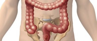
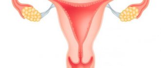
This procedure, regardless of its name and type, is prescribed to study the patency of the fallopian tubes. The fallopian tubes are a paired organ of the female reproductive system. They play the role of a kind of corridors through which a fertilized egg passes from the ovaries to the uterus. If there are adhesions, scars, crimps or constrictions along the path of the future embryo, the conception process will not be completed. It is to clarify the circumstances that impede pregnancy that the doctor prescribes HSG. Diagnosis is made by filling the fallopian tubes and uterus with a special substance. X-rays or ultrasound waves are then passed through the inflated organs. The procedure does not pose a threat to health, since the proportion of radiation from x-rays in modern devices is very small, and ultrasound, in principle, is not dangerous.
The choice of X-ray radiation or ultrasonic waves is determined by the purpose of the study and entails a fundamental difference in the methodology of the procedure.
- During ultrasound HSG, sterile saline is used as a contrast fluid, and ultrasound waves are sent through the vagina (transvaginally). The result of ultrasound passing through organs filled with saline solution is an image that is broadcast on a computer screen. The research is carried out in real time.
- When performing an X-ray HSG, the patient is also given a water-soluble drug that reflects X-ray radiation due to the content of iodine compounds. Then, using a special device, one or more photographs of the uterine cavity are taken.
Each method has its own advantages. EchoHSG does not cause such unpleasant sensations in the patient as X-ray, but has an error. For example, if during the procedure, due to a spasm, one wall of the pipe is pressed against the other, the image on the monitor will look like a commissure. But this type of diagnosis, according to doctors, produces an additional therapeutic effect. During the examination, the liquid puts pressure on minor adhesions inside the tubes and breaks them, thereby increasing the patency of the fallopian tubes. The likelihood of pregnancy increases. X-ray examination is a much more effective method than ultrasound, both in terms of the accuracy of the results and in terms of functionality: images after HSG using X-rays remain on the carrier and can be viewed by several doctors.
Note! Before any type of HSG procedure, during the period from the onset of the last menstruation until the procedure itself, you should not have sex, even protected sex. After an HSG with x-ray has been performed, conception is not allowed during one menstrual cycle, however, this rule does not apply to ultrasound examination.
The cost of ultrasound HSG in our Center is 2000 rubles, x-ray HSG is 3500 rubles. X-ray HSG is possible with the use of anesthesia (additional payment - 4500 rubles). You can sign up for studies on this website in the “ Make an appointment ” section (you leave a request and phone number, the registrars will contact you and offer to choose a convenient time) or at the reception desk of the medical appointment department by phone 8 (3902) 242-008, 242- 048.
A little more about ultrasound HSG.
Indications for carrying out One of the necessary conditions for pregnancy is the normal patency of the fallopian (or fallopian) tubes in a woman. After all, it is through these channels that the fertilized egg penetrates the uterus. If the patency is impaired, then the woman is diagnosed with infertility. In case of partial obstruction, a life-threatening condition may occur - an ectopic pregnancy. To protect a woman from such problems and assess her chances of conceiving a child, the doctor prescribes an ultrasound scan of the patency of the fallopian tubes.
Brief characteristics of the examination To assess the patency of the fallopian tubes, the patient is prescribed an ultrasound of the patency of the fallopian tubes, or, in medical parlance, hysterosalpingosonography (HSG). This is a special diagnostic test that, using an injected contrast agent, allows you to examine the female genital area. Unfortunately, conventional ultrasound is not able to provide complete information about the patency of the tubes. That is why doctors resort to a special technique that detects unpleasant pathologies. Ultrasound (HSG) of the fallopian tubes can be performed in two ways: transvaginally (by insertion into the vagina); when using an external sensor. This examination method is completely safe and very effective.
Indications for the study Ultrasound HSG of the fallopian tubes is recommended for women who have an irregular menstrual cycle (irregularity or absence of menstruation); infertility; constant pain in the lower abdomen. In such conditions, examination may be prescribed after treatment. This allows you to determine the effectiveness of the prescribed therapy and assess the woman’s condition. Ultrasound of fallopian tube patency can be performed repeatedly. After all, such a study is painless and does not harm women’s health.
Dates (*the doctor will tell you the exact date at the appointment) To get the most reliable clinical picture, you need to choose the right days. It is very important. It is recommended to conduct the study from the 6th day of the cycle to the 21st (with a 28-day cycle). It is preferable to perform an ultrasound before ovulation, from the 7th to the 12th day of the cycle. Why are these particular dates considered the most optimal? During these periods, the cervix is maximally dilated. The endometrium after menstruation is minimally thick. These features allow the study to be carried out as accurately as possible. Preparation for an ultrasound examination of the fallopian tubes for patency is a chance to identify pathology at an early stage and begin adequate therapy in a timely manner. However, in order to correctly diagnose a woman’s condition, it is necessary not only to select the appropriate timing for the procedure, but also to properly prepare for the study. The gynecologist will definitely tell you about all the necessary measures.
The preparatory stage for the examination usually includes the following activities: • Taking a smear for the vaginal microflora. • Diet 3 days before the examination. During the event, it is necessary to exclude the presence of gases in the intestines. That is why it is important to adhere to a diet for 3 days aimed at reducing fermentation in the gastrointestinal tract. Avoid starchy, sweet, and fermented milk foods. Do not consume vegetables, fruits, carbonated drinks, legumes. • Ensuring vaginal cleanliness. A week before the ultrasound, it is recommended to stop using vaginal sprays, suppositories, and tablets. Avoid douching. • Taking an antispasmodic. 20 minutes before the start of the procedure, the patient is recommended to take an antispasmodic medicine (“Spazmalgon”, “No-Shpa”). This medication will relax the smooth muscles and prevent reflex contractions of the uterus.
Carrying out an ultrasound HSG examination The procedure consists of several stages: 1. Diagnosis of the fallopian tubes begins with preliminary ultrasound monitoring of the condition of the pelvic organs. Such a study is carried out to exclude the woman’s pregnancy and the presence of inflammation. 2. The patient sits comfortably in the gynecological chair. A disposable catheter is inserted intravaginally into the cervix. Through it, a contrast agent enters the organ cavity, preheated to a comfortable temperature (37 degrees). The solution fills the uterus and travels up the fallopian tubes. The contrast agent is then moved into the abdominal cavity. 3. It is at the third stage that the analysis of pipe patency begins. Free fluid localized in the pelvis indicates satisfactory patency. It is much worse if the substance does not penetrate the peritoneum. Liquid that has not left the pipes clearly indicates channel obstruction.
The duration of the study is on average 25-30 minutes.
Examination results During the procedure, a special sensor records the presence of fluid in the peritoneum. The doctor begins the examination with the uterus. Then it determines the condition of the fallopian tubes. After this, if necessary, the examination is completed by examining the ovaries.
Ultrasound of the patency of the fallopian tubes reveals: • congenital anomalies • fibroids, polyps, endometriosis, fibroids; • adhesive processes (at the same time clearly determines the localization of pathology); • pipe contours; • location of the uterine canals.
Contraindications to ultrasound for the patency of the fallopian tubes - a fairly simple procedure. It does not involve invasive intervention and does not require complex preparation for implementation.
It is strictly forbidden to perform an ultrasound: • during uterine bleeding; • gynecological diseases occurring in acute or chronic form; violations of the vaginal microflora; • inflammatory diseases of the pelvic organs; • pregnancy; • tumors (benign, malignant) localized in the pelvis; infectious pathologies during exacerbation. Advantages of the study HSG has a number of advantages in comparison with other diagnostic methods for the patency of the fallopian tubes.
The main advantages of ultrasound are: • The procedure does not require hospitalization. • The injected liquid separates the fused areas and washes away the derivative. • During the examination, many uterine pathologies are simultaneously identified. The advancement of the egg is ensured. • The patient does not require anesthesia. • The study is carried out quickly enough, and the results are visible immediately. There is no need for peritoneal punctures.
During ultrasound, a saline solution is injected into the uterus, which has an additional therapeutic effect, for example, breaking small adhesions. That is why, after ultrasound HSG, a long-awaited pregnancy often occurs if the problem was only the presence of minor pathologies.
If you notice the first warning symptoms, do not delay your visit to the gynecologist. Be sure to seek help from specialists who, if necessary, will recommend you an effective and safe ultrasound.
Evaluation of results
The doctor must interpret the results based on the images obtained.
Signs of various diseases on an x-ray picture are as follows:
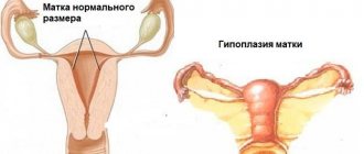
- Noticeable asymmetry of the uterus is a diagnosis of “unicornuate uterus.”
- An elongated cervix and a pronounced decrease in the volume of its cavity is a diagnosis of “infantile uterus.”
- Short, long, or asymmetrical fallopian tubes are diagnosed as “congenital tubal obstruction.”
- The presence of a flask-shaped expansion in the tubes is a possible diagnosis of “tubal adhesions”, or “sactosalpinx”, or a combination of these two diagnoses.
- The presence of light areas in the tubes is “fallopian tube adhesions.”
- The shape of the fallopian tubes, reminiscent of the shape of smoking pipes, the presence of flask-shaped extensions in them, a decrease in the volume of the uterine cavity - the diagnosis of “genital tuberculosis”.
- Uneven contours of the uterus, detection of oval or other shaped defects - diagnosis of “uterine polyps” or “endometrial hyperplasia”.
Of course, only the most obvious and common signs of certain diseases were listed. Only the attending physician can make a final diagnosis, assessing the entire range of results obtained in the course of various studies.
Interpretation of results
The doctor who performed the HSG will give the patient not only an x-ray, but also a conclusion with an interpretation (this is a transcript of the results), which in medicine is no less important than the analysis itself. With normal patency of the oviducts, the uterus on the radiograph looks like a regular triangle with the apex at the bottom and the oviducts in the form of ribbons, from which the injected liquid emerges, similar to smoke in the picture.
If the pipes are impassable, the cause of this pathology should be visible. With an x-ray and a conclusion, you need to contact your doctor, he will explain everything and prescribe treatment.
Cons of GHA
The disadvantages of the procedure are the following:
- A woman receives a dose of radiation, albeit a small one.
- The likelihood of an allergic reaction to the administered contrast agent. Particular caution should be exercised by women with a history of bronchial asthma, as well as allergic patients.
- There is a risk of mechanical damage to the epithelial layer of the uterus, which leads to the appearance of bloody discharge.
Should you be afraid of HSG?
No. And again no! There is nothing special about this procedure for a woman! Women will endure everything, survive everything. Well, they injected it in the wrong place and in the wrong place, so what? We won't survive, or what? It hurts, but not for long. And the pain is still not remembered. Those who have given birth will not even think about it; those who have not given birth, let them consider it a training session (I didn’t give birth, they had a Caesarean section). Somewhere I read a reasonable thought: this pain differs not in strength, but in localization. Cutting your hand is painful, but understandable, but there the pain is scary on a natural level, probably.
How to check the patency of the fallopian tubes?
Checking the patency of the fallopian tubes can be done in several ways:
- diagnostic laparoscopy (checking the patency of the fallopian tubes is usually carried out during surgery to remove adhesions - laparoscopy is usually not prescribed just to check the tubes);
- HSG (hysterosalpingography, MSG, metrosalpingography - other names);
- hydrosonography (ultrasound);
- fertiloscopy (a method similar to laparoscopy; often combined with it). The difference between fertiloscopy and laparoscopy is that the instruments are inserted not through the abdominal wall, but through the vagina.
How to check the patency of the fallopian tubes, which method to choose?
Taking into account the fact that both laparoscopy and fertiloscopy are traumatic methods, and ultrasound does not give a clear “picture of what is happening”, HSG in most cases is the optimal method.
Laparoscopy. How to check the patency of the fallopian tubes with its help?
Laparoscopy is a surgical method for assessing the patency of the fallopian tubes. Through punctures in the abdominal wall, internal organs are examined using optical instruments. If you are scheduled for laparoscopy, the forum can help you choose a clinic or even a surgeon.
Laparoscopy in gynecology is a method of treating and diagnosing various pathologies of the pelvic organs. Laparoscopy is one of the modern methods of surgery with minimal intervention and damage to the skin. Laparoscopies are performed for both diagnostic and therapeutic purposes.
Laparoscopy can be performed to clarify various diagnoses. If you experience pain after laparoscopy, contact the clinic where you had the operation.
Diagnostic laparoscopy is an operative research technology in which the doctor examines the abdominal organs without making large incisions on the abdominal wall. Most often, two small incisions are made. To increase the field of vision, a small amount of gas is injected into the abdominal cavity.
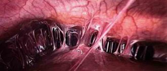
A device called a laparoscope is inserted into one incision - a thin tube with a lens at one end and an eyepiece at the other (the other end can also be connected to a video camera unit, which transmits the image to a screen). A manipulator is inserted into another incision, with the help of which the doctor displaces the abdominal organs, carefully examines them and makes a diagnosis.
Diagnostic laparoscopy is performed to assess the condition of the outer surface of the fallopian tubes and pelvic organs, as well as to identify their pathologies.
The most common operations:
- ovarian laparoscopy;
- fallopian tube laparoscopy;
- laparoscopy of the abdominal organs.
After laparoscopy:
- As a rule, the patient stays in the hospital for no more than a day: doctors monitor her condition and perform an ultrasound. After 2-3 days you can return to work.
- It is not recommended to consume alcohol or eat heavy meals in the next 2-3 weeks after surgery. — Sex should be postponed for 2–3 weeks to avoid infection.
- Physical activity should be increased evenly. It is better to start with walking and gradually increase its duration. You should not lift anything heavy after surgery.
Decoding the results
After the HSG has been done and the doctor has the pictures in his hands, conclusions can be drawn. If the pictures clearly show how the coloring agent has filled the fallopian tubes, then the patency is good. If there are adhesions in the tubes, this will certainly be reflected in the images. Also, with the help of this study, the doctor receives more information about the structure of the uterus itself. According to medical observations, after the procedure the likelihood of becoming pregnant increases several times.
View gallery
Contraindications
HSG is contraindicated:
- for general infectious processes occurring in the body (flu, rhinitis, sore throat, furunculosis, thrombophlebitis, etc.);
- hyperthyroidism;
- severe kidney and (or) liver diseases;
- insufficiency of the cardiovascular system;
- acute inflammation in the uterus, ovaries and oviducts;
- infectious inflammation of the large gland in the vestibule of the vagina;
- cervicitis (inflammation of the cervix);
- poor blood and (or) urine tests.
Absolute contraindications are pregnancy and hypersensitivity to iodine.